I have confirmed the Sembulingam chapter structure (Chapters 133-164 for nervous system) and extracted key content. The book is not in the digital library, but I have confirmed chapter numbers, topics, and some direct text extracts. Let me now compile the full notes precisely structured to Sembulingam's format and chapter organization.
NERVOUS SYSTEM - PHYSIOLOGY
As per Sembulingam's Essentials of Medical Physiology (7th/8th Edition)
CHAPTER 133 - ORGANIZATION OF THE NERVOUS SYSTEM
Central Nervous System (CNS)
The CNS consists of the brain and spinal cord.
Brain - Developmental Divisions:
| Division | Subdivision | Structures |
|---|
| Prosencephalon (Forebrain) | Telencephalon | Cerebral hemispheres, basal ganglia, hippocampus, amygdaloid nucleus |
| Diencephalon | Thalamus, hypothalamus, metathalamus, subthalamus |
| Mesencephalon (Midbrain) | - | Midbrain (corpora quadrigemina, cerebral peduncles) |
| Rhombencephalon (Hindbrain) | Metencephalon | Pons + Cerebellum |
| Myelencephalon | Medulla oblongata |
Brainstem = Midbrain + Pons + Medulla oblongata
Spinal Cord:
- Extends from foramen magnum to lower border of L1 vertebra
- Ends as conus medullaris; below this: cauda equina (horse's tail)
- Has 31 segments: 8 cervical, 12 thoracic, 5 lumbar, 5 sacral, 1 coccygeal
- Contains grey matter (H-shaped, neurons) and white matter (tracts)
Peripheral Nervous System (PNS)
Formed by neurons and processes outside CNS:
- Cranial nerves (12 pairs) - arise from brain
- Spinal nerves (31 pairs) - arise from spinal cord
PNS Subdivisions:
- Somatic nervous system - voluntary control of skeletal muscles + somatic sensory input
- Autonomic nervous system (ANS) - involuntary control of viscera, glands, smooth and cardiac muscle
- Sympathetic division (thoracolumbar)
- Parasympathetic division (craniosacral)
FUNCTIONS OF THE NERVOUS SYSTEM
According to Sembulingam, the three main functions are:
- Sensory function - Collection of information from internal and external environment via sensory receptors
- Integrative function - Processing and interpretation of sensory information; decision-making (mainly by brain)
- Motor function - Sending commands to muscles and glands to bring about appropriate responses
CHAPTER 140 - SYNAPSE
Definition
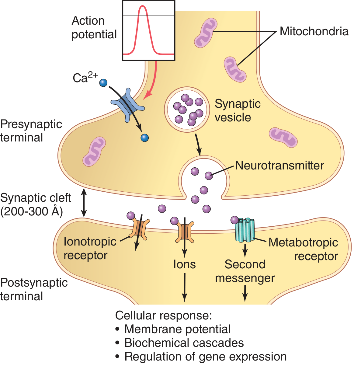
A synapse is the structural and functional junction between two neurons where information is transmitted from one neuron (presynaptic) to the next (postsynaptic).
The term "synapse" was coined by Sir Charles Sherrington (1897).
Functional Anatomy of a Synapse
Components:
- Presynaptic terminal (synaptic knob/bouton):
- Expanded end of axon of presynaptic neuron
- Contains mitochondria (energy supply) and synaptic vesicles (neurotransmitter)
- Presynaptic membrane has active zones for vesicle docking and fusion
- Synaptic cleft:
- Gap of 20-30 nm between pre- and postsynaptic membranes
- Postsynaptic membrane:
- Has receptor proteins specific for the neurotransmitter
- Subsynaptic web beneath postsynaptic membrane
Classification of Synapses
A. Based on the part of the neuron involved:
- Axodendritic - axon terminal contacts dendrite (most common)
- Axosomatic - axon terminal contacts cell body (soma)
- Axoaxonic - axon terminal contacts another axon terminal (modulates neurotransmitter release)
- Dendrodendritic - dendrite to dendrite contact
B. Based on the mechanism of transmission:
- Chemical synapse - uses neurotransmitters; most common in CNS; one-way transmission
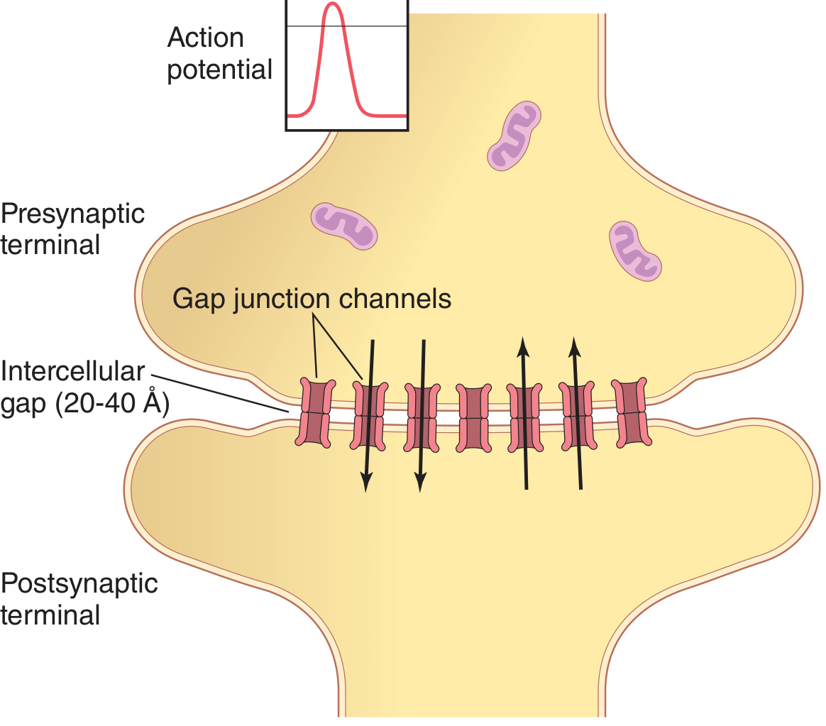
- Electrical synapse - gap junctions; ions pass directly; bidirectional; faster; found in cardiac muscle, smooth muscle, some brain areas
C. Based on the effect produced:
- Excitatory synapse - produces EPSP (Excitatory Postsynaptic Potential) → depolarization
- Inhibitory synapse - produces IPSP (Inhibitory Postsynaptic Potential) → hyperpolarization
Synaptic Transmission (Chemical Synapse - Steps)
- Arrival of action potential at presynaptic terminal
- Depolarization opens voltage-gated Ca²⁺ channels in presynaptic membrane
- Ca²⁺ influx into terminal (from ECF)
- Fusion of synaptic vesicles with presynaptic membrane → exocytosis
- Neurotransmitter release into synaptic cleft
- Diffusion of NT across cleft (20-30 nm gap)
- Binding of NT to specific receptors on postsynaptic membrane
- Ion channels open/close → change in postsynaptic membrane potential
- If depolarization → EPSP → if threshold reached → action potential
- If hyperpolarization → IPSP → inhibition
- Termination of NT action by:
- Re-uptake into presynaptic terminal (e.g., norepinephrine, dopamine)
- Enzymatic degradation (e.g., acetylcholinesterase degrades ACh)
- Diffusion away from cleft
Properties of Synapses (Sembulingam lists these specifically)
-
One-way conduction (Unidirectional transmission): Impulse travels only from presynaptic → postsynaptic (because neurotransmitter is only in presynaptic terminal and receptors only on postsynaptic membrane). This ensures nerve impulses do not travel backward.
-
Synaptic delay (Latent period): Time taken for transmission across synapse = 0.5 ms (0.3-0.5 ms). This delay is due to time needed for Ca²⁺ influx, vesicle movement, exocytosis, diffusion, and receptor binding.
-
Summation: Weak stimuli can be added together to generate an action potential:
- Spatial summation - simultaneous stimulation of many presynaptic terminals
- Temporal summation - repeated stimuli in quick succession at same synapse
-
Facilitation: Subthreshold stimuli make the postsynaptic neuron more excitable (partial depolarization). Does not produce action potential alone but lowers threshold for subsequent stimuli.
-
Occlusion: When two groups of fibers separately cause maximal excitation, their combined stimulation produces less effect than expected (sharing of postsynaptic neurons).
-
Subliminal fringe: Neurons near the discharge zone that are facilitated but not yet fired; represent the "reserve" for summation.
-
Post-tetanic potentiation: After a tetanic (rapid, repetitive) stimulus, subsequent stimuli produce greater postsynaptic response. Due to accumulation of Ca²⁺ in presynaptic terminal.
-
Fatigue: Prolonged stimulation at high frequency → decreased synaptic response due to depletion of neurotransmitter from vesicles.
-
Low oxygen susceptibility: Synaptic transmission is impaired by hypoxia because the process requires energy (ATP).
-
Effect of drugs: Synapses are sensitive to drugs:
- Strychnine - blocks inhibitory synapses (glycine antagonist)
- Caffeine - increases excitability
- Benzodiazepines - enhance GABA → inhibitory
-
Convergence and Divergence:
- Convergence: Many presynaptic neurons synapse on one postsynaptic neuron
- Divergence: One presynaptic neuron synapses on many postsynaptic neurons
CHAPTER 139 - SENSORY RECEPTORS
Definition
Sensory receptors are specialized structures at the peripheral end of afferent nerve fibers that detect changes in the internal or external environment and convert these stimuli into electrical signals (action potentials) - process called transduction.
Classification of Sensory Receptors
A. Based on the location (Sherrington's classification):
- Exteroceptors: Located on body surface; respond to external stimuli
- Touch, pressure, temperature, pain, light, sound, smell, taste
- Interoceptors (Visceroceptors): Located in visceral organs; respond to internal stimuli
- Detect pressure, stretch, temperature, pain from organs
- Proprioceptors: Located in muscles, tendons, joints; detect body position and movement
- Muscle spindles, Golgi tendon organs, joint receptors, labyrinthine receptors
B. Based on the stimulus (type/modality):
- Mechanoreceptors - respond to mechanical deformation (touch, pressure, stretch, vibration)
- Thermoreceptors - respond to temperature changes
- Nociceptors (Pain receptors) - respond to painful stimuli (tissue damage)
- Chemoreceptors - respond to chemical stimuli (taste, smell, O₂, CO₂, pH)
- Photoreceptors - respond to light (rods and cones of retina)
- Osmoreceptors - respond to osmotic pressure changes
C. Based on the structure:
- Free nerve endings (non-encapsulated): Bare dendrites; detect pain, crude touch, temperature
- Encapsulated nerve endings:
- Meissner's corpuscles - light touch, two-point discrimination; fingertips, lips
- Pacinian (Lamellar) corpuscles - vibration, deep pressure; rapidly adapting
- Merkel's disc (Tactile disc) - sustained touch, pressure; slowly adapting
- Ruffini endings - skin stretch, joint position; slowly adapting
- Krause's end bulbs - cold temperature (some authors question this classification)
- Specialized receptors: Hair cells (cochlea, vestibular), rods/cones, taste buds
Properties (Characteristics) of Sensory Receptors
-
Specificity (Adequate stimulus): Each receptor is maximally sensitive to one type of stimulus (e.g., Pacinian corpuscle for vibration). The specific stimulus that a receptor is most sensitive to is called its adequate stimulus. This is explained by the Labeled Line Principle.
-
Generator potential (Receptor potential): When a receptor is stimulated, a local, graded, non-propagated change in membrane potential is produced = generator potential. The magnitude is proportional to stimulus intensity. When generator potential reaches threshold at the first node of Ranvier, an action potential is generated.
-
Receptor threshold: Minimum intensity of stimulus needed to activate the receptor. Low threshold = high sensitivity.
-
Adaptation (Accommodation): Decrease in frequency of firing of a receptor despite constant stimulus application.
- Rapidly adapting receptors (phasic): Fire at onset and offset of stimulus; detect change in stimulus intensity (e.g., Meissner's, Pacinian corpuscles, hair follicle endings)
- Slowly adapting receptors (tonic): Fire throughout stimulus duration; detect sustained stimuli (e.g., Merkel's disc, Ruffini endings, pain receptors, muscle spindles)
-
Receptive field: The area of skin (or tissue) from which a single sensory unit can be activated. Smaller receptive field = greater spatial resolution/acuity (e.g., fingertip has small receptive fields and high acuity).
CHAPTER 144 - SOMATOSENSORY SYSTEM AND SENSORY PATHWAYS
Types of Somatic Sensations (Sembulingam's Classification)
Basic (Primary) Sensations:
- Touch (crude and fine/tactile)
- Pressure
- Temperature (hot and cold)
- Pain
Synthetic (Cortical) Sensations - require cortical processing/integration:
- Tactile localization - ability to identify exact spot touched
- Two-point discrimination (Tactile discrimination) - ability to distinguish two separate points simultaneously (normal fingertip: 2-3 mm; back: 40-70 mm)
- Stereognosis - ability to recognize a 3D object by touch alone (without vision)
- Vibratory sensation - detection of vibrating stimuli (tuning fork at 128 Hz or 256 Hz)
- Graphesthesia - ability to recognize letters/numbers written on skin
The Ascending (Sensory) Tracts
1. Posterior Column Tract (Dorsal Column-Medial Lemniscal Pathway)
Sensations conveyed: Fine touch, tactile localization, two-point discrimination, vibration, proprioception, stereognosis
| Neuron | Cell body | Fiber | Pathway |
|---|
| 1st order neuron | Dorsal root ganglion | Enters dorsal column ipsilaterally | Fasciculus gracilis (lower limb, sacral to T6) + Fasciculus cuneatus (upper limb, T6 to C2) |
| 1st synapse | Nucleus gracilis + Nucleus cuneatus (medulla oblongata) | | |
| 2nd order neuron | Nucleus gracilis/cuneatus → axons cross as internal arcuate fibers → form medial lemniscus | Decussates in medulla | Ascends as medial lemniscus |
| 2nd synapse | VPL nucleus of thalamus | | |
| 3rd order neuron | VPL thalamus | Via internal capsule (posterior limb) | Postcentral gyrus (Areas 3, 1, 2) = Primary somatosensory cortex |
Clinical note: Lesion of posterior column → ipsilateral loss of fine touch, vibration, proprioception below the lesion. Examples: Tabes dorsalis (syphilis), Subacute combined degeneration (Vit B12 deficiency)
2. Lateral Spinothalamic Tract
Sensations conveyed: Pain and Temperature
| Neuron | Cell body | Pathway |
|---|
| 1st order neuron | Dorsal root ganglion | Enters spinal cord → synapse in Substantia Gelatinosa of Rolando (Lamina I and II) |
| 2nd order neuron | Dorsal horn (Laminae I, II, V) | Crosses to opposite side in anterior white commissure (within 1-2 spinal cord segments above entry) → ascends in lateral funiculus |
| 2nd synapse | VPL nucleus of thalamus | |
| 3rd order neuron | VPL thalamus | Via posterior limb of internal capsule → Postcentral gyrus (Areas 3, 1, 2) |
Note: Fibers cross 1-2 segments above entry level - important clinically (syringomyelia causes cape-like loss of pain/temperature).
Clinical: Unilateral lesion → contralateral loss of pain and temperature below the lesion (1-2 segments below site of lesion).
3. Anterior Spinothalamic Tract
Sensations conveyed: Crude touch and Pressure (Light touch)
| Neuron | Cell body | Pathway |
|---|
| 1st order neuron | Dorsal root ganglion | Synapse in dorsal horn (Laminae III, IV) |
| 2nd order neuron | Dorsal horn | Crosses anterior commissure → ascends in anterior funiculus |
| 2nd synapse | VPL thalamus | |
| 3rd order neuron | VPL thalamus | Postcentral gyrus |
Note: Because crude touch has bilateral representation, lesion of one anterior spinothalamic tract does NOT completely abolish crude touch.
Sensory Pathway of Trigeminal Nerve
Sensations conveyed: All somatic sensations from face, scalp, anterior two-thirds of tongue, teeth, gums, nasal mucosa, oral mucosa, meninges, cornea
1st order neuron: Trigeminal (Gasserian) ganglion cells
Modality-specific nuclei in brainstem:
| Sensation | Brainstem Nucleus |
|---|
| Fine touch + Pressure (from face) | Principal (Chief) sensory nucleus - in pons |
| Pain + Temperature | Spinal nucleus of trigeminal nerve - extends from pons to C2 (most inferior part = subnucleus caudalis = homologous to dorsal horn) |
| Proprioception (jaw muscles) | Mesencephalic nucleus - unique: cell bodies of 1st order neurons lie within CNS |
2nd order neurons from all nuclei: Cross midline → ascend as trigeminal lemniscus (trigeminothalamic tract) → VPM (Ventral Posteromedial) nucleus of thalamus
3rd order neuron: VPM thalamus → internal capsule → face area of postcentral gyrus (S1)
SENSORY CORTEX AND SOMATIC SENSATIONS
Primary Somatosensory Cortex (S1)
- Location: Postcentral gyrus of parietal lobe (Brodmann Areas 3, 1, 2)
- Area 3a - receives proprioception from muscle spindles (Ia fibers via VPL)
- Area 3b - receives cutaneous mechanoreception (Meissner's and Merkel's)
- Area 1 - receives rapidly adapting cutaneous receptors (texture)
- Area 2 - receives pressure and joint position
Sensory Homunculus: Somatotopic map of the body on the cortex. Areas with highest receptor density (hands, lips, tongue, face) have largest cortical representation.
Secondary Somatosensory Cortex (S2):
- Located in parietal operculum (upper bank of lateral sulcus)
- Receives input from S1 and from thalamus
- Bilateral representation
- Important for learning by touch and tactile memory
KINESTHETIC SENSATIONS
Definition (Sembulingam): Kinesthesia or kinesthetic sensation is the sense of movement or position of different parts of the body. Also called proprioception or muscle sense.
Sources of kinesthetic information:
- Muscle spindles (annulospiral endings - Ia, flower spray endings - II) - sense muscle length and rate of change of length
- Golgi tendon organs (GTOs) - Ib fibers - sense muscle tension
- Joint receptors (Ruffini, Pacinian) - sense joint angle and rate of movement
- Cutaneous receptors (Meissner's, Ruffini) - sense skin stretch during movement
Pathway: Dorsal columns → VPL thalamus → Area 3a (primary) + Area 5 (parietal association cortex)
Conscious proprioception → posterior column pathway
Unconscious proprioception → spinocerebellar tracts (anterior and posterior) → cerebellum
Applied: Romberg's test - patient stands with feet together and eyes closed. Positive Romberg's (swaying/falling with eyes closed) indicates loss of proprioception. Distinguish from cerebellar ataxia (ataxia present even with eyes open).
CHAPTER 145 - PHYSIOLOGY OF PAIN
Definition
Pain is an unpleasant sensory and emotional experience associated with actual or potential tissue damage (IASP definition). Sembulingam describes it as a protective mechanism alerting the body to tissue damage.
Pain Receptors (Nociceptors)
- Free nerve endings distributed throughout the body
- Respond to: mechanical, thermal, and chemical (polymodal) painful stimuli
- Algogenic (pain-producing) substances: Bradykinin (most potent), prostaglandins, histamine, serotonin, K⁺ ions, H⁺ ions, substance P, lactic acid
Cutaneous Pain
Fast Pain (First Pain, Pricking Pain):
- Fiber: Aδ (Type A delta) - myelinated, small diameter, 6-30 m/s conduction velocity
- Quality: Sharp, pricking, well-localized
- Onset: Immediate
- Pathway: Neospinothalamic tract → VPL thalamus → somatosensory cortex (conscious localization)
- Function: Acute warning; triggers immediate withdrawal reflex
Slow Pain (Second Pain, Burning/Aching Pain):
- Fiber: C fibers - unmyelinated, very small, 0.5-2 m/s conduction velocity
- Quality: Burning, aching, poorly localized, throbbing
- Onset: Delayed (0.5-1 second after fast pain)
- Pathway: Paleospinothalamic tract → intralaminar nuclei of thalamus → limbic system, hypothalamus, reticular formation, periaqueductal gray (PAG)
- Function: Prolonged suffering; drives autonomic and emotional responses
Double pain sensation: When you stub your toe, you feel the initial sharp pain (fast - Aδ) followed by a throbbing/burning pain (slow - C fibers).
Mechanism of Pain Transmission (Sembulingam)
- Gate Control Theory (Melzack and Wall, 1965):
- Substantia gelatinosa (SG) of dorsal horn acts as a "gate"
- Large diameter (Aβ) fibers carrying touch CLOSE the gate → inhibit pain transmission
- Small diameter (Aδ, C) fibers carrying pain OPEN the gate
- This explains why rubbing an injured area reduces pain (activates Aβ fibers)
- Descending control from brain can also modulate the gate
Hyperalgesia
Definition: Increased pain sensitivity - a lower threshold for pain or exaggerated pain response to noxious stimuli.
Primary Hyperalgesia:
- At the site of injury
- Due to peripheral sensitization - damaged tissue releases prostaglandins, bradykinin → lower threshold of nociceptors; sensitize C fibers
- Mediated by increased prostaglandin synthesis → therapeutic target of NSAIDs/aspirin
Secondary Hyperalgesia:
- In the area surrounding the injury (uninjured tissue)
- Due to central sensitization in dorsal horn neurons
- Mechanism: "Wind-up" - repeated C fiber activation → NMDA receptor activation → spinal cord neurons become hypersensitive
Deep Somatic Pain
- Origin: Muscles, bones, tendons, periosteum, joints
- Character: Diffuse, dull, aching, poorly localized; difficult to distinguish from visceral pain
- Accompanied by: Nausea, sweating, fall in blood pressure, muscle spasm (reflex muscle guarding)
- Pathway: Mainly C fibers
Visceral Pain
- Origin: Hollow viscera (distension, spasm, ischemia); NOT cutting or burning
- Character: Dull, cramping, poorly localized (visceral afferents = fewer in number, widely spaced)
- Pathway: Sympathetic afferents (most viscera); pelvic organs via parasympathetic (pelvic splanchnic)
- Associated with: Autonomic responses (nausea, vomiting, sweating, changes in BP and HR)
Referred Pain
Definition: Pain felt at a location (usually skin) distant from the actual site of pathology (usually a viscus).
Mechanism - Convergence-Projection Theory (most accepted):
- Visceral afferents and somatic afferents from the referred zone converge on the same second-order neurons in the dorsal horn
- The brain cannot distinguish between visceral and somatic input → interprets the signal as coming from the somatic (skin) area → pain is "referred" to skin
Important examples:
| Organ | Referred Area | Spinal segments |
|---|
| Heart (angina) | Left arm, left jaw, left shoulder | T1-T4 |
| Gallbladder | Right shoulder tip | C3-C5 (phrenic nerve) |
| Appendix (early) | Periumbilical region | T10 |
| Kidney | Loin to groin (ipsilateral) | T10-L1 |
| Diaphragm | Shoulder tip (C3-C5) | C3-C5 |
| Ureter | Groin, inner thigh | L1 |
MOTOR CORTEX AND DESCENDING TRACTS
Motor Cortex (Sembulingam - Chapter 152)
Primary Motor Cortex (M1):
- Location: Precentral gyrus of frontal lobe - Brodmann Area 4
- Contains giant Betz cells (giant pyramidal cells, 60-80 μm) - largest neurons in CNS
- Arranged as motor homunculus - inverted map; hand and face have disproportionately large area
- Controls contralateral voluntary movements
Premotor Cortex:
- Brodmann Area 6 (anterior to M1)
- Lateral premotor area (PMC): Plans movements guided by external sensory cues
- Supplementary Motor Area (SMA) - medial Area 6: Plans self-initiated movements; active before voluntary movement begins (Bereitschaftspotential/readiness potential)
Other motor areas:
- Frontal Eye Fields (Area 8): Voluntary conjugate eye movements (saccades)
- Posterior parietal cortex (Areas 5, 7): Sensorimotor integration
DESCENDING TRACTS
A. Pyramidal Tracts (Corticospinal + Corticobulbar)
Definition: Motor tracts that pass through the medullary pyramids.
Corticospinal Tract
Origin:
- 60% from primary motor cortex (Area 4)
- 20% from premotor/SMA (Area 6)
- 20% from somatosensory cortex (Areas 3, 1, 2)
Course:
- Corona radiata (white matter of cerebral hemisphere)
- Posterior limb of internal capsule (anterior two-thirds - genu and posterior limb)
- Basis pedunculi (cerebral peduncles) of midbrain - middle three-fifths
- Pons - scattered into small bundles by transverse pontine fibers
- Reunite in medulla to form pyramids
- At junction of medulla and spinal cord: Pyramidal decussation (decussation of pyramids)
- ~85% of fibers cross → Lateral corticospinal tract (LCS)
- ~15% do not cross → Anterior corticospinal tract (ACS) - eventually cross at spinal level
Lateral Corticospinal Tract: Crossed; descends in lateral funiculus; controls distal limb muscles (fine, skilled, voluntary movements - hands, fingers); direct monosynaptic connection to alpha motor neurons
Anterior Corticospinal Tract: Initially uncrossed; descends in anterior funiculus; crosses at spinal levels; controls proximal and axial muscles (trunk, shoulder, hip)
Termination: Alpha motor neurons of ventral horn (directly or via interneurons)
Corticobulbar Tract
- Fibers from motor cortex → brainstem nuclei of cranial nerves (V, VII, IX, X, XI, XII)
- Most cranial nerve nuclei receive bilateral corticobulbar input
- Exception: Lower face (CN VII) and tongue (CN XII) → predominantly contralateral supply
- Clinical: Unilateral UMN lesion spares upper face (bilateral supply) but causes contralateral lower face weakness
B. Extrapyramidal Tracts
Tracts that do NOT pass through medullary pyramids; originate from brainstem nuclei.
| Tract | Origin | Crosses? | Termination | Function |
|---|
| Rubrospinal | Red nucleus (midbrain tegmentum) | Crosses immediately | Ventral horn (cervical/thoracic) | Flexor muscle tone; limb movements |
| Pontine (Medial) Reticulospinal | Pontine reticular formation | Uncrossed | Ventral horn (all levels) | Facilitates extensor tone; activates antigravity muscles |
| Medullary (Lateral) Reticulospinal | Medullary reticular formation | Bilateral (mostly uncrossed) | Ventral horn | Inhibits extensor tone; inhibits muscle tone |
| Lateral Vestibulospinal | Lateral vestibular nucleus (Deiters') | Uncrossed | Ipsilateral ventral horn (all levels) | Activates extensors; inhibits flexors; antigravity posture |
| Medial Vestibulospinal | Medial vestibular nucleus | Bilateral | Cervical cord | Head/neck posture; VOR |
| Tectospinal | Superior colliculus (midbrain) | Crosses | Cervical ventral horn | Orients head/eyes toward visual/auditory stimuli |
Decerebrate Rigidity: Lesion between midbrain and pons → removes cortical inhibition + removes rubrospinal (facilitates flexors) → pontine reticulospinal + vestibulospinal overactivate extensors → rigid extension of all limbs.
UPPER MOTOR NEURON (UMN) vs LOWER MOTOR NEURON (LMN)
Upper Motor Neuron: All neurons above the anterior horn cell = motor cortex + corticospinal/corticobulbar tracts
Lower Motor Neuron: Anterior horn cell of spinal cord (or cranial nerve motor nucleus) + its axon to muscle = "Final Common Pathway" (Sherrington)
| Feature | UMN Lesion | LMN Lesion |
|---|
| Tone | Hypertonia (spasticity - velocity-dependent, clasp-knife reflex) | Hypotonia/Flaccidity |
| Power | Weakness/paralysis | Weakness/paralysis |
| Reflexes | Hyperreflexia | Hyporeflexia/Areflexia |
| Babinski sign | Positive (extensor plantar response) | Absent |
| Clonus | Present | Absent |
| Wasting | Mild (disuse atrophy only) | Marked wasting and fasciculations |
| Distribution | Affects groups of muscles | May affect individual muscles |
Paralysis
| Type | Definition | Common Site of Lesion |
|---|
| Monoplegia | Paralysis of one limb | Cortex (small motor cortex lesion) or spinal nerve/root |
| Hemiplegia | Paralysis of one arm and leg on same side | Contralateral internal capsule, cerebral cortex, upper brainstem |
| Paraplegia | Paralysis of both lower limbs | Thoracic/lumbar spinal cord; bilateral |
| Quadriplegia (Tetraplegia) | Paralysis of all four limbs | Cervical spinal cord (C1-C8); C3-C5 also impairs diaphragm |
| Diplegia | Paralysis of same region on both sides (e.g., both arms) | Bilateral cortical lesions |
MUSCLE TONE
Definition (Sembulingam)
Muscle tone (tonus) is defined as the state of partial contraction of a muscle at rest or the resistance offered by a muscle to passive stretching.
Mechanism of Muscle Tone
Maintained by the myotatic (stretch) reflex arc:
- Gamma motor neurons (fusimotor neurons) from ventral horn → intrafusal muscle fibers of muscle spindle
- Gamma activation maintains spindle sensitivity
- Ia afferents from muscle spindle → dorsal root → alpha motor neurons
- Alpha MN → extrafusal muscle fibers → slight contraction = tone
Supraspinal regulation of tone:
- Facilitatory areas (increase tone): Pontine reticular formation + lateral vestibulospinal tract
- Inhibitory areas (decrease tone): Medullary reticular formation + cerebellum
Properties/Types of Altered Tone
Hypotonia: Decreased muscle tone
- Flaccidity; muscle feels soft and doughy
- Causes: LMN lesion, cerebellar lesion, acute spinal shock, deep anesthesia
- Features: Decreased resistance to passive movement, pendular reflexes
Atonia: Complete absence of muscle tone
- Causes: Severe LMN destruction, complete spinal shock
- Muscles completely flaccid, no response to stretch
Hypertonia: Increased muscle tone - two types:
- Spasticity (Pyramidal/UMN): Velocity-dependent resistance to passive stretch; clasp-knife phenomenon (gives way suddenly like a penknife); affects antigravity muscles (flexors of arm, extensors of leg); deep reflexes brisk
- Rigidity (Extrapyramidal/Basal ganglia): Resistance to passive stretch that is NOT velocity-dependent; present throughout range of movement; two types:
- Lead-pipe rigidity: Uniform resistance throughout
- Cogwheel rigidity: Ratchet-like, jerky resistance (Parkinson's disease = rigidity + tremor superimposed)
SPINAL CORD LESIONS (Sembulingam - Chapter 143)
Complete Transection (Complete Cord Injury)
Immediate effects - SPINAL SHOCK:
- Complete flaccid paralysis below level of lesion (bilateral)
- Complete loss of all sensory modalities below lesion (bilateral)
- Loss of all spinal reflexes (areflexia) below lesion
- Urinary retention (atonic bladder)
- Bowel retention (paralytic ileus)
- Loss of vasomotor control → hypotension
- Priapism (paradoxical - loss of sympathetic)
- Duration: Days to 3-6 weeks (longer in humans than animals)
Recovery after spinal shock:
- Return of spinal reflexes (exaggerated → hyperreflexia)
- Spasticity develops
- Babinski sign becomes positive
- Automatic (reflex) bladder develops
- Mass reflex (triple flexion reflex) → powerful noxious stimulus below lesion triggers: bilateral flexion of lower limbs + evacuation of bladder and bowel + profuse sweating
- Autonomic dysreflexia (lesions above T6) → massive sympathetic discharge below lesion triggered by stimuli (full bladder, etc.) → hypertensive crisis, bradycardia (dangerous)
Brown-Séquard Syndrome (Hemisection of Spinal Cord)
Unilateral cord lesion (e.g., stab wound, multiple sclerosis plaque)
| Finding | Side | Explanation |
|---|
| UMN signs (spasticity, hyperreflexia, Babinski) below lesion | Ipsilateral | Lateral corticospinal tract (uncrossed) |
| Loss of fine touch, vibration, proprioception below lesion | Ipsilateral | Posterior column (uncrossed below medulla) |
| Loss of pain and temperature below lesion | Contralateral (1-2 levels below) | Lateral spinothalamic tract (crossed) |
| LMN signs (flaccidity, areflexia, wasting) | Ipsilateral at the level of lesion | Anterior horn cells |
| Complete sensory loss at level of lesion | Ipsilateral | Entering dorsal root fibers |
CHAPTER 150 - CEREBELLUM
Functions (Sembulingam)
- Coordination of voluntary movements - ensures smooth, accurate, purposeful movements (timing, sequencing, coordination)
- Regulation of muscle tone - via connections with reticulospinal and vestibulospinal tracts (generally decreases tone)
- Maintenance of posture and equilibrium - vestibulocerebellum; coordinates with vestibular apparatus
- Coordination of eye movements
- Motor learning - stores learned motor skills; climbing fiber-LTD
Divisions
| Division | Phylogenetic name | Main Input | Function |
|---|
| Flocculonodular lobe | Archicerebellum (oldest) | Vestibular | Balance, equilibrium, eye movements |
| Vermis + intermediate zone | Paleocerebellum | Spinocerebellar tracts | Muscle tone, posture, gait, limb coordination |
| Lateral hemispheres | Neocerebellum (newest) | Pontocerebellar (from cerebral cortex) | Planning and initiation of complex voluntary movements |
Cerebellar peduncles (connections):
- Superior cerebellar peduncle (brachium conjunctivum): Main OUTPUT - dentate nucleus → contralateral red nucleus and VL thalamus → motor cortex
- Middle cerebellar peduncle (brachium pontis): Input from contralateral pontine nuclei (corticopontocerebellar)
- Inferior cerebellar peduncle (restiform body): Input from spinal cord (spinocerebellar tracts), inferior olivary nucleus (climbing fibers), vestibular nuclei
Cerebellar Cortex Layers
| Layer | Position | Cell types | Key function |
|---|
| Molecular layer | Outermost | Outer stellate cells, basket cells, Purkinje cell dendrites, parallel fibers | Modulation of Purkinje cell activity |
| Purkinje cell layer | Middle | Purkinje cells | ONLY output cell of cerebellar cortex; always inhibitory (GABA) → deep nuclei |
| Granular layer | Innermost | Granule cells, Golgi II cells, glomeruli | Granule cells receive mossy fiber input; axons form parallel fibers |
Deep cerebellar nuclei (from medial to lateral): Fastigial, Globose, Emboliform, Dentate (largest, main output of neocerebellum)
Cerebellar Ataxia (Signs of Cerebellar Lesion)
Sembulingam uses mnemonic DANISH or lists these features:
- Dysmetria: Inability to judge distance; past-pointing (overshot or undershoot target) in finger-nose-finger test
- Ataxia: Incoordination; wide-based, staggering, "drunken" gait (truncal ataxia = vermis lesion; limb ataxia = hemisphere lesion)
- Nystagmus: Involuntary rhythmic eye oscillations; towards side of lesion
- Intention tremor: Tremor that appears/worsens during voluntary movement (not at rest); worsens as limb approaches target
- Dysdiadochokinesia: Inability to perform rapid alternating movements (e.g., pronation-supination)
- Scanning (staccato) dysarthria: Slurred, explosive, irregular speech
- Hypotonia: Decreased muscle tone; pendular knee jerk
- Rebound phenomenon (Loss of check reflex): Cannot halt a movement when resistance is suddenly removed (forearm flexion against resistance - when resistance removed, forearm hits the face)
- Romberg test: Negative in cerebellar ataxia (ataxia present even with eyes OPEN; does not significantly worsen with eye closure). This distinguishes from sensory/posterior column ataxia (Romberg positive = worse with eyes closed).
CHAPTER 157 - POSTURE AND EQUILIBRIUM
Postural Reflexes Classification (Sembulingam's Classification)
1. Spinal Postural Reflexes (spinal cord level; persist after spinal transection)
- Stretch reflex (Myotatic reflex): Maintains tone against gravity; monosynaptic
- Positive supporting reaction: Pressure on sole → extension of lower limb (supports weight)
- Crossed extensor reflex: Flexion of one limb → extension of opposite limb
2. Medullary (Brainstem) Postural Reflexes
- Tonic neck reflexes (TNR):
- Asymmetric TNR: Rotation of head → extension of limbs on side toward which face turns (jaw limbs extend; skull limbs flex)
- Symmetric TNR: Flexion of neck → forelimb flexion + hindlimb extension; extension of neck → reverse
- Tonic labyrinthine reflexes: Changes in head position in space → changes in extensor tone (via otolith organs)
- Righting reflexes (medullary): Labyrinthine righting reflex → orients head upright; body righting reflex acting on head
3. Midbrain Postural Reflexes
- Complete righting reflexes - full body righting; need intact midbrain + vestibular apparatus + vision
- Optical righting reflex - visual cues orient head/body
- Placing and hopping reactions
4. Cortical Reflexes
- Voluntary postural adjustments - learned, anticipatory
- Cortical placing reaction - visual-guided placement of limb
Equilibrium (Balance):
Maintained by integration of 3 systems:
- Vestibular system (semicircular canals for angular acceleration; utricle and saccule for linear acceleration and gravity)
- Proprioceptive system (muscle spindles, joint receptors)
- Visual system
CHAPTER 147 - THALAMUS
Nuclei and Functions
| Nuclear Group | Nuclei | Relay for |
|---|
| Specific Relay (Sensory) | VPL (Ventral Posterolateral) | Body sensation (pain, temp, touch, proprioception) from spinothalamic + medial lemniscus |
| VPM (Ventral Posteromedial) | Face sensation from trigeminal lemniscus |
| LGN (Lateral Geniculate Nucleus) | Vision → visual cortex (Area 17) |
| MGN (Medial Geniculate Nucleus) | Hearing → auditory cortex (Area 41, 42) |
| Specific Relay (Motor) | VL (Ventral Lateral) | Cerebellum + basal ganglia output → motor cortex |
| VA (Ventral Anterior) | Basal ganglia → premotor cortex |
| Association | Pulvinar | Multimodal sensory association; visual attention |
| LP (Lateral Posterior) | Parietal association cortex |
| MD (Mediodorsal) | Prefrontal cortex; cognition, emotion, working memory |
| AN (Anterior nuclei) | Limbic circuit (Papez); memory |
| Intralaminar | CM (Centromedian), PF (Parafascicular) | Arousal; diffuse cortical activation; pain |
| Midline | Various | Hypothalamic/visceral functions; interoception |
| Reticular nucleus | TRN | Gate-keeping; modulates thalamic output (inhibitory) |
Thalamus as relay station: All sensory pathways except olfaction relay in thalamus before reaching cortex.
Thalamic Syndrome (Dejerine-Roussy Syndrome)
- Cause: Thalamic infarction (most commonly posterior thalamus - thalamogeniculate artery territory)
- Features:
- Initial contralateral hemisensory loss (all modalities)
- Followed by thalamic pain - severe, persistent, burning, spontaneous contralateral pain; disproportionate to stimulus; hyperpathia (exaggerated response) and allodynia (pain to non-painful stimuli)
- Mild contralateral hemiplegia
- Hemiataxia
- Choreoathetosis (involuntary movements)
- Mechanism: Loss of normal sensory gating → thalamus generates spontaneous pain signals
CHAPTER 149 - HYPOTHALAMUS
Nuclei and Functions (Sembulingam)
| Nucleus/Area | Function |
|---|
| Anterior nucleus | Heat loss mechanisms (sweating, vasodilation); parasympathetic control; lesion → hyperthermia |
| Posterior nucleus | Heat conservation (vasoconstriction, shivering); sympathetic control; lesion → inability to regulate body temperature |
| Lateral hypothalamic area | Hunger/feeding center (lesion → anorexia, weight loss) |
| Ventromedial nucleus (VMN) | Satiety center (lesion → hyperphagia → obesity) |
| Supraoptic nucleus (SON) | Synthesizes ADH (Vasopressin) → released from posterior pituitary; lesion → Diabetes Insipidus |
| Paraventricular nucleus (PVN) | Synthesizes Oxytocin → released from posterior pituitary |
| Suprachiasmatic nucleus (SCN) | Biological clock - circadian rhythms; receives photic input from retina |
| Arcuate nucleus | Dopamine (inhibits prolactin); GnRH; NPY (feeding) |
| Mammillary bodies | Memory (Papez circuit); lesion → Wernicke-Korsakoff syndrome |
| Preoptic area | Thermoregulation; sexual behavior; sleep |
Hypothalamic Functions (Summary - Sembulingam's 5 D's):
- Defense reaction (rage, fear - via limbic connections)
- Drinking (thirst center in lateral hypothalamus)
- Diet (feeding and satiety)
- Dynamo (ANS control - sympathetic and parasympathetic)
- Diuresis (ADH regulation, water balance)
- Also: Pituitary control (releasing/inhibiting hormones), thermoregulation, sleep-wake cycle, circadian rhythms, sexual behavior
CHAPTER 154 - RETICULAR FORMATION
Components
Diffuse network of neurons in the core of the brainstem (medulla, pons, midbrain) extending up to the thalamus (intralaminar nuclei).
Ascending Reticular Activating System (ARAS):
- All sensory pathways send collateral branches to ARAS
- ARAS sends diffuse projections to the entire cerebral cortex via thalamus (intralaminar nuclei)
- Maintains wakefulness and alertness (consciousness)
Descending Reticular Formation:
- Pontine (medial) RF → facilitates spinal motor neurons (extensors)
- Medullary (lateral) RF → inhibits spinal motor neurons
Functions of Reticular Formation
- Arousal and consciousness: ARAS maintains wakefulness; damage → coma
- Sleep-wake cycle: Interacts with hypothalamic sleep center
- Control of muscle tone: Facilitatory and inhibitory centers
- Pain modulation: Periaqueductal gray (PAG) + Nucleus Raphe Magnus → descending inhibition of pain via enkephalins and serotonin (endogenous analgesia system)
- Cardiovascular control: Vasomotor center (medullary RF) - pressor and depressor areas
- Respiratory control: Respiratory rhythm generation (pre-Bötzinger complex in medulla)
- Vomiting: Emetic center in medullary RF
- Habituation and attention
CHAPTER 153 - LIMBIC SYSTEM
Components (Broca's "Le Grand Lobe Limbique" - ring of cortex)
Cortical structures:
- Cingulate gyrus (above corpus callosum)
- Parahippocampal gyrus
- Hippocampus + dentate gyrus (archicortex)
- Entorhinal cortex (area 28)
- Olfactory cortex (piriform cortex)
Subcortical structures:
- Amygdala (amygdaloid complex)
- Septal nuclei
- Anterior thalamic nuclei
- Hypothalamus (mammillary bodies)
- Fornix (major white matter pathway)
Papez Circuit (memory and emotion):
Hippocampus → Fornix → Mammillary bodies → Anterior thalamic nucleus → Cingulate gyrus → Entorhinal cortex → Hippocampus
Functions of Limbic System (Sembulingam)
- Emotional behavior: Fear, rage, aggression, pleasure (amygdala is key)
- Memory: Hippocampus essential for consolidation of declarative memory (short-term → long-term memory)
- Olfaction (piriform cortex)
- Autonomic and somatic responses to emotion (via hypothalamus)
- Feeding behavior and appetite
- Sexual behavior and reproduction
- Motivation and reward (nucleus accumbens - mesolimbic dopamine)
- Sleep-wake cycle modulation
Klüver-Bucy Syndrome (bilateral temporal lobectomy/amygdala destruction):
- Psychic blindness (visual agnosia)
- Hypersexuality
- Hyperorality (puts everything in mouth)
- Emotional blunting (placidity, tameness - no fear or aggression)
- Dietary changes (dietary indiscretion)
CHAPTER 151 - BASAL GANGLIA
Components (Sembulingam)
- Corpus striatum:
- Neostriatum = Caudate nucleus + Putamen (main INPUT - receive from cortex)
- Paleostriatum = Globus pallidus (internal GPi + external GPe) (main OUTPUT)
- Subthalamic nucleus (STN) - diencephalon
- Substantia nigra - Pars compacta (SNc) + Pars reticulata (SNr)
- Amygdaloid nucleus (some classifications include)
Circuits
Direct pathway (net excitatory to motor cortex → facilitates movement):
Cortex → Striatum (GABA, D1 receptors) → GPi/SNr (inhibited) → Thalamus (disinhibited/more active) → Cortex → ↑ Movement
Indirect pathway (net inhibitory to motor cortex → suppresses unwanted movement):
Cortex → Striatum (GABA, D2 receptors, inhibits) → GPe (disinhibited, inhibits) → STN (disinhibited, excitatory) → GPi/SNr (overactive, inhibits) → Thalamus (inhibited) → Cortex → ↓ Movement
Dopamine (from SNc): Acts on D1 receptors = excite direct pathway; D2 receptors = inhibit indirect pathway → overall facilitates movement
Functions of Basal Ganglia (Sembulingam)
- Control and execution of voluntary movements (especially learned, automatic, sequential)
- Regulation of automatic associated movements (e.g., arm swing during walking)
- Control of muscle tone
- Postural control
- Cognitive functions (via caudate-prefrontal circuit)
Parkinson's Disease
Pathology: Degeneration of dopaminergic neurons of substantia nigra pars compacta → ↓ dopamine in striatum → Indirect pathway overactivated + direct pathway underactivated → Hypokinesia
Microscopy: Lewy bodies (eosinophilic intraneuronal inclusions containing α-synuclein)
Features (TRAP):
| Feature | Description |
|---|
| Tremor | Resting tremor ("pill-rolling"); 4-6 Hz; diminishes with voluntary movement; worsens at rest and with stress |
| Rigidity | Cogwheel or lead-pipe; affects all muscles; due to loss of dopamine influence on basal ganglia → excess GPi inhibition of thalamus → excess thalamic inhibition |
| Akinesia/Bradykinesia | Slowness and poverty of movement; reduced arm swing; shuffling gait; masked (expressionless) facies (hypomimia); micrographia; hypophonia |
| Postural instability | Loss of postural reflexes; festinating gait (accelerating small steps trying to keep up with shifted center of gravity); falls |
Other features: Depression, dementia (late), sleep disturbances, constipation, urinary problems, orthostatic hypotension, drooling
Treatment: Levodopa + carbidopa; dopamine agonists (pramipexole, ropinirole); MAO-B inhibitors (selegiline); COMT inhibitors; anticholinergics; deep brain stimulation (subthalamic nucleus or GPi)
CHAPTER 152 - CEREBRAL CORTEX
Lobes and Key Areas
| Lobe | Separated by | Key areas |
|---|
| Frontal | Central sulcus (anteriorly), lateral sulcus (inferiorly) | Motor cortex (4), Premotor (6), SMA (6), Broca's (44, 45), FEF (8), Prefrontal cortex |
| Parietal | Central sulcus (posteriorly), lateral sulcus, parietooccipital sulcus | Somatosensory (3,1,2), Angular gyrus (39), Supramarginal gyrus (40) |
| Temporal | Lateral sulcus | Auditory (41, 42), Wernicke's (22), Memory (hippocampus via medial temporal lobe) |
| Occipital | Parietooccipital sulcus | Primary visual (17), Visual association (18, 19) |
| Insular (5th lobe) | Lateral sulcus | Visceral/interoceptive sensation, taste, pain integration |
Brodmann's Areas and Functions (Sembulingam)
| Area | Location | Function |
|---|
| 4 | Precentral gyrus | Primary motor cortex - voluntary movement |
| 6 | Premotor/SMA | Motor planning and programming |
| 8 | Frontal eye fields | Voluntary eye movements |
| 44, 45 | Inferior frontal gyrus (Broca) | Speech production (dominant hemisphere) |
| 3, 1, 2 | Postcentral gyrus | Primary somatosensory cortex |
| 5, 7 | Superior parietal lobule | Somatosensory association; spatial orientation |
| 39 | Angular gyrus | Reading, writing, calculation; word recognition |
| 40 | Supramarginal gyrus | Language processing; tactile recognition |
| 17 | Calcarine cortex (occipital) | Primary visual cortex (striate cortex) |
| 18, 19 | Occipital association | Visual association; form, color, motion recognition |
| 41, 42 | Heschl's gyrus (temporal) | Primary auditory cortex |
| 22 | Superior temporal gyrus (Wernicke) | Speech comprehension (dominant hemisphere) |
| 9, 10, 11 | Prefrontal cortex | Executive function, working memory, judgment, personality |
| 24, 23 | Cingulate gyrus | Emotion, attention, pain modulation |
Higher Functions
Learning and Memory (Chapter 162)
Types of Memory:
Short-term (working) memory:
- Temporary storage (seconds to minutes)
- Maintained by reverberating circuits (self-re-exciting loops)
- Limited capacity (7 ± 2 items, Miller's Law)
Long-term memory:
- Declarative (Explicit):
- Semantic memory (facts, knowledge)
- Episodic memory (personal experiences)
- Requires hippocampus for consolidation
- Non-declarative (Implicit):
- Procedural memory (skills, habits) - basal ganglia, cerebellum
- Conditioning - amygdala (emotional conditioning), cerebellum (motor conditioning)
Mechanism (Synaptic Plasticity):
- Long-Term Potentiation (LTP): Repeated stimulation → NMDA receptor activation → Ca²⁺ influx → kinase activation → insertion of more AMPA receptors → strengthened synapse → basis of learning and memory
- Long-Term Depression (LTD): Weakening of synaptic connections (important for cerebellar motor learning)
Speech
| Area | Location | Lesion effect |
|---|
| Broca's area (44, 45) | Inferior frontal gyrus, left hemisphere | Broca's (Motor/Expressive) Aphasia: Non-fluent, effortful, telegraphic speech; comprehension intact; patient knows what they want to say but cannot express it ("I know but I cannot say") |
| Wernicke's area (22) | Posterior superior temporal gyrus, left | Wernicke's (Sensory/Receptive) Aphasia: Fluent but meaningless speech (jargon, paraphrasias, neologisms); impaired comprehension |
| Arcuate fasciculus | Connects Broca's to Wernicke's | Conduction Aphasia: Fluent speech, intact comprehension, unable to repeat words; poor repetition |
| Global aphasia | Large dominant hemisphere lesion | All aspects of language impaired |
Dominant hemisphere (left in 95% right-handers and 70% left-handers): Language, analytical thinking, calculation
Non-dominant hemisphere (right): Visuospatial tasks, face recognition, music, emotional tone of language (prosody), attention
CHAPTER 163 - CEREBROSPINAL FLUID (CSF)
Formation
- Site: Choroid plexus of the lateral, third, and fourth ventricles (70-80%); remainder from ependymal cells
- Rate: 500 mL/day formed; total volume = ~150 mL (turnover 3-4 times/day)
- Mechanism: Active secretion - Na⁺-K⁺-ATPase pumps Na⁺ into ventricle → Cl⁻ and HCO₃⁻ follow → water follows osmotically
Composition (Sembulingam's Table)
| Constituent | CSF | Plasma |
|---|
| Appearance | Clear, colorless ("like rock water") | Yellow |
| Specific gravity | 1.005 | 1.025 |
| pH | 7.33 | 7.4 |
| Pressure (lateral recumbent) | 70-180 mm H₂O (7-18 cm H₂O) | - |
| Protein | 20-45 mg/dL | 6000-8000 mg/dL |
| Glucose | 50-80 mg/dL (60-80% plasma) | 80-120 mg/dL |
| Chloride | Higher than plasma (~125 mEq/L) | 100 mEq/L |
| Na⁺ | ~140 mEq/L | ~140 mEq/L |
| WBC | 0-5 lymphocytes/mm³ | 4000-11000/mm³ |
| RBC | Nil | Present |
Circulation of CSF (Sembulingam's Flow)
Lateral ventricles
        ↓ (via Foramen of Monro = Interventricular foramen)
Third ventricle
        ↓ (via Aqueduct of Sylvius = Cerebral aqueduct)
Fourth ventricle
        ↓ (via Foramen of Magendie - median + Foramina of Luschka - 2 lateral)
Subarachnoid space (around brain and spinal cord)
        ↓
Arachnoid villi/granulations (Pacchionian granulations) → Superior sagittal sinus → Venous blood
Functions of CSF
- Protection (mechanical): Buoyancy - reduces effective weight of brain from 1400 g to 25 g; cushions brain from jolts
- Nutrition: Carries glucose and amino acids; removes metabolic waste (CO₂, lactic acid)
- Maintenance of uniform environment: Stable ionic composition for neurons
- Regulation of intracranial pressure
- Neurohumoral communication
- Lymphatic equivalent for CNS (immune surveillance via lymphocytes)
Lumbar Puncture (LP)
Site: L3-L4 or L4-L5 interspace (conus medullaris ends at L1-L2; so below this, only cauda equina is present - safe zone)
Position: Patient in lateral decubitus (fetal position, spine maximally flexed) or sitting leaning forward
Layers pierced (Sembulingam lists in order):
- Skin
- Subcutaneous fat
- Supraspinous ligament
- Interspinous ligament
- Ligamentum flavum
- Epidural (extradural) space
- Dura mater
- Subdural space (potential)
- Arachnoid mater
- Subarachnoid space ← CSF obtained here
Clinical Significance:
- Measure CSF pressure (normal 70-180 mm H₂O)
- Diagnose meningitis (↑ cells, ↓ glucose, ↑ protein; organisms on culture)
- Diagnose viral encephalitis (↑ lymphocytes, normal glucose)
- Diagnose subarachnoid hemorrhage (xanthochromia - yellow CSF; RBCs)
- Diagnose TB meningitis (very low glucose, high protein, lymphocytes, fibrin clot)
- Diagnose MS (oligoclonal bands, ↑ IgG, myelin basic protein)
- Diagnose Guillain-Barré Syndrome (albuminocytologic dissociation: ↑↑ protein, normal cells)
- Spinal anesthesia/intrathecal drug administration
- Queckenstedt's test: compress jugular vein → rise in CSF pressure (normal); no rise = spinal block
Contraindications: Raised ICP with papilledema (risk of tonsillar/uncal herniation), bleeding disorders, local infection at puncture site
Blood-Brain Barrier (BBB)
Formed by: Tight junctions between cerebral capillary endothelial cells + astrocyte foot processes (perivascular end-feet)
Functions:
- Maintains stable ionic environment for neuronal function
- Prevents entry of harmful substances, large molecules, pathogens
- Selective - allows: O₂, CO₂, glucose (GLUT-1), lipid-soluble substances, water, ethanol
- Blocks: Proteins, polar molecules, many drugs, most antibiotics (exception: chloramphenicol, rifampicin, metronidazole)
Areas without BBB (Circumventricular organs):
- Area postrema (vomiting center - "senses" emetogenic substances in blood)
- Subfornical organ, organum vasculosum of lamina terminalis (osmoreception)
- Neurohypophysis, pineal gland, adenohypophysis
Hydrocephalus
Definition: Excessive accumulation of CSF causing dilation of ventricles and raised ICP.
| Type | Mechanism | Common Causes |
|---|
| Non-communicating (Obstructive) | Block within ventricular system | Aqueduct stenosis, tumors at foramen of Monro, 4th ventricle tumors |
| Communicating | Block outside ventricular system (at arachnoid villi) | Post-meningitis (fibrosis of arachnoid villi), post-SAH, meningeal carcinomatosis |
| Normal Pressure Hydrocephalus (NPH) | Enlarged ventricles, normal LP pressure | Triad: Dementia + Urinary incontinence + Gait apraxia ("Wet, Wacky, Wobbly") - treatable with shunt |
| Hydrocephalus ex vacuo | Ventricular enlargement due to brain atrophy (not increased pressure) | Alzheimer's, old age |
CHAPTER 164 - AUTONOMIC NERVOUS SYSTEM (ANS)
Sympathetic Nervous System
Origin: Thoracolumbar (T1-L2) - preganglionic neurons in lateral horn (intermediolateral cell column)
Ganglia:
- Paravertebral (sympathetic chain) ganglia - 22 pairs
- Prevertebral ganglia - celiac, superior/inferior mesenteric, aorticorenal
Neurotransmitters:
- Preganglionic: Acetylcholine (nicotinic N₁ receptors)
- Postganglionic: Norepinephrine (adrenergic α and β receptors)
- Exception: Sweat glands = cholinergic (muscarinic); adrenal medulla releases epinephrine + NE
Actions:
| Organ | Sympathetic Effect | Receptor |
|---|
| Heart | ↑ HR, ↑ force, ↑ conduction velocity | β1 |
| Blood vessels (skin, viscera) | Vasoconstriction | α1 |
| Blood vessels (skeletal muscle) | Vasodilation | β2 |
| Bronchi | Bronchodilation | β2 |
| GI tract | ↓ Motility, ↓ secretion; sphincter contraction | α1, β2 |
| Bladder (detrusor) | Relaxation | β2 |
| Bladder (sphincter) | Contraction | α1 |
| Eye (iris - dilator muscle) | Pupil dilation (mydriasis) | α1 |
| Eye (ciliary muscle) | Relaxation → far vision | β2 |
| Salivary glands | Thick viscous saliva | α1 |
| Sweat glands | Secretion | Muscarinic (cholinergic) |
| Liver | Glycogenolysis, gluconeogenesis | β2, α |
| Adipose | Lipolysis | β3 |
| Adrenal medulla | Epinephrine + NE secretion | Nicotinic |
Parasympathetic Nervous System
Origin: Craniosacral:
- Cranial - CN III (Edinger-Westphal nucleus → ciliary ganglion), CN VII (superior salivatory nucleus → pterygopalatine + submandibular ganglia), CN IX (inferior salivatory nucleus → otic ganglion), CN X (dorsal motor nucleus → ganglia in/near target organs)
- Sacral - S2, S3, S4 → pelvic splanchnic nerves
Ganglia: Near or within target organs (long preganglionic, short postganglionic)
Neurotransmitters: Both pre- and postganglionic = Acetylcholine; target organ receptors = Muscarinic (M1-M5)
Actions:
| Organ | Parasympathetic Effect | Receptor |
|---|
| Heart | ↓ HR (bradycardia), ↓ AV conduction | M2 |
| Bronchi | Bronchoconstriction, ↑ secretion | M3 |
| GI tract | ↑ Motility, ↑ secretion; sphincter relaxation | M3 |
| Bladder | Detrusor contraction; sphincter relaxation → urination | M3 |
| Eye (iris - sphincter) | Pupil constriction (miosis) | M3 |
| Eye (ciliary muscle) | Contraction → accommodation (near vision) | M3 |
| Salivary glands | Copious watery saliva | M3 |
| Lacrimal glands | Tear secretion | M3 |
| Genitalia | Erection (vasodilation via NO, VIP) | M3 |
Comparison - Sympathetic vs. Parasympathetic (Sembulingam's Table)
| Feature | Sympathetic | Parasympathetic |
|---|
| Origin | Thoracolumbar (T1-L2) | Craniosacral (CN III, VII, IX, X; S2-4) |
| Preganglionic neuron (location) | Lateral horn of spinal cord | Brainstem nuclei / sacral cord (S2-4) |
| Ganglion location | Near spinal cord (chain) or prevertebral | Near/within target organ |
| Preganglionic fiber | Short | Long |
| Postganglionic fiber | Long | Short |
| Preganglionic NT | Acetylcholine (Nicotinic N₁) | Acetylcholine (Nicotinic N₁) |
| Postganglionic NT | Norepinephrine (adrenergic) | Acetylcholine (Muscarinic) |
| Effect | Catabolic; fight/flight/fright | Anabolic; rest/digest |
| Distribution | Widespread (through chain ganglia) | Localized to specific organs |
| Ratio preganglionic: postganglionic | 1:10-20 (divergence) | 1:1-2 (discrete) |
EFFECTS OF EXERCISE ON BODY SYSTEMS
Effects on the Cardiovascular System
Acute effects:
- ↑ Heart rate (HR): Sympathetic activation (↑ NE/epinephrine) + withdrawal of vagal tone
- ↑ Stroke volume (SV): Increased venous return → Frank-Starling mechanism + increased inotropy (↑ catecholamines)
- ↑ Cardiac output (CO = HR × SV): From 5 L/min (rest) → up to 20-25 L/min (maximal exercise)
- ↑ Systolic BP: ↑ CO; diastolic minimally changed or falls (peripheral vasodilation in muscles)
- Redistribution of blood: ↑ to skeletal muscle (15-20% at rest → up to 85% with exercise) and heart; ↓ to viscera, kidneys, skin (early); ↑ to skin later for heat dissipation
- ↑ Oxygen consumption (VO₂): Can increase 15-20 times above resting
Chronic (training) effects:
- Athlete's heart:
- Resting bradycardia (↑ vagal tone + ↑ SV)
- Eccentric LV hypertrophy (increased volume)
- ↑ Stroke volume at rest and exercise
- ↑ Blood volume (plasma volume first, then RBC mass)
- ↑ Maximum cardiac output (↑ VO₂ max)
- Improved lipid profile (↑ HDL, ↓ LDL, ↓ triglycerides)
- ↓ Resting BP (beneficial in hypertension)
Effects on the Respiratory System
Acute effects:
- ↑ Rate and depth of breathing → ↑ Minute ventilation (VE): From 6-8 L/min (rest) → up to 100-150 L/min (maximal)
- ↑ VO₂ (O₂ uptake) and VCO₂ (CO₂ output)
- Stimuli for increased ventilation: ↑ CO₂ (main), ↓ pH, ↑ K⁺, ↑ body temperature, proprioceptive input from exercising muscles, central command
- At anaerobic threshold (lactate threshold): Anaerobic glycolysis → lactic acid → metabolic acidosis → excess CO₂ → steep rise in ventilation (ventilatory anaerobic threshold/VT)
- Improved O₂ diffusion: ↑ pulmonary capillary recruitment → ↑ surface area
Chronic effects:
- ↑ Respiratory muscle strength
- ↑ Pulmonary diffusing capacity (DLCO)
- Lower ventilation at same workload (efficiency)
- ↑ Tidal volume (predominantly, not rate)
Effects on the Musculoskeletal System
Acute effects:
- ↑ Local blood flow (metabolic vasodilation)
- ↑ Temperature → right shift of O₂-Hb dissociation curve (Bohr effect) → ↑ O₂ release to tissues
- Glycogenolysis for rapid ATP; at higher intensities: anaerobic glycolysis → lactic acid
- DOMS (Delayed Onset Muscle Soreness) 24-72 hours post-exercise
Chronic (training) effects:
Aerobic (endurance) training:
- ↑ Mitochondrial density and size (most important adaptation)
- ↑ Oxidative enzymes (citrate synthase, succinate dehydrogenase)
- ↑ Capillary density (better O₂ delivery)
- ↑ Myoglobin content (O₂ storage)
- Selective hypertrophy of Type I (slow-twitch) fibers
- Glycogen sparing (increased fat oxidation)
- ↑ Lactate threshold and ↑ VO₂ max
Resistance (strength) training:
- Hypertrophy of Type II (fast-twitch) fibers
- ↑ Myofibrillar proteins (actin, myosin)
- ↑ Muscle strength, power, and endurance
- Neural adaptation (improved motor unit recruitment and synchronization)
- ↑ Bone mineral density (Wolff's law - mechanical load stimulates osteoblasts)
- ↑ Tendon and ligament tensile strength
These notes follow the chapter structure of Sembulingam's Essentials of Medical Physiology (7th/8th Edition), incorporating the specific terminology, classifications, and emphasis characteristic of that textbook. Chapters referenced: 133 (Organization), 139 (Receptors), 140 (Synapse), 141 (Neurotransmitters), 143 (Spinal cord), 144 (Somatosensory/Somatomotor), 145 (Pain), 147 (Thalamus), 149 (Hypothalamus), 150 (Cerebellum), 151 (Basal Ganglia), 152 (Cerebral Cortex), 153 (Limbic System), 154 (Reticular Formation), 157 (Posture and Equilibrium), 163 (CSF), 164 (ANS).