
Short note answer 5 marks mbbs on decorticate and decrebrate rigidity
decorticate decerebrate rigidity MBBS neurology short note
Decorticate and Decerebrate Rigidity
Definition
Historical Background
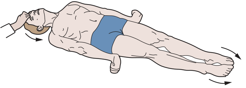
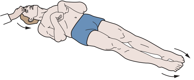
1. Decorticate Rigidity (Abnormal Flexion Posturing)
- Upper extremities: flexion of arm, wrist, and fingers + adduction at shoulder
- Lower extremities: extension, internal rotation, and plantar flexion
- Head may be extended
2. Decerebrate Rigidity (Abnormal Extension Posturing)
- Upper extremities: extension, pronation of forearms, fingers flexed
- Lower extremities: extension, internal rotation, plantar flexion (toes pointed inward)
- Opisthotonus (neck and back extended), jaw clenching
Key Differences at a Glance
| Feature | Decorticate | Decerebrate |
|---|---|---|
| Lesion level | Above red nucleus (rostral midbrain/forebrain) | At/below red nucleus (lower midbrain-upper pons) |
| Arms | Flexed, adducted | Extended, pronated |
| Legs | Extended | Extended |
| Prognosis | Serious but less severe | More ominous |
| Mechanism | Rubrospinal tract intact | Rubrospinal also disrupted |
Diagram (Ganong's Physiology)
Etiology (Common Causes)
- Traumatic brain injury (TBI) - most common
- Cerebral hemorrhage or infarct
- Uncal herniation from supratentorial mass (tumor, hematoma)
- Anoxic brain injury / hypoglycemia
- Hepatic coma (rare)
- Severe drug/alcohol intoxication (rare)
Clinical Significance
- Rostrocaudal deterioration: Progression from decorticate -> decerebrate posturing indicates downward herniation and worsening brainstem compromise.
- Decerebrate -> flaccidity: If posturing gives way to complete flaccidity, the lesion has descended below the vestibular nuclei, indicating impending brain death.
- Both responses are ominous in the setting of head injury, carrying high mortality.
- GCS scoring: Decorticate = M3 (abnormal flexion); Decerebrate = M2 (extension) on the motor component of the Glasgow Coma Scale.
- Metabolic causes (anoxia, hypoglycemia, hepatic coma) can produce either pattern and may be reversible.
Stages i Of spinal shock
stages of spinal shock Ditunno 4 phases MBBS neurology
https://www.ncbi.nlm.nih.gov/books/NBK448163
https://en.wikipedia.org/wiki/Spinal_shock
Stages of Spinal Shock
Definition
Classical Description: Two Stages (Adams & Victor)
Stage 1 - Stage of Spinal Shock (Areflexia)
- Flaccid paralysis below the level of lesion
- Complete loss of all deep tendon reflexes (areflexia)
- Loss of all sensation below the lesion
- Bladder: atonic detrusor, contracted sphincter → overflow incontinence
- Bowel: paralytic ileus, fecal retention
- Autonomic: hypotension (BP may drop to 40 mmHg), loss of vasomotor tone, loss of sweating and piloerection below lesion
- Genital reflexes abolished (bulbocavernosus, cremasteric)
- Priapism may occur
Stage 2 - Stage of Heightened Reflex Activity (Spasticity)
- Spasticity gradually replaces flaccidity
- Deep tendon reflexes return and become hyperactive (hyperreflexia)
- Extensor plantar response (Babinski sign)
- Mass reflex (flexor spasms in response to cutaneous stimuli)
- Bladder and bowel become spastic (automatic/reflex bladder)
- Autonomic dysreflexia may develop (in lesions above T6)
Modern 4-Phase Model (Ditunno et al., 2004)
| Phase | Timing | Examination Finding | Underlying Mechanism |
|---|---|---|---|
| Phase 1 | 0-1 day | Areflexia / Hyporeflexia | Loss of descending facilitation |
| Phase 2 | 1-3 days | Initial reflex return (polysynaptic first) | Denervation supersensitivity (↑ receptor expression) |
| Phase 3 | 1-4 weeks | Initial hyperreflexia | Axon-supported synapse growth (shorter axons, interneurons) |
| Phase 4 | 1-12 months | Hyperreflexia + Spasticity | Soma-supported synapse growth (longer axons, growth factors) |
Phase 1 - Areflexia (0-1 day)
- All reflexes below SCI are absent or markedly reduced
- Neurons lose supraspinal excitatory input → hyperpolarization → unresponsive to stimuli
- Flaccid paralysis, sensory loss, autonomic dysfunction
Phase 2 - Return of Polysynaptic Reflexes (1-3 days)
- Polysynaptic reflexes return first (e.g., bulbocavernosus reflex)
- Monosynaptic DTRs not yet restored
- Mechanism: denervation supersensitivity - upregulation of neurotransmitter receptors on post-synaptic neurons
- Recovery is polysynaptic → monosynaptic (NOT rostral to caudal as previously believed)
Phase 3 - Hyperreflexia Begins (1-4 weeks)
- Monosynaptic DTRs return and become exaggerated
- Interneuron axon sprouting creates new synapses below the injury
- Early spasticity appears
Phase 4 - Established Spasticity (1-12 months)
- Full hyperreflexia and spasticity
- Soma-mediated sprouting: longer axons with soma-transported growth factors form new synaptic connections
- Autonomic dysreflexia may appear in high thoracic/cervical lesions
Order of Reflex Return (Guyton & Hall)
- Stretch reflexes (DTRs) - first monosynaptic reflexes
- Flexor reflexes
- Postural antigravity reflexes
- Rudimentary stepping reflexes
Key Points for Exam
- Spinal shock is NOT neurogenic shock (do not confuse)
- Duration is longer in humans and higher primates than in lower animals
- The end of spinal shock is marked by return of the bulbocavernosus reflex
- Incomplete or slow-developing lesions produce little or no spinal shock
- Permanent spinal shock (no reflex recovery) occurs in ~5/29 cases (Kuhn) - suggests secondary vascular injury to cord segments below the lesion
- Above T6 injuries: neurogenic shock + spinal shock can coexist acutely; autonomic dysreflexia in the chronic phase
Classical decerevrate and ischemic rigidity
classical decerebrate rigidity vs ischemic rigidity alpha rigidity gamma rigidity difference physiology
classical decerebrate rigidity ischemic rigidity Sherrington gamma loop alpha rigidity dorsal root section physiology
Classical Decerebrate Rigidity vs. Ischemic (Alpha) Rigidity
Background: Normal Motor Control
| Pathway | Origin | Effect on Extensors |
|---|---|---|
| Pontine reticulospinal tract | Pons | Excitatory (++) |
| Lateral vestibulospinal tract (Deiters' nucleus) | Lateral vestibular nucleus | Excitatory (++) |
| Medullary reticulospinal tract | Medullary RF | Inhibitory (--) |
| Corticospinal, Rubrospinal, Corticoreticular | Cortex / Red nucleus | Drive inhibitory medullary RF |
Classical Decerebrate Rigidity (Gamma Rigidity)
Experimental Basis
Mechanism
- Corticospinal tract
- Corticobulbar tract
- Rubrospinal tract
- Corticoreticular fibers to medullary inhibitory RF
- Pontine excitatory reticulospinal tract
- Lateral vestibulospinal tract (Deiters' nucleus)
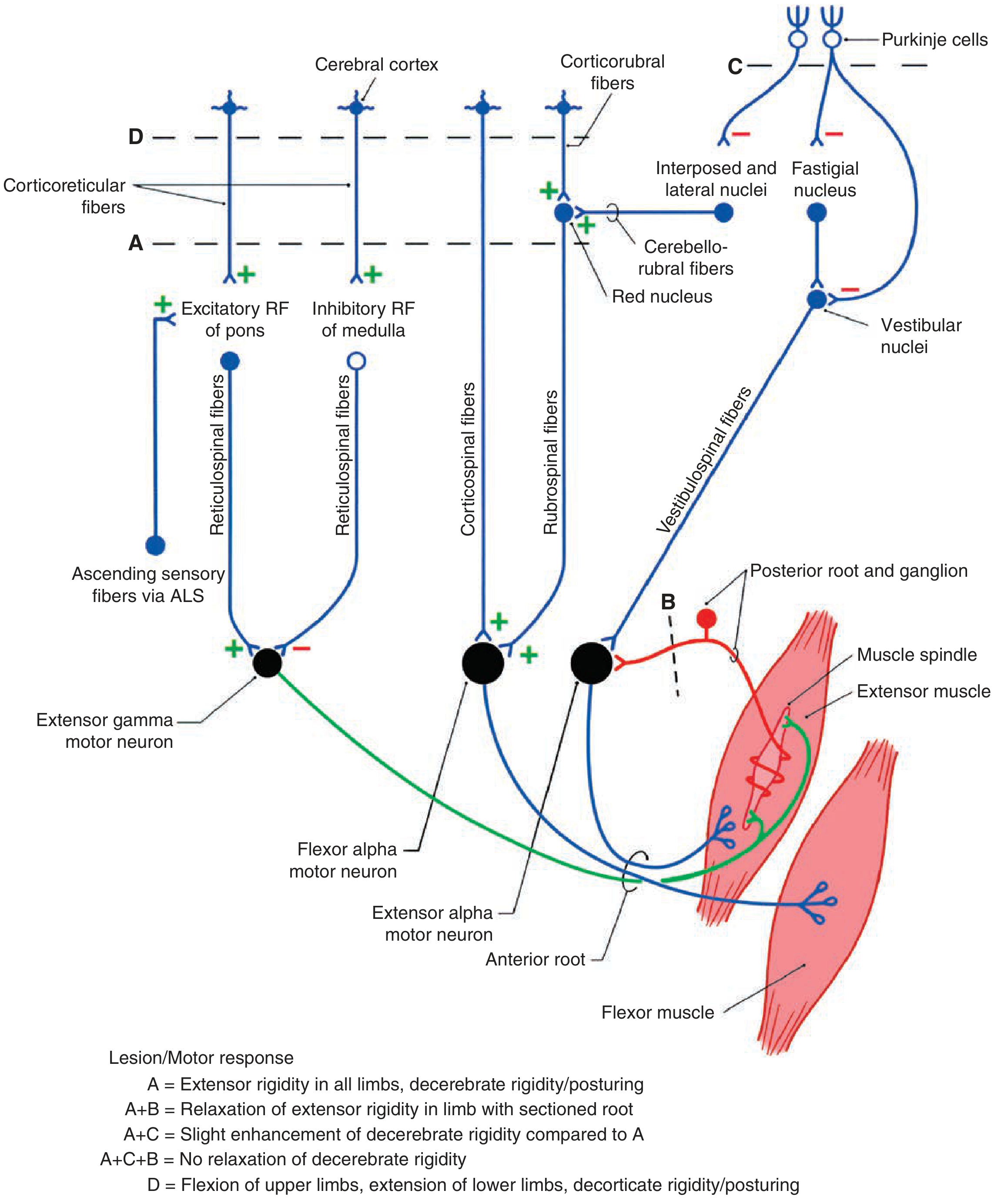
The Gamma Loop (Key Mechanism)
- Cutting the dorsal roots (section B in diagram) of a limb in a midcollicular decerebrate animal immediately abolishes the rigidity in that limb
- This is because dorsal root section interrupts the Ia afferent spindle feedback, breaking the gamma loop
- Therefore classical decerebrate rigidity is GAMMA rigidity - it depends on the intact gamma loop
Features of Classical Decerebrate Rigidity
| Feature | Detail |
|---|---|
| Lesion site | Intercollicular (between superior and inferior colliculi) |
| Antigravity muscles | Affected - neck, trunk, limb extensors |
| Abolished by | Dorsal root section, deafferentation |
| Dependent on | Gamma loop (γ → Ia spindle → α) |
| Inhibitory RF | Nonfunctional (lost cortical/rubral drive) |
| Type | Gamma (γ) rigidity |
Ischemic (Alpha) Rigidity - Decerebellate Rigidity
Experimental Basis
Mechanism
- Purkinje cell inhibitory output to the fastigial nucleus
- This disinhibits the fastigial nucleus → increased excitatory output → vestibular nuclei (Deiters') become hyperexcited
- This enormously enhances excitation of α-motor neurons directly (not via gamma loop)
- Subsequent dorsal root section (B after A+C) does NOT reverse the rigidity
- This proves it is independent of the gamma loop - alpha motor neurons are being activated directly
- Therefore ischemic/decerebellate rigidity is ALPHA rigidity
Features of Ischemic (Alpha) Rigidity
| Feature | Detail |
|---|---|
| Lesion | Anterior cerebellar lobe removal / global brainstem ischemia |
| Mechanism | Direct α-motor neuron activation (independent of spindle) |
| Abolished by | Dorsal root section does NOT abolish it |
| Dependent on | Direct drive to α-motor neurons |
| Vestibular nuclei | Greatly hyperexcited |
| Type | Alpha (α) rigidity |
Key Comparison Table
| Feature | Classical Decerebrate Rigidity | Ischemic (Alpha) Rigidity |
|---|---|---|
| Also called | Gamma rigidity | Alpha rigidity / Decerebellate rigidity |
| Transection level | Intercollicular (mid-collicular) | Intercollicular + anterior cerebellar lobe removed (or global ischemia) |
| Mechanism | γ-motor neuron → Ia spindle afferent → α-motor neuron (gamma loop) | Direct α-motor neuron activation |
| Dorsal root section | Abolishes rigidity | Does NOT abolish rigidity |
| Severity | Less severe | More severe (exaggerated rigidity) |
| Cerebellar influence | Anterior lobe intact (inhibits fastigial → partially dampens rigidity) | Anterior lobe removed → fastigial disinhibited → Deiters' nucleus hyperactive |
| Clinical correlate | Intercollicular lesions, uncal herniation | Global anoxia, severe ischemia, phenytoin toxicity (rare) |
Summary Mnemonic
"Gamma - Gone with dorsal root section" (classical decerebrate - gamma rigidity is abolished by cutting dorsal roots) "Alpha - Always stays" (ischemic/alpha rigidity persists even after dorsal root section)
Clinical Points
- Both types produce the same posture: neck extended (opisthotonus), jaws clenched, arms and legs extended, forearms pronated, feet plantar flexed
- Classical decerebrate rigidity clinically correlates with midbrain/upper pontine lesions, herniation, or metabolic causes
- Ischemic/alpha rigidity is seen after cardiac arrest, severe hypoxia/anoxia and carries an extremely poor prognosis
- The distinction is experimentally important for understanding stretch reflex physiology and the role of the gamma loop

Ans for 5 marks
professional qualities roles of a physician MBBS medical professionalism empathy patient encounters 5 marks
https://abimfoundation.org/wp-content/uploads/2015/12/Medica…
Professional Qualities and Roles of a Physician; Empathy in Patient Encounters
I. Professional Qualities of a Physician
1. Knowledge and Competence
- Thorough grounding in medical science - anatomy, physiology, pathology, pharmacology, and clinical medicine
- Commitment to lifelong learning and updating knowledge
- Clinical skills: accurate history-taking, physical examination, investigation, and management
2. Communication Skills
- Ability to communicate clearly, respectfully, and empathetically with patients, families, and colleagues
- Use of simple language - avoiding jargon when speaking to patients
- Active listening - allowing the patient to express themselves fully
3. Ethical Integrity
- Honesty and transparency in clinical practice
- Maintaining confidentiality of patient information
- Obtaining informed consent before procedures
- Avoiding conflicts of interest
4. Empathy and Compassion
- Understanding the patient's emotional and physical suffering
- Responding with compassion without losing clinical objectivity
- Treating all patients with dignity regardless of background
5. Responsibility and Accountability
- Being reliable, punctual, and thorough
- Acknowledging errors and working to correct them
- Accepting scrutiny and participating in self-regulation
6. Respect for Diversity
- Non-judgmental attitude toward patients of all races, religions, genders, and socioeconomic backgrounds
- Commitment to social justice and equitable healthcare
7. Teamwork and Collaboration
- Working effectively with nurses, paramedics, specialists, and allied health professionals
- Placing patient welfare above personal or institutional interests
II. Roles of a Physician
| Role | Description |
|---|---|
| Medical Expert | Core role - applying clinical knowledge and skills to patient care |
| Communicator | Effective communication with patients, families, and team |
| Collaborator | Working within health teams |
| Leader/Manager | Resource allocation, managing healthcare delivery |
| Health Advocate | Promoting patient and community health |
| Scholar | Lifelong learning, teaching, research |
| Professional | Ethical, accountable practice |
III. Empathy in Patient Encounters
Definition
Empathy is NOT the same as sympathy.
- Sympathy = sharing the feeling ("I feel sad too")
- Empathy = understanding the feeling ("I understand how difficult this must be for you")
Why Empathy Matters
- Builds trust in the doctor-patient relationship
- Improves patient satisfaction and treatment adherence
- Leads to better clinical outcomes - patients share more information, leading to accurate diagnosis
- Reduces patient anxiety and distress
How to Demonstrate Empathy in Clinical Encounters
- Maintain eye contact; do not interrupt
- Use open-ended questions: "Tell me how you have been feeling"
- Verbal acknowledgment: "I can see this has been very worrying for you"
- Normalize emotions: "It is understandable to feel scared about this diagnosis"
- Appropriate facial expression, nodding
- Sitting at the same level as the patient (not standing over them)
- Appropriate gentle touch when culturally appropriate
- Ask about the patient's beliefs and concerns: "What do you think is causing this?"
- Involve the patient in decision-making
- Avoid dismissing concerns
- Avoid rushing consultations
- Avoid using phones or computers during the encounter without explanation
Empathy vs. Professional Boundaries
- While empathy is essential, a physician must maintain professional detachment - over-involvement can impair clinical judgment
- The balance is called "detached concern" - caring for the patient without being emotionally overwhelmed
Key Points (for exam recall)
- Medical professionalism = knowledge + ethics + communication + empathy + accountability
- Empathy improves patient satisfaction, adherence, and outcomes
- Empathy ≠ sympathy - it is cognitive + emotional understanding
- The CanMEDS model defines 7 physician roles
- Demonstrated through active listening, verbal acknowledgment, and appropriate non-verbal cues
Moleculer motors
molecular motors kinesin dynein myosin MBBS biochemistry cell biology
Molecular Motors
Definition
Classification
| Superfamily | Track | Direction | Primary Function |
|---|---|---|---|
| Kinesin | Microtubules | Minus → Plus (anterograde) | Intracellular transport, mitosis |
| Dynein | Microtubules | Plus → Minus (retrograde) | Retrograde transport, cilia, flagella |
| Myosin | Actin filaments | Toward (+) barbed end | Muscle contraction, cell motility |
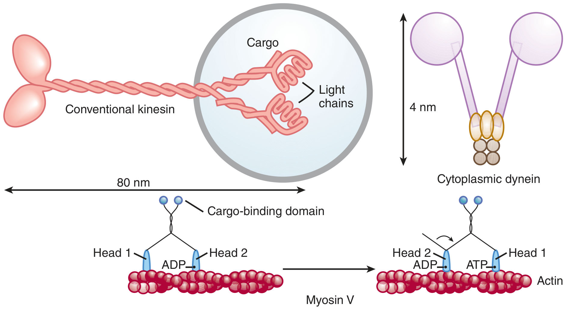
1. Kinesin
- Double-headed (two globular heads, or motor domains)
- Long coiled-coil tail that attaches to cargo (vesicles, organelles)
- Associated light chains help bind cargo
- One head binds the microtubule and bends its neck region (power stroke)
- The other head swings forward and binds the next tubulin dimer
- Produces nearly continuous, hand-over-hand "walking" powered by ATP hydrolysis at each head
- Moves toward the plus (+) end of microtubules (cell periphery / axon tips)
- Anterograde fast axonal transport in neurons: moves vesicles from cell body centrosome (minus end) to axon terminal (plus end) - e.g., neurotransmitter vesicles, synaptic proteins
- Mitosis and meiosis: spindle formation, chromosome segregation
- Transport of organelles (endosomes, lysosomes, Golgi-derived vesicles)
- Some kinesins (minus-end directed) move in the opposite direction
2. Dynein
- Axonemal (ciliary) dynein - found in cilia and flagella
- Cytoplasmic dynein - found in the cell cytoplasm
- Two heads with neck pieces embedded in a complex multi-subunit protein structure
- Large and complex compared to kinesin (~4 nm stalks connecting to microtubule-binding domain)
- ATP hydrolysis causes conformational change (bending motion) in the dynein molecule
- In cilia: dynein arms connect adjacent outer tubules of the 9+2 axoneme; when dynein on one outer tubule pulls on the adjacent tubule, the entire axoneme bends → ciliary beating motion
- Nexin links restrict excessive sliding, converting the sliding force into a coordinated bending wave
- Cytoplasmic dynein: retrograde transport (plus → minus direction) - brings endosomes, autophagosomes, and signaling molecules back toward the cell body/nucleus
- Axonemal dynein: powers beating of cilia (respiratory epithelium, fallopian tube) and flagella (sperm motility)
- Chromosome movement during cell division
3. Myosin
- Globular heads (motor domain) that bind actin + hydrolyze ATP
- Neck region (lever arm / hinge region) amplifies conformational change
- Tail region for cargo binding or thick filament formation
- 18 classes - most important are:
- Myosin II (conventional muscle myosin) - forms thick filaments, responsible for muscle contraction
- Myosin V - "walks" along actin filaments carrying organelles; vesicle and melanosome transport
- ATP binds to myosin head → myosin detaches from actin
- ATP hydrolysis (ADP + Pi) → myosin head cocks into high-energy "cocked" position
- Myosin head binds actin (cross-bridge formation)
- Pi release → power stroke - myosin head pivots 45° at hinge → actin filament slides
- ADP released → rigor state (tight binding)
- New ATP binds → cycle repeats
- Myosin II: skeletal, cardiac, and smooth muscle contraction; cytokinesis (contractile ring)
- Myosin V: transports organelles, vesicles, and melanosomes along actin filaments (hand-over-hand walking, toward barbed + end)
- Cell motility, lamellipodia formation, phagocytosis
Comparison Summary
| Feature | Kinesin | Dynein | Myosin |
|---|---|---|---|
| Track | Microtubule | Microtubule | Actin filament |
| Direction | Anterograde (+ end) | Retrograde (- end) | Toward barbed (+) end |
| Size | ~100 kDa | Large complex | Varies (230 kDa heavy chain) |
| Energy source | ATP | ATP | ATP |
| Key role | Axonal anterograde transport | Retrograde transport; cilia/flagella | Muscle contraction; cell motility |
| Found in | Neurons, dividing cells | Cilia, neurons, all cells | Muscle, all cells |
Clinical Relevance
| Disease | Motor Involved | Mechanism |
|---|---|---|
| Kartagener syndrome (Primary Ciliary Dyskinesia) | Axonemal dynein (absent dynein arms) | Immotile cilia → bronchiectasis, situs inversus, male infertility |
| Charcot-Marie-Tooth disease | Kinesin (KIF1B mutation) | Impaired axonal transport |
| Alzheimer's disease | Kinesin/Dynein | Disrupted axonal transport → amyloid accumulation |
| Muscle myopathies | Myosin heavy chain mutations | Impaired cross-bridge cycling |
| Vitiligo/Griscelli syndrome | Myosin Va | Defective melanosome transport to keratinocytes |
Key Points
- All three superfamilies are ATPases - they use ATP hydrolysis to generate movement
- Kinesin = anterograde (cell periphery / axon tip); Dynein = retrograde (cell centre / cell body)
- Myosin = actin-based; Kinesin and Dynein = microtubule-based
- The "head" domain binds the cytoskeletal track and contains the ATPase site
- The "tail" domain binds the cargo
- Dynein arms in cilia cause microtubule sliding → converted to bending by nexin cross-links
Short note on gibbs donan equilibrium
Gibbs Donnan equilibrium MBBS physiology short note membrane impermeant ions
Gibbs-Donnan Equilibrium
Definition
Prerequisites / Conditions
- A semipermeable membrane must separate two compartments
- One side must contain a non-diffusible charged ion (impermeant ion - usually a large negatively charged protein)
- The diffusible ions must be free to move across the membrane
- Electroneutrality must be maintained in each compartment at all times
Basic Example / Setup
| Compartment X (e.g., plasma / inside cell) | Compartment Y (e.g., ISF / outside cell) |
|---|---|
| K⁺, Cl⁻, Prot⁻ (impermeant protein anion) | K⁺, Cl⁻ only |
| Membrane permeable to K⁺ and Cl⁻, but NOT to Prot⁻ |
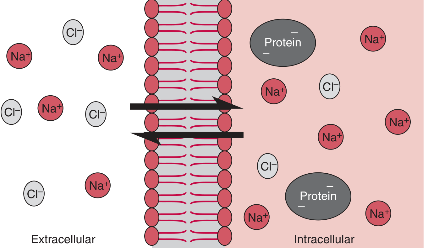
The Gibbs-Donnan Equation
- This is the product rule: the product of diffusible cation and anion concentrations is equal on both sides
- This holds for any pair of monovalent cation and anion
- The ratio r = [Na⁺]outside / [Na⁺]inside = [Cl⁻]inside / [Cl⁻]outside is called the Donnan ratio
- In plasma vs. interstitial fluid, the Gibbs-Donnan ratio is approximately 0.95 for both Na⁺ and Cl⁻
Three Key Consequences
1. Asymmetric Distribution of Diffusible Ions
- Cations (Na⁺, K⁺) are slightly higher on the protein-containing side (X)
- Anions (Cl⁻, HCO₃⁻) are slightly lower on the protein-containing side (X)
- Electrical neutrality is maintained in each compartment individually
- Plasma has impermeant proteins (albumin, globulins - negatively charged)
- Result: [Na⁺]plasma slightly > [Na⁺]interstitial fluid
- Result: [Cl⁻]plasma slightly < [Cl⁻]interstitial fluid
- Example: Cl⁻ in CSF is ~15% higher than plasma (blood-brain barrier excludes proteins, so CSF side must have higher Cl⁻ to balance)
2. Osmotic Pressure Difference (Oncotic Pressure)
- The protein-containing side (X) has more total osmotically active particles than side Y
- This is because cations are attracted in, adding to the particles already present
- This contributes to the colloid osmotic pressure (oncotic pressure) of plasma
- The Gibbs-Donnan effect adds approximately 1 mOsm/L extra to plasma oncotic pressure beyond just the protein molecules themselves - this is the Donnan osmotic effect
[K⁺_X] + [Cl⁻_X] + [Prot⁻_X] > [K⁺_Y] + [Cl⁻_Y] Side X has more osmotically active particles
3. Electrical Potential Difference Across the Membrane
- Because diffusible ions are asymmetrically distributed, an electrical potential difference develops across the membrane
- Side X (protein-containing) becomes electrically negative relative to side Y
- The charges line up along the membrane - the electrical gradient exactly balances the concentration gradient for each ion (satisfying the Nernst equation for each diffusible ion)
- This Donnan potential contributes to the resting membrane potential of cells
Gibbs-Donnan Effect and Cell Volume (Critical Concept)
- These intracellular anions attract cations (Na⁺, K⁺) inward
- More osmotically active particles accumulate inside the cell
- Water enters osmotically → cell swells → eventually bursts (osmotic lysis)
- The Na-K pump continuously pumps 3 Na⁺ out and 2 K⁺ in per cycle
- This actively excludes NaCl from the intracellular space, counteracting Donnan-driven swelling
- The cell exists in a steady state, NOT true equilibrium - it requires constant energy expenditure
- If the Na-K pump is inhibited (e.g., hypoxia, ouabain):
- Na⁺ enters, Cl⁻ follows (to maintain electroneutrality)
- Intracellular osmolarity rises → water enters → cell swells and may rupture
Key concept: Normal cell volume depends on the Na⁺/K⁺-ATPase doing "osmotic work" to counteract Donnan forces. NaCl acts as a functionally impermeant extracellular solute that offsets intracellular negative charges.
Physiological Applications
| Situation | Application of Gibbs-Donnan Principle |
|---|---|
| Plasma vs. Interstitial fluid | Higher cations, lower anions in plasma due to plasma proteins (albumin) |
| Plasma vs. CSF | Cl⁻ ~15% higher in CSF (blood-brain barrier keeps proteins out) |
| Resting membrane potential | Donnan forces contribute to negative intracellular potential |
| Cell volume regulation | Na-K pump counteracts Donnan-driven swelling |
| Oncotic pressure | Donnan effect adds to colloid osmotic pressure of plasma |
| Red blood cells | Chloride shift (hamburger phenomenon) - HCO₃⁻/Cl⁻ exchange maintains Donnan equilibrium |
| Capillary fluid exchange (Starling forces) | Plasma proteins create oncotic pressure partly through Donnan effect, retaining fluid in capillaries |
Key Points for Exam
- Gibbs-Donnan equilibrium: unequal distribution of diffusible ions due to impermeant charged ion on one side
- Equation: [cation_X × anion_X] = [cation_Y × anion_Y]
- Side with impermeant anion: more cations, fewer anions, more total osmotic particles, electrically negative
- Three effects: asymmetric ion distribution, osmotic pressure difference, electrical potential
- Animal cells must use Na-K pump to prevent Donnan-driven swelling - it is a steady state, not true equilibrium
- Donnan ratio (r) between plasma and ISF ≈ 0.95 for Na⁺ and Cl⁻
Difference between Na and K channels
difference between voltage gated sodium and potassium channels structure gating kinetics action potential physiology
Differences Between Voltage-Gated Na⁺ and K⁺ Channels
1. Structure
| Feature | Voltage-gated Na⁺ Channel (Nav) | Voltage-gated K⁺ Channel (Kv) |
|---|---|---|
| Functional unit | Single large α-subunit (pseudotetramer) | Tetramer of 4 identical or similar subunits |
| α-subunit domains | 1 α-subunit with 4 homologous domains (I-IV), each containing 6 transmembrane segments (S1-S6) | Each subunit contains 6 transmembrane segments (S1-S6); 4 subunits assemble around a central pore |
| Total TM segments | 24 (4 domains × 6 segments) | 24 (4 subunits × 6 segments) |
| Auxiliary subunits | β₁ and β₂ subunits (modulate gating and expression) | β subunits (Kvβ) in some types |
| Voltage sensor | S4 segment in each domain (positively charged Arg/Lys residues) | S4 segment in each of the 4 subunits |
| Selectivity filter | DEKA motif (Asp-Glu-Lys-Ala) in the P-loops of the 4 domains - Na⁺ selective | TVGYG motif (Thr-Val-Gly-Tyr-Gly) - K⁺ selective |
| Inactivation particle | III-IV linker loop (intracellular) acts as "ball and chain" inactivation gate (h gate) | Some Kv channels have N-type inactivation ("ball and chain" from N-terminus); slow C-type inactivation also exists |
| Gene family | SCN1A-SCN11A (Nav1.1-Nav1.9) | KCNA-KCNS (Kv1-Kv12 families), >40 subtypes |
2. Gating States
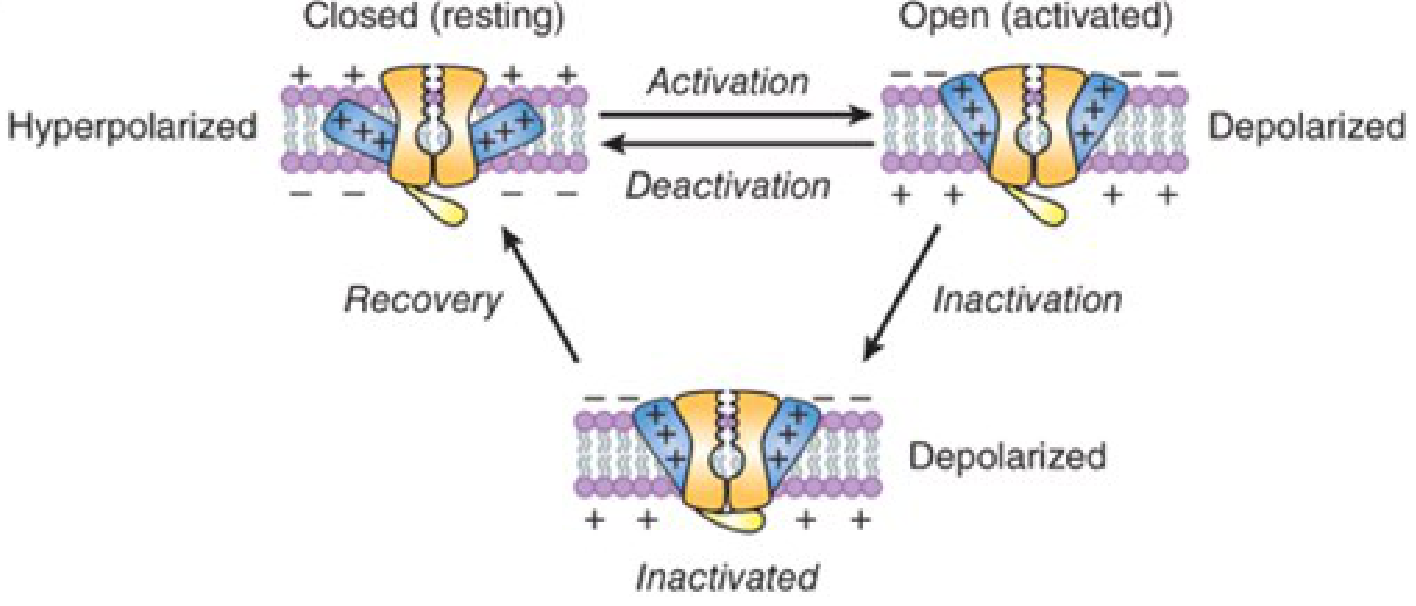
Na⁺ Channel - THREE States
-
Closed (Resting) - at resting membrane potential (-70 mV)
- Activation gate (m gate) = CLOSED
- Inactivation gate (h gate) = OPEN
- Channel is available to open
-
Open (Activated) - at threshold/during depolarization
- Activation gate (m gate) = OPEN
- Inactivation gate (h gate) = OPEN
- Na⁺ rushes in (upstroke of AP)
- Duration: only ~1 ms (extremely brief)
-
Inactivated - rapidly after opening
- Activation gate (m gate) = OPEN
- Inactivation gate (h gate) = CLOSED (III-IV linker "plugs" the pore)
- Channel cannot re-open → absolute refractory period
- Returns to closed (resting) state only after repolarization
K⁺ Channel (Delayed Rectifier) - TWO States
-
Closed - at resting potential
- Activation gate (n gate) = CLOSED
- No separate inactivation gate in most delayed rectifier Kv channels
-
Open - after delayed activation during depolarization
- Activation gate (n gate) = OPEN
- K⁺ flows out (repolarization)
- Returns to closed upon repolarization - does NOT inactivate (in most delayed rectifier types)
Key difference: Na⁺ channels have a fast inactivation mechanism; delayed rectifier K⁺ channels do not inactivate (or inactivate very slowly).
3. Kinetics (Speed)
| Feature | Na⁺ Channel | K⁺ Channel (Delayed Rectifier) |
|---|---|---|
| Activation speed | Very fast (opens in < 1 ms after threshold) | Slow (opens after 1-5 ms delay) |
| Inactivation | Fast (closes within 1-2 ms of opening) | None (delayed rectifier) or very slow |
| Recovery from inactivation | Requires repolarization (~1-2 ms) | Simple deactivation upon repolarization |
| Hodgkin-Huxley notation | m³h (3 m-gates + 1 h-gate) | n⁴ (4 n-gates) |
- Na⁺ channel opens fast → rapid depolarization (upstroke)
- Na⁺ channel inactivates fast → AP cannot sustain
- K⁺ channel opens slowly → repolarization (falling phase)
- K⁺ channel does not inactivate → may cause brief hyperpolarization (after-hyperpolarization)
4. Role in the Action Potential
| Phase | Channel | Event |
|---|---|---|
| Resting potential (-70 mV) | Both closed | Leak K⁺ channels maintain resting potential |
| Threshold (-55 mV) | Na⁺ channels begin opening | Threshold depolarization reached |
| Upstroke / Depolarization (+30 mV) | Na⁺ channels open rapidly | Na⁺ influx → rapid depolarization (Eₙₐ = +60 mV) |
| Peak (+30 mV) | Na⁺ channels inactivate; K⁺ channels open | Na⁺ inflow stops; K⁺ outflow begins |
| Repolarization | K⁺ channels open fully | K⁺ efflux → membrane repolarizes |
| After-hyperpolarization | K⁺ channels slowly close | Brief dip below resting potential |
| Refractory period | Na⁺ channels inactivated | No new AP can be generated (absolute refractory period) |
5. Pharmacology and Blockers
| Drug / Toxin | Na⁺ Channel | K⁺ Channel |
|---|---|---|
| Tetrodotoxin (TTX) | Blocks (extracellular pore) | No effect |
| Local anesthetics (lidocaine, bupivacaine) | Block Na⁺ channels (use-dependent) | No direct effect |
| Antiepileptics (phenytoin, lamotrigine, carbamazepine) | Block (bind to inactivated state) | No effect |
| Tetraethylammonium (TEA) | No effect | Blocks K⁺ channels |
| 4-Aminopyridine (4-AP) | No effect | Blocks voltage-gated K⁺ channels |
| Scorpion/sea anemone toxins | Delay inactivation | - |
| Class I antiarrhythmics (quinidine, flecainide) | Block Na⁺ channels | - |
| Class III antiarrhythmics (amiodarone, sotalol) | Some Na⁺ block | Block K⁺ channels (prolong repolarization) |
6. Summary Comparison Table
| Feature | Voltage-gated Na⁺ Channel | Voltage-gated K⁺ Channel |
|---|---|---|
| Ion selectivity | Na⁺ (inward current) | K⁺ (outward current) |
| Structure | 1 α-subunit, 4 domains, pseudotetramer | 4 separate subunits, true tetramer |
| Activation threshold | ~ -55 mV | ~ -40 mV (opens after Na⁺ channel) |
| Activation speed | Very fast (< 1 ms) | Slow (1-5 ms delay) |
| Inactivation | Fast (h gate, III-IV linker) | No inactivation (delayed rectifier) |
| States | 3 states: Closed / Open / Inactivated | 2 states: Closed / Open |
| Direction of ion flow | Inward (Na⁺ in) | Outward (K⁺ out) |
| Role in AP | Upstroke (depolarization) | Repolarization |
| Refractory period | Responsible for absolute RP | Contributes to relative RP |
| Selectivity filter | DEKA motif | TVGYG motif |
| Toxin | Blocked by TTX | Blocked by TEA / 4-AP |
| Gene | SCN family | KCNA-KCNS family |
Clinical Relevance
- Mutations in Nav1.4 (SCN4A): Hyperkalemic periodic paralysis, paramyotonia congenita (impaired inactivation → sustained Na⁺ influx → membrane depolarization)
- Mutations in Nav1.1 (SCN1A): Dravet syndrome (epilepsy)
- Mutations in Kv1.1 (KCNA1): Episodic ataxia type 1
- Long QT syndrome: Mutations in cardiac K⁺ channels (KCNQ1, KCNH2) or Na⁺ channels (SCN5A) - impaired repolarization → prolonged AP → arrhythmia
- Local anesthetics: Block Na⁺ channels in their inactivated state (use-dependent block) → prevent nerve conduction
Physiological basis of st elevation
physiological basis ST elevation current of injury ischemia ECG mechanism diastolic systolic current
https://pmc.ncbi.nlm.nih.gov/articles/PMC4116460
Physiological Basis of ST Elevation
What is the ST Segment?
Core Concept: The "Current of Injury"
"Severe, acute ischemia lowers the resting membrane potential and shortens the duration of the action potential. Such changes cause a voltage gradient between normal and ischemic zones. As a consequence, current flows between those regions. These currents of injury are represented on the surface ECG by deviation of the ST segment." — Harrison's Principles of Internal Medicine 22e
Cellular Effects of Ischemia on the Action Potential
| Effect | Mechanism |
|---|---|
| Resting membrane potential rises (less negative, partial depolarization) | ATP depletion → Na⁺/K⁺-ATPase fails → Na⁺ accumulates inside → K⁺ leaks out → reduced electronegativity |
| Action potential duration shortens | Ischemia accelerates repolarization (shortens Phase 2 plateau) |
| AP amplitude decreases | Reduced Na⁺ channel availability at the depolarized resting potential |
| Loss of epicardial AP dome | Ischemia particularly shortens the epicardial action potential plateau (Phase 2 dome lost) |
Two Mechanisms of ST Elevation
1. Diastolic Current of Injury (TQ Depression)
- Ischemic cells are partially depolarized (resting Vm less negative, e.g., -60 mV instead of -90 mV) → relatively positive compared to normal
- Normal cells are fully repolarized (-90 mV) → relatively negative
- This depresses the TQ baseline (the true zero reference) in leads facing the ischemic zone
- Because ECG machines are AC-coupled and use the TP segment as their baseline reference, this TQ depression is interpreted as apparent ST elevation
2. Systolic Current of Injury (True ST Elevation)
- Normal cells are fully depolarized and at plateau potential → relatively positive
- Ischemic cells have an abbreviated/shortened action potential (early repolarization, shortened plateau) → repolarize earlier → relatively negative during systole
- The ischemic zone is relatively negative during systole → the ST vector points away from the ischemic zone in normal leads, but the epicardial ischemia creates a net outward ST vector pointing toward overlying leads
- This produces true ST elevation in leads overlying the transmural/epicardial ischemia
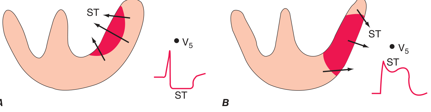
Vector Basis: Transmural vs. Subendocardial Ischemia
| Type | ST Vector Direction | ECG Appearance |
|---|---|---|
| Transmural (epicardial) ischemia | Directed outward (toward epicardium / away from cavity) | ST elevation in overlying leads; reciprocal ST depression in opposite leads |
| Subendocardial ischemia | Directed inward (toward endocardium / into cavity) | ST depression in precordial leads; ST elevation in aVR |
Additional Ionic Mechanisms
-
ATP-sensitive K⁺ channels (I_KATP) open - ischemia depletes ATP → I_KATP channels activate → K⁺ efflux → shortens AP plateau → creates transmural voltage gradient
-
Accumulation of extracellular K⁺ - K⁺ exits ischemic cells → [K⁺]outside rises → depolarizes resting Vm of ischemic cells → potentiates the diastolic injury current
-
Loss of epicardial AP dome - Ischemia predominantly affects the epicardial cells (shorter AP plateau) while endocardial cells retain their dome → this transmural gradient directly generates the systolic current of injury producing ST elevation
-
Na⁺ channel inactivation - Partial depolarization of ischemic cells inactivates fast Na⁺ channels → reduced AP amplitude → reduced excitability in epicardium → systolic transmural gradient
ECG Evolution in STEMI (Time Course)
| Time | ECG Change | Mechanism |
|---|---|---|
| Minutes | Hyperacute T waves (tall, broad, peaked) | Earliest sign; increased T amplitude due to focal ischemia |
| Minutes-hours | ST elevation (injury current) | Transmural ischemia/injury - current of injury |
| Hours-days | T-wave inversion | Repolarization abnormality in peri-infarct zone |
| Hours-days | Pathological Q waves | Necrosis → loss of depolarization forces from infarcted tissue |
| Days-weeks | ST normalizes | Resolution of injury; Q waves and T-inversion may persist |
Localization of ST Elevation
| Leads with ST elevation | Region of ischemia | Likely artery |
|---|---|---|
| V1-V4 | Anterior wall | LAD (Left Anterior Descending) |
| II, III, aVF | Inferior wall | RCA (Right Coronary Artery) |
| I, aVL, V5-V6 | Lateral wall | LCx (Left Circumflex) |
| V1-V3 (ST depression - "posterior equivalent") | Posterior wall | RCA / LCx |
| V1, V4R (right-sided leads) | Right ventricle | Proximal RCA |
Other Causes of ST Elevation (Non-Ischemic)
- Acute pericarditis - diffuse, saddle-shaped ST elevation in multiple leads + PR depression
- Vasospastic (Prinzmetal's) angina - transient, reversible ST elevation without infarction
- Benign early repolarization - concave upward ST elevation in young individuals, notching at J-point
- Left ventricular aneurysm - persistent ST elevation after old MI
- Brugada syndrome - coved-type ST elevation in V1-V3
- Hyperkalemia - may cause ST changes
- Takotsubo syndrome - transient ST elevation mimicking STEMI
Summary
- Diastolic: Partially depolarized ischemic cells are relatively positive → current flows toward normal cells → true TQ baseline depression (appears as ST elevation on AC-coupled ECG)
- Systolic: Ischemic cells have shortened APs and repolarize early → are relatively negative during systole → current flows from normal to ischemic zone → true ST elevation
Clinical Case: 60-year-old Male, Left Hemiplegia + MCA Blockage
a) Probable Diagnosis (1 mark)
- Left-sided weakness (upper and lower limb) → contralateral to lesion → right hemisphere
- Drowsiness → cortical involvement
- Difficulty speaking (dysphasia/aphasia) → though patient is 60M, if dominant hemisphere (usually left) were involved this would be classic Broca's aphasia; here the "uncommunicative behaviour / difficulty speaking" with LEFT hemiplegia suggests right MCA, though dysarthria is also possible
- Left facial paralysis → upper motor neuron type (central), right hemisphere corticobulbar tract involvement
- CT scan confirms blockage of MCA
- Increased tone in left limbs → UMN lesion pattern
b) Pathophysiology of Ischemic Stroke (2 marks)
Step 1: Occlusion
Step 2: Energy Failure
- Normal cerebral blood flow (CBF) = 50-55 mL/100g/min
- CBF < 20 mL/100g/min → neuronal dysfunction (electrical failure) → symptoms begin
- CBF < 10 mL/100g/min → neuronal death (infarction core) within minutes
Step 3: Ionic Cascade
- Na⁺ accumulates inside neurons → osmotic water entry → cytotoxic edema (cell swelling)
- Membrane depolarization → voltage-gated Ca²⁺ channels open → massive Ca²⁺ influx (most damaging step)
Step 4: Excitotoxicity
- Ischemic neurons release excessive glutamate
- Glutamate binds NMDA and AMPA receptors → further Ca²⁺ influx → excitotoxic neuronal death
Step 5: Downstream Damage
- Ca²⁺ overload activates:
- Phospholipases → membrane breakdown → free fatty acids + arachidonic acid
- Proteases (calpain) → cytoskeletal destruction
- Endonucleases → DNA fragmentation
- Nitric oxide synthase (NOS) → free radical (ROS) generation → oxidative stress
- Mitochondrial dysfunction → cytochrome C release → apoptosis
Step 6: Ischemic Penumbra
- Surrounding the infarct core is the ischemic penumbra - a zone of functionally impaired but structurally viable tissue that can be salvaged if reperfusion occurs within the therapeutic window (4.5 hours for IV tPA)
Step 7: Secondary Injury
- Vasogenic edema (blood-brain barrier breakdown, hours to days)
- Inflammatory cell infiltration
- If large infarct: cerebral herniation
c) Hemiplegia and Paraplegia (2 marks)
Hemiplegia
- Definition: Complete paralysis of one side of the body (upper AND lower limb on the same side), often with facial palsy on the same side
- Cause: Unilateral lesion of the corticospinal/corticobulbar tract above the pyramidal decussation (i.e., in cerebral hemisphere, internal capsule, or upper brainstem)
- Side: Contralateral to the lesion (because the corticospinal tract crosses at the pyramidal decussation in the medulla)
- Features (UMN type):
- Increased tone (spasticity) - clasp-knife rigidity
- Hyperreflexia (exaggerated deep tendon reflexes)
- Extensor plantar response (Babinski sign)
- Initial flaccidity → later spasticity
- No significant muscle wasting
- Example: Right MCA stroke → Left hemiplegia (as in this case)
Paraplegia
- Definition: Paralysis of both lower limbs (legs) only - upper limbs are spared
- Cause: Lesion of the spinal cord at thoracic level (T1-L1), affecting both corticospinal tracts bilaterally; or bilateral lesion affecting both lower limb areas of the motor cortex (parasagittal meningioma, superior sagittal sinus thrombosis)
- Features (UMN type - if spinal cord lesion):
- Spastic paraplegia (increased tone, brisk reflexes)
- If below conus medullaris: LMN features (flaccid paraplegia)
- Bladder and bowel involvement common
- Sensory level may be present
- Examples: Traumatic spinal cord injury, multiple sclerosis, spinal cord tumor, transverse myelitis
| Feature | Hemiplegia | Paraplegia |
|---|---|---|
| Limbs affected | One side (arm + leg + face) | Both lower limbs only |
| Lesion site | Above pyramidal decussation (hemisphere / IC / upper brainstem) | Bilateral spinal cord (thoracic) or bilateral parasagittal cortex |
| Side affected | Contralateral to hemispheric lesion | Bilateral |
| Common cause | Stroke, brain tumor, head injury | Spinal cord injury, MS, tumor |
d) Origin, Course, and Termination of Corticospinal Tract (4 marks)
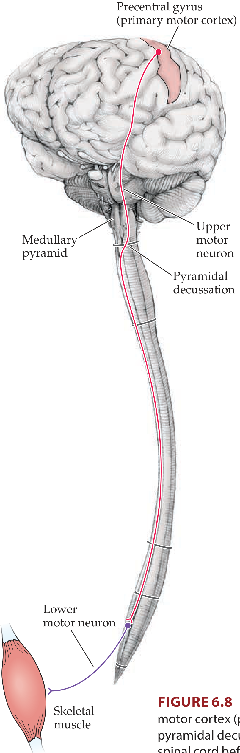
Origin
| Region | % of fibers |
|---|---|
| Primary motor cortex (Brodmann area 4) - precentral gyrus | >50% |
| Premotor and supplementary motor areas (area 6) | ~30% |
| Parietal somatosensory cortex (areas 3, 1, 2, 5, 7) | ~20% |
- ~3% are Betz cells (giant pyramidal neurons, layer V) - the largest neurons in the human nervous system
- Somatotopic arrangement: face (medial) → arm → leg (lateral, near longitudinal fissure)
Course
- Axons descend from cortex into the corona radiata (fan-shaped white matter above the internal capsule)
- Fibers converge into the posterior limb of the internal capsule
- Somatotopic order: face fibers most anterior (near genu), arm fibers intermediate, leg fibers most posterior
- Compact arrangement here - a small lesion (lacunar infarct) can cause complete contralateral hemiplegia
- Pass through the middle one-third of the basis pedunculi (cerebral peduncles)
- Arrangement: face (medial) → arm → leg (lateral)
- Corticopontine fibers occupy the medial and lateral thirds
- Fibers become scattered as fascicles (separated by transverse pontine fibers and pontine nuclei)
- Corticobulbar fibers leave here to synapse on cranial nerve motor nuclei
- Fibers re-converge on the ventral surface of the medulla to form the medullary pyramids
- This is why the corticospinal tract is also called the pyramidal tract
- At the junction of medulla and spinal cord (foramen magnum level), ~85% of fibers cross to the opposite side → pyramidal decussation (decussation of the pyramids)
- ~15% do NOT cross → continue as anterior (ventral) corticospinal tract
After Decussation
- Crossed fibers form the lateral corticospinal tract in the dorsolateral white matter of the lateral funiculus
- Present throughout the length of the spinal cord
- Somatotopic: upper limb fibers medial, lower limb fibers lateral within the tract
- Uncrossed 15% form the anterior (ventral) corticospinal tract in the anterior funiculus
- Most of these cross at the spinal cord segmental level before terminating
Termination
- Corticospinal fibers synapse (directly or via interneurons) on lower motor neurons (LMN) in the anterior (ventral) horn of the spinal cord gray matter (Rexed laminae VI, VII, IX)
- Direct monosynaptic connections are most important for fine, skilled movements (especially hand and finger movements)
- Corticobulbar fibers terminate on cranial nerve motor nuclei in the brainstem (III, IV, V, VI, VII, IX, X, XI, XII)
Key Rule - Laterality
- Lesion above pyramidal decussation (cortex, IC, midbrain, pons, medulla above decussation) → contralateral weakness
- Lesion below pyramidal decussation (spinal cord) → ipsilateral weakness
e) Line of Treatment (1 mark)
Acute Management (within 4.5 hours = "Golden Window")
- IV Alteplase (tPA) 0.9 mg/kg - if within 4.5 hours of symptom onset, no contraindications (standard of care for ischemic stroke)
- Mechanical thrombectomy - if large vessel occlusion (MCA), within 6-24 hours
Immediate Supportive Care
- ABC - Airway, Breathing, Circulation
- Aspirin 300 mg (if tPA not given) - antiplatelet
- Oxygen if SpO₂ < 94%
- IV fluids (Normal saline) - maintain euvolemia
- Blood pressure control - allow permissive hypertension up to 220/120 mmHg acutely (unless thrombolysis given, then < 180/105)
- Blood glucose control - target 140-180 mg/dL
- Statin therapy - atorvastatin 40-80 mg
- DVT prophylaxis - heparin/LMWH after 24-48 hours
Rehabilitation
- Physiotherapy - limb strengthening, gait training
- Speech therapy - for aphasia/dysarthria
- Occupational therapy
Secondary Prevention
- Antiplatelet therapy (aspirin + clopidogrel or clopidogrel alone long-term)
- Statin (atorvastatin)
- Antihypertensives (if hypertensive - ACE inhibitor + diuretic preferred)
- Anticoagulation if atrial fibrillation is the cause (warfarin / NOAC)
- Carotid endarterectomy if significant carotid stenosis (>70%) identified
Walleriaj degeneration in Short guyton with flowchart
Wallerian degeneration Guyton physiology nerve injury axon degeneration regeneration steps
Wallerian Degeneration
Definition
Prerequisites
- Requires axonotmesis (grade II) or higher injury - simple neurapraxia (conduction block) does NOT cause Wallerian degeneration
- The cell body (perikaryon) must remain intact for regeneration to occur
- The endoneurial tubes (Schwann cell basement membrane) must ideally be preserved for guided regeneration
Diagram
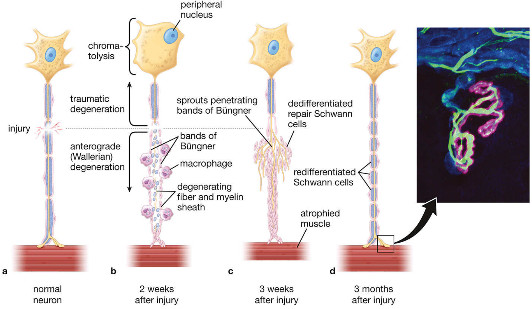
Flowchart
NERVE INJURY (axonotmesis / neurotmesis)
│
▼
┌──────────────────────────────────────────┐
│ IMMEDIATE (0-24 hrs) │
│ - Axoplasm leaks at injury site │
│ - Ca²⁺ and Na⁺ influx through │
│ disrupted axonal membrane │
│ - Axoplasmic flow interrupted │
└──────────────┬───────────────────────────┘
│
┌─────────┴──────────┐
▼ ▼
DISTAL TO INJURY PROXIMAL TO INJURY
(Wallerian / (Retrograde changes)
Anterograde
degeneration)
│ │
▼ ▼
┌─────────────────┐ ┌────────────────────┐
│ 8-24 hrs: │ │ CELL BODY: │
│ Axonal swelling │ │ CHROMATOLYSIS │
│ │ │ - Nissl bodies │
│ │ │ disperse/dissolve │
│ │ │ - Nucleus moves to │
│ │ │ cell periphery │
│ │ │ - Cell swells │
│ │ │ - Switch from │
│ │ │ maintenance to │
│ │ │ regeneration mode │
└────────┬────────┘ └────────────────────┘
│
▼
┌─────────────────────┐
│ DAY 1-3: │
│ Granular │
│ disintegration of │
│ axonal cytoskeleton │
│ - Microtubules │
│ disassemble │
│ - Neurofilaments │
│ break down │
│ - Axon fragments │
└────────┬────────────┘
│
▼
┌─────────────────────┐
│ DAY 3-7: │
│ SCHWANN CELL │
│ CHANGES │
│ - Retract from │
│ nodes of Ranvier │
│ - Dedifferentiate │
│ → Repair Schwann │
│ cells │
│ - Downregulate │
│ myelin proteins │
│ - Upregulate c-Jun │
│ - Myelin autophagy │
│ (myelin breakdown) │
└────────┬────────────┘
│
▼
┌─────────────────────┐
│ DAY 3-14: │
│ MACROPHAGE │
│ INFILTRATION │
│ - Schwann cells │
│ secrete cytokines │
│ - Macrophages │
│ recruited │
│ - Phagocytosis of │
│ axon debris + │
│ myelin fragments │
│ (myelin = "debris") │
└────────┬────────────┘
│
▼
┌─────────────────────┐
│ ~1 WEEK: │
│ BANDS OF BÜNGNER │
│ - Repair Schwann │
│ cells proliferate │
│ - Align along empty │
│ endoneurial tubes │
│ - Form hollow │
│ cellular "tubes" │
│ - Secrete NGF, BDNF, │
│ GGFs (neurotrophins│
│ guide regeneration)│
└────────┬────────────┘
│
▼
┌─────────────────────────────────────────────┐
│ REGENERATION PHASE │
│ │
│ Day 4 onwards: Proximal stump sends sprouts │
│ Growing axon cone penetrates Bands of Büngner│
│ Rate: 0.5-3 mm/day (avg ~1 mm/day) │
│ Target organ reinnervated │
│ Schwann cells redifferentiate → remyelinate │
│ New myelin = thinner, shorter internodes │
└─────────────────────────────────────────────┘
Detailed Steps (Guyton-style)
A. Distal Changes - Wallerian (Anterograde) Degeneration
| Time | Event |
|---|---|
| 0-24 hours | Axonal swelling; Ca²⁺ influx activates proteases; loss of anterograde/retrograde transport |
| Day 1-3 | Axon fragments and disintegrates; granular disintegration of cytoskeleton (microtubules, neurofilaments break down) |
| Day 3 | Schwann cells retract from nodes of Ranvier; dedifferentiate into repair Schwann cells; myelin autophagy begins |
| Day 3-14 | Macrophages infiltrate; phagocytose axon fragments and myelin debris |
| ~1 week | Complete Wallerian degeneration of distal stump; endoneurial tubes emptied |
| 1-2 weeks | Proliferating repair Schwann cells form Bands of Büngner (linear columns inside endoneurial tubes) |
B. Proximal Changes - Traumatic (Retrograde) Degeneration
- Limited degeneration proximal to injury - up to the first node of Ranvier
- More extensive with proximal injuries
C. Cell Body Changes - Chromatolysis
- Chromatolysis = dissolution of Nissl bodies (rough ER - ribosomes)
- Nucleus moves to cell periphery (eccentric displacement)
- Cell body swells
- Switch from neurotransmitter synthesis → structural protein synthesis (actin, tubulin for regeneration)
- Transcription factors upregulated (c-Jun, ATF3, Sox2)
- If injury is very proximal → cell body may undergo apoptosis (cell death, no regeneration)
D. Nerve Regeneration
- Day 4: Proximal stump sends multiple axonal sprouts (growth cones) into the endoneurial tube
- Sprouts attracted by neurotrophins (NGF, BDNF, GDNF) secreted by repair Schwann cells
- Growth cones penetrate Bands of Büngner and advance at 0.5-3 mm/day
- On reaching target organ: new neuromuscular junctions form
- Schwann cells redifferentiate → remyelinate regenerated axon
- New myelin is thinner with shorter internodes than original
PNS vs CNS - Why PNS Regenerates But CNS Does Not
| Feature | PNS | CNS |
|---|---|---|
| Supporting cells | Schwann cells → dedifferentiate, support regeneration | Oligodendrocytes → undergo apoptosis after axon loss |
| Debris clearance | Efficient (macrophages + repair Schwann cells) | Inefficient (microglia, astrocytes fail to clear myelin) |
| Myelin inhibitors | Absent | Myelin-associated glycoprotein (MAG), Nogo-A inhibit axon growth |
| Tubes | Endoneurial tubes guide growth | No such guiding structure |
| Neurotrophins | Secreted by repair Schwann cells | Limited production |
| Result | Regeneration possible | No functional regeneration |
Clinical Significance
| Aspect | Detail |
|---|---|
| Denervation atrophy | Muscle supplied by injured nerve atrophies (seen from ~3 weeks); irreversible after ~1-2 years if reinnervation doesn't occur |
| Nerve conduction studies | Distal CMAP amplitude drops after day 3-5 (Wallerian degeneration complete); useful to differentiate neurapraxia from axonotmesis |
| Tinel's sign | Tingling on percussion over regenerating nerve tip - tracks progression of regeneration |
| Rate of recovery | Can calculate expected recovery time = distance from injury to target muscle ÷ 1 mm/day |
| Surgical repair | End-to-end or graft repair within 6-12 months for best outcome before target organs become refractory |
Key Points (Exam Summary)
- Wallerian degeneration = anterograde (distal) degeneration after axon injury
- First sign: axonal swelling at 8-24 hours
- Complete degeneration: ~1 week
- Cell body shows chromatolysis (retrograde reaction)
- Schwann cells → Bands of Büngner (guide regeneration)
- Regeneration rate: 1 mm/day
- New myelin is thinner with shorter internodes
- PNS regenerates; CNS does not (due to Nogo, MAG inhibitors + no Schwann cells)