
BPH etiology and pathogenesis
benign prostatic hyperplasia BPH pathology histology zones
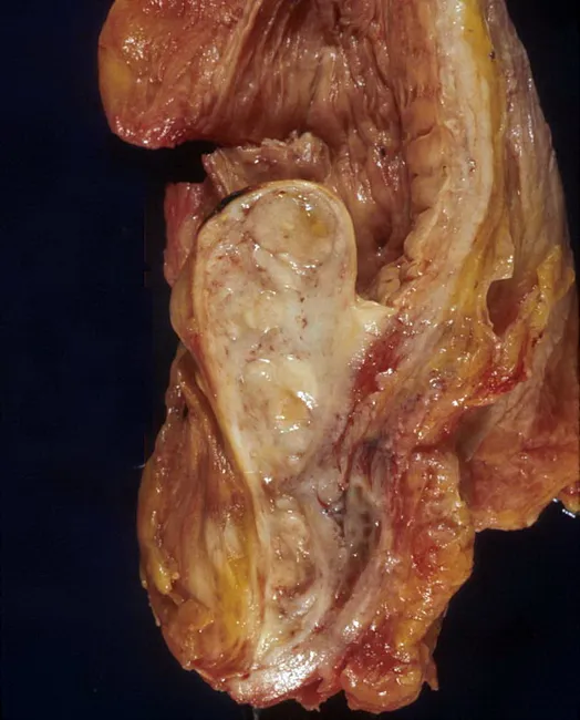
Gross pathology photograph of an autopsy prostatic specimen demonstrating benign prostatic hyperplasia with median lobe enlargement projecting into the bladder neck. The tissue appears tan‑brown and nodular, with a prominent midline lobe extending anteriorly toward the bladder outlet and creating a ball‑valve type obstruction at the bladder neck. The surrounding capsule and adjacent prostatic zones show variable, fibromuscular hyperplasia consistent with nodular hyperplasia rather than malignant invasion. This gross appearance reproduces the classic mechanism of lower urinary tract symptoms in aging men: progressive prostatic enlargement compresses the urethral channel, especially at the bladder outlet, producing obstructive and irritative voiding disorders. The autopsy context confirms a non‑malignant process, though histology would be required to definitively exclude focal carcinoma. Notable features include bilateral symmetry of nodular growth and conspicuous midline protrusion, which can be associated with reduced urinary flow, incomplete emptying, and increased postvoid residuals. Clinically, these findings underpin diagnostic considerations such as benign prostatic hyperplasia and differential diagnoses including prostatic adenocarcinoma or prostatitis; radiologic or endoscopic correlation may help stage obstruction. Educationally, this image serves as a reference for urology trainees and pathology learners to understand gross anatomy, angioarchitecture, and the anatomic basis of bladder outlet obstruction in BPH.
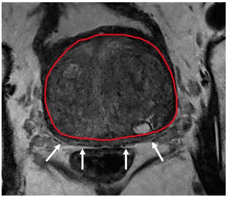
This diagnostic axial MRI scan (T2-weighted) of the prostate illustrates severe benign prostatic hyperplasia (BPH) with transition zone hypertrophy. The transition zone is segmented with a red outline, occupying the vast majority of the prostatic volume and exhibiting a heterogeneous, predominantly medium-gray signal intensity. Due to this significant expansion, the peripheral zone is severely compressed and displaced posteriorly, visible only as a thin, crescent-shaped sliver of lower signal intensity, indicated by white arrows. This image demonstrates a common challenge in prostatic zonal segmentation and radiological assessment: the effacement of anatomical landmarks between the transition and peripheral zones. The anatomical region focuses on the mid-gland level of the prostate. Educational focus includes understanding prostatic zonal anatomy, the pathophysiological effect of transition zone expansion on surrounding tissues, and the clinical imaging characteristics of severe hypertrophy in the context of urological diagnostics and radiotherapy planning.
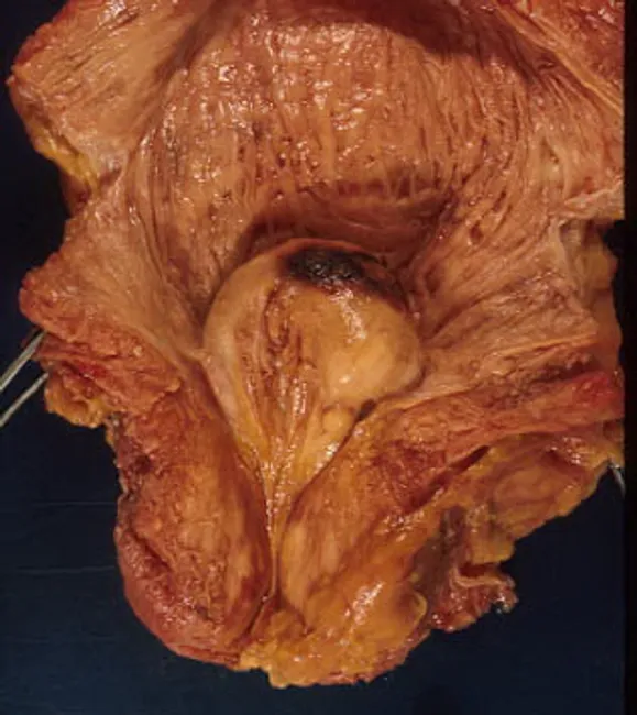
Gross pathology photograph of an autopsy prostate demonstrating benign prostatic hyperplasia with median lobe enlargement. The image captures in situ tissue within the pelvic cavity, with the enlarged median lobe protruding into the bladder neck and producing a ball-valve type obstruction of the prostatic urethra. The prostatic tissue is tan to yellow-brown, with nodular surfaces and a heterogeneous but non-neoplastic appearance consistent with nodular hyperplasia. The bladder neck region appears narrowed by the midline lobulation, while surrounding adenomatous prostatic tissue maintains identifiable lobular architecture. This specimen illustrates the anatomic basis of lower urinary tract symptoms in aging men, including urinary hesitancy, weak stream, and nocturia, attributable to outlet obstruction caused by BPH. The description emphasizes the median lobe as a dominant contributor to obstructive physiology and demonstrates how intravesical protrusion can impede urine flow. While autopsy findings may not reflect functional dynamics in vivo, they correlate with clinical presentations and surgical targets such as monopolar or holmium laser prostatectomy. Educational relevance includes urology, anatomy, pathology, and radiology correlation, providing a tangible example of bladder outlet obstruction mechanisms and the relationship between prostatic zones and the bladder. Useful for teaching, case review, and surgical planning.
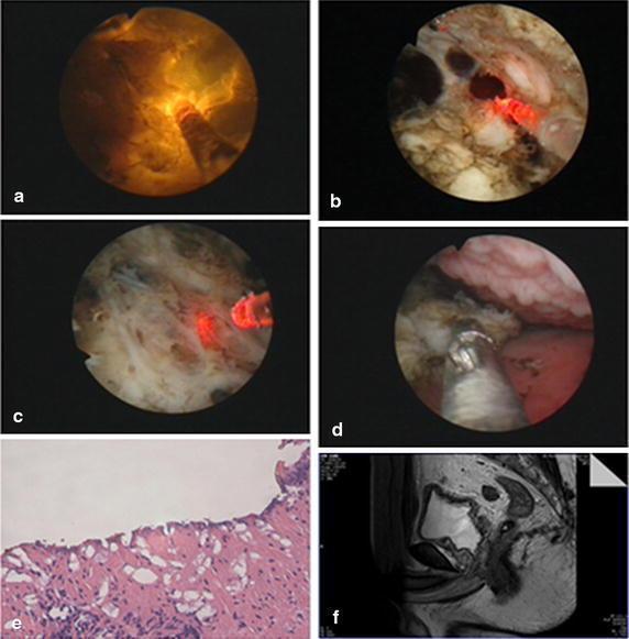
A multi-panel figure illustrating the surgical management and pathology of Benign Prostatic Hyperplasia (BPH) using Plasma-Resection Stripping-Vaporization of the Prostate (PRSVP). Panels (a-d) provide endoscopic views of the procedure: (a) demonstrates a straight beam LBO (green) laser in operation; (b) shows the mechanical stripping of hyperplastic prostate tissue; (c) visualizes the surgical plane at the level of the fibrous prostatic capsule; and (d) depicts the use of endoscopic forceps to retrieve tissue specimens. Panel (e) is a light microscopy image (H&E stain, 10x magnification) of the harvested specimen, confirming prostatic hyperplasia with characteristic glandular and stromal proliferation. Panel (f) is a sagittal T2-weighted MRI of the pelvis, showing the post-operative anatomical results with a widened prostatic urethra and surgical cavity. The collection serves as an educational resource for urological surgery, correlating intraoperative endoscopic findings with histopathology and post-operative radiological assessment.
"benign prostatic hyperplasia" pathogenesis etiology
Benign Prostatic Hyperplasia (BPH): Etiology and Pathogenesis
Definition and Overview
Anatomical Site of Origin
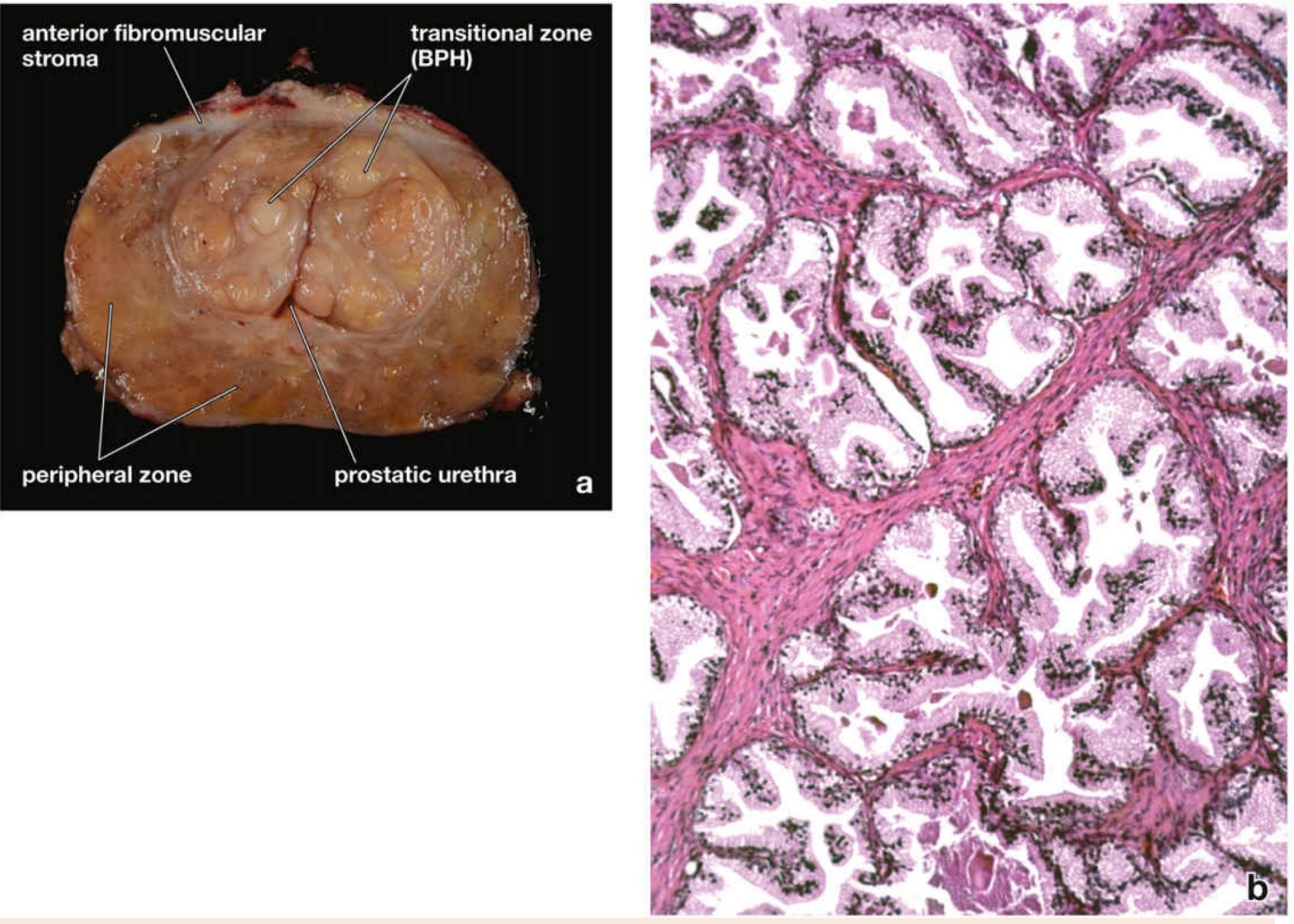
Etiology: Major Contributing Factors
1. Aging
2. Functioning Testes (Androgen Dependence)
3. Hormonal Milieu: The Testosterone-Estrogen Imbalance with Aging
- Testosterone levels fall progressively
- Estrogen levels remain unchanged or rise (due to peripheral aromatization of androgens in adipose tissue)
- DHT levels in prostatic tissue remain relatively high despite falling serum testosterone
Pathogenesis: Central Molecular Mechanism
The DHT Hypothesis
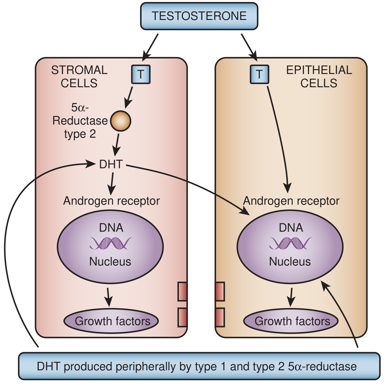
- Circulating testosterone enters prostatic stromal and epithelial cells via passive diffusion.
- In the stromal cell, testosterone is converted to DHT by 5α-reductase type 2 (the predominant prostatic isoenzyme, localized primarily in stromal cells by immunohistochemistry).
- DHT binds to nuclear androgen receptors (AR) - which also bind testosterone, though with lower affinity.
- The DHT-AR complex translocates to the nucleus and activates androgen-regulated genes, stimulating production of growth factors.
- These growth factors act in a paracrine fashion - DHT-induced stromal growth factors drive both stromal cell proliferation and epithelial cell survival (reduced apoptosis).
| Feature | Type 1 | Type 2 |
|---|---|---|
| Main tissue distribution | Skin, liver | Prostate (stromal cells) |
| Role in BPH | Minor/unclear | Critical |
| Inhibitor sensitivity | Dutasteride (not finasteride) | Finasteride + dutasteride |
| Clinical relevance in BPH | Limited | Primary target of 5ARI therapy |
Growth Factors Implicated
- FGF-7 (KGF - Keratinocyte Growth Factor): Produced by stromal cells, acts on epithelial cells to stimulate proliferation.
- EGF (Epidermal Growth Factor) and TGF-α: Promote epithelial and stromal proliferation.
- TGF-β: Has complex roles - generally inhibitory but may promote stromal fibrosis.
- IGF (Insulin-like Growth Factor): Promotes cell survival.
Stromal-Epithelial Interactions
Morphological Changes
- Prostate weight increases 3- to 5-fold (normal ~20 g; BPH often 60-100 g or greater)
- Enlarged nodules arise in the transition zone
- The prostatic urethra is compressed to a slitlike orifice
- Cut surface shows nodules that vary in color/consistency: solid (fibromuscular-predominant) or cystic (glandular-predominant with dilated lumina)
- Nodules contain variable proportions of:
- Proliferating glandular (epithelial) elements
- Fibromuscular stroma
- Hyperplastic glands are lined by two cell layers: inner tall columnar secretory cells + outer layer of flattened basal cells (key distinction from malignant glands in prostate cancer, which lack basal cells)
- Glandular lumina often contain corpora amylacea - laminated proteinaceous secretory concretions
- Robbins & Kumar Basic Pathology, p. 659
Pathophysiology of Symptoms (LUTS)
- Bladder wall hypertrophy (detrusor hypertrophy) from sustained increased intravesical pressure
- Detrusor instability and overactive bladder - producing irritative symptoms (urgency, frequency, nocturia)
- Incomplete bladder emptying → residual urine → medium for bacterial colonization → recurrent UTIs
- Prolonged severe obstruction → hydronephrosis and renal impairment
- Storage symptoms: urgency, frequency, nocturia
- Voiding symptoms: hesitancy, weak/intermittent stream, straining
- Post-void symptoms: dribbling, sensation of incomplete emptying
Additional Etiologic Factors
Metabolic Syndrome and Inflammation
- Hyperinsulinemia and IGF-1 signaling: promoting prostatic cell growth
- Chronic low-grade inflammation: activating NF-κB pathways, promoting stromal fibrosis
- Adipokine dysregulation (leptin, adiponectin): modulating prostatic growth
- Sympathetic nervous system overactivation in obesity → increased α-adrenergic tone in the prostate
Vascular Contributions
Summary: Key Pathogenetic Concepts
| Concept | Detail |
|---|---|
| Required conditions | Age + functioning testes |
| Key hormone | DHT (10× more potent than testosterone) |
| Key enzyme | 5α-reductase type 2 (in stromal cells) |
| Primary cell model | Stromal-epithelial paracrine signaling |
| Net cellular effect | ↑ stromal proliferation + ↓ epithelial apoptosis |
| Zone affected | Periurethral transition zone |
| Morphology | Mixed fibroglandular nodules; two-cell-layer glands; corpora amylacea |
| Symptom mechanisms | Static obstruction + dynamic α1-adrenergic smooth muscle tone |
| Other risk factors | Age, metabolic syndrome, inflammation, altered androgen-to-estrogen ratio |
- Robbins & Kumar Basic Pathology, pp. 659-660
- Robbins, Cotran & Kumar Pathologic Basis of Disease, p. 906
- Campbell-Walsh-Wein Urology 3-Volume Set, p. 4347
- Goldman-Cecil Medicine, pp. 1316-1318
- Sabiston Textbook of Surgery, p. 2824
- Histology: A Text and Atlas, p. 2145
- Fu X et al. (2024). Association between metabolic syndrome and BPH: The underlying molecular connection. Life Sciences. PMID 39488266
Pathogenesis flowchart