
Chronic periodontitis according to carranza in detail
Carranza chronic periodontitis classification clinical features pathogenesis treatment detailed
https://www.slideshare.net/slideshow/chronic-periodontitis-2…
chronic periodontitis clinical features periodontal pocket bone loss
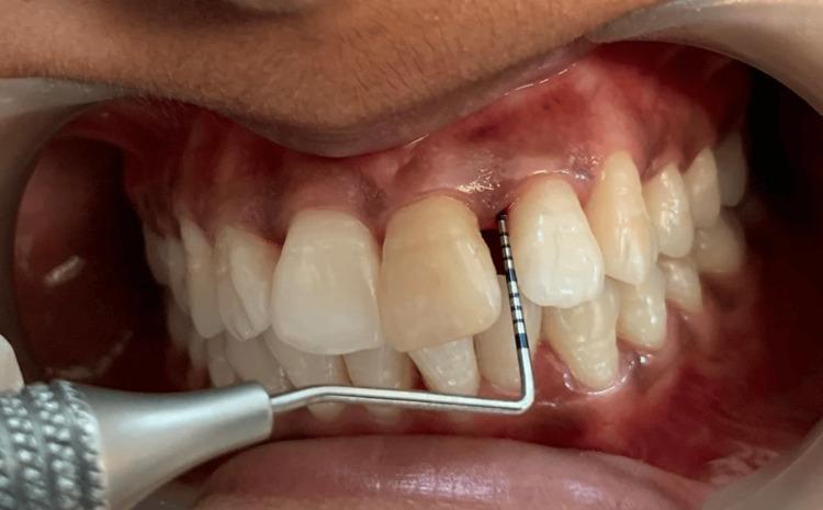
An intraoral clinical photograph showing a periodontal assessment of the maxillary left lateral incisor (tooth 22). A calibrated metal periodontal probe is inserted into the gingival sulcus on the mesiobuccal aspect to measure clinical attachment loss (CAL). The probe features visible millimeter markings, indicating a significant pocket depth. The surrounding gingiva exhibits signs of chronic periodontitis, including erythema, edema of the interdental papillae, and gingival recession. The image demonstrates the clinical procedure for diagnosing periodontal disease severity by measuring the distance from the cementoenamel junction to the base of the periodontal pocket. The teeth are visible in occlusion, and a lip retractor is utilized for optimal visualization of the maxillary arch. This visual is typical for dental educational materials focusing on periodontology and clinical examination techniques.
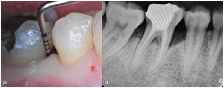
This composite figure illustrates a case of chronic periodontitis presenting as an infrabony periodontal defect. (a) Clinical photograph showing a baseline examination of the mesio-buccal aspect of the first mandibular right molar. A UNC-15 periodontal probe is inserted into a deep periodontal pocket, measuring approximately 9 mm. The surrounding gingival tissue appears erythematous and edematous, with evidence of bleeding on probing (BOP) visible as a small hemorrhagic spot on the gingival margin. (b) Preoperative intraoral periapical radiograph of the same region. The imaging reveals significant vertical bone loss forming a deep infrabony (angular) defect at the mesial aspect of the mandibular first molar. The tooth exhibits a radiopaque coronal restoration and well-defined root canals. The radiograph highlights the reduction in alveolar bone density and the steep vertical orientation of the bone loss relative to the root surface. This case demonstrates the clinical and radiographic correlation required for diagnosing complex periodontal attachment loss and planning regenerative surgical interventions.
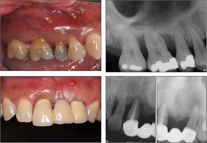
This composite figure presents clinical and diagnostic imaging of chronic periodontitis. The left panels contain intraoral clinical photographs showing generalized gingival inflammation, characterized by erythema, edema, and blunting of the interdental papillae. Notable findings include dental crowns with visible margins, attrition, and a localized erythematous gingival lesion (parulis or abscess) in the lower left quadrant. The right panels display periapical radiographs demonstrating moderate-to-severe horizontal and vertical alveolar bone resorption. Radiographic features include significantly reduced interdental bone levels, radiopaque dental restorations (fillings and crowns), and a fixed dental bridge. The imaging highlights the clinical and radiographic manifestations of periodontal disease, specifically the loss of supporting alveolar bone and concomitant soft tissue inflammation. This material is suitable for dental students and periodontology residents studying disease progression and diagnostic features of inflammatory periodontal conditions.
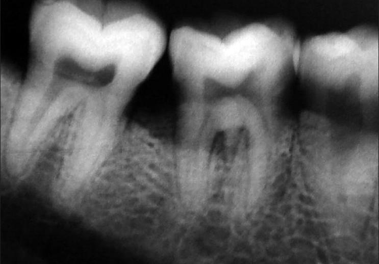
This diagnostic image is a periapical intraoral radiograph of the mandibular posterior region, demonstrating multiple molars and their supporting structures. The radiographic features highlight significant horizontal and vertical alveolar bone loss, with the alveolar crest positioned significantly apical to the cementoenamel junction (CEJ), characteristic of localized chronic periodontitis. The interproximal bone shows blunting and loss of the crestal lamina dura. Visible pathology includes a large radiolucent area in the crown of the first molar, consistent with a deep carious lesion or restoration with recurrent decay. The root morphology exhibits clear root canals and visible periodontal ligament spaces, though there is evidence of furcation involvement. Of educational note is the potential presence of a cervical enamel projection (CEP) extending into the furcation area, which acts as a predisposing factor for localized periodontal destruction. This radiograph serves as a teaching tool for identifying periodontal bone loss patterns, furcation defects, and the radiographic presentation of dental caries in a clinical periodontics context.
Chronic Periodontitis - According to Carranza's Clinical Periodontology
Definition
"An infectious disease resulting in inflammation within the supporting tissues of the teeth, progressive attachment loss, and bone loss."
Historical Classification Context
| Year (AAP) | Classification |
|---|---|
| 1989 | Adult periodontitis, Early onset periodontitis (prepubertal, juvenile), Necrotizing ulcerative, Refractory |
| 1999 | Chronic periodontitis (localized/generalized), Aggressive periodontitis, Periodontitis as manifestation of systemic disease |
| 2017 (new staging) | Periodontitis Stage I-IV, Grade A/B/C (replaces "chronic" terminology) |
Classification (Carranza)
Based on Disease Distribution:
- Localized chronic periodontitis - less than 30% of the teeth show attachment and bone loss
- Generalized chronic periodontitis - 30% or more of the teeth are affected
Based on Disease Severity:
| Severity | Clinical Attachment Loss (CAL) |
|---|---|
| Slight (Mild) | 1-2 mm |
| Moderate | 3-4 mm |
| Severe | ≥ 5 mm |
Prevalence
- Most common cause of tooth loss in adults over 35-40 years of age
- Affects approximately 35% of adults (30-90 years) in the US with at least one site having CAL ≥ 3 mm and probing depth ≥ 4 mm
- Prevalence increases with age; can occur at any age but is predominantly seen in adults
Etiology and Pathogenic Microorganisms
- Porphyromonas gingivalis (key periodontal pathogen; produces extracellular proteases)
- Treponema denticola (binds serum factors, interferes with complement deposition)
- Aggregatibacter actinomycetemcomitans (formerly Actinobacillus)
- Prevotella intermedia
- Tannerella forsythia (Bacteroides forsythus)
- Fusobacterium nucleatum
- Treponema vincentii
Pathogenesis
Step 1: Plaque Accumulation and Biofilm Formation
Step 2: Host Inflammatory Response
- Bacterial products (LPS, proteases, collagenases) trigger a host immune response
- Polymorphonuclear leukocytes (PMNs), lymphocytes, and plasma cells infiltrate the gingival connective tissue
- Collagen is lost from the inflamed connective tissue adjacent to the sulcular epithelium
- Toll-like receptors (TLRs) on host cells recognize bacterial components, amplifying inflammation
Step 3: Tissue Destruction
- Bacteria do not directly invade tissues in large numbers (at least in early stages)
- Indirect tissue destruction via:
- Bacterial enzymes (collagenases, hyaluronidase)
- Host-derived matrix metalloproteinases (MMPs)
- Prostaglandins (especially PGE2) stimulating bone resorption
- Cytokines: IL-1β, TNF-α, IL-6 activate osteoclasts
- Junctional epithelium migrates apically along the root surface, deepening the gingival pocket into a periodontal pocket
Step 4: Bone and Attachment Loss
- Alveolar bone resorption occurs via osteoclast activation
- Periodontal ligament fibers are destroyed
- Progressive apical migration leads to loss of clinical attachment (CAL)
- Disease progresses as a series of acute episodes separated by quiescent periods of variable duration (burst theory)
Risk Factors (Carranza)
Local Factors:
- Dental plaque and calculus (primary)
- Calculus acts as a reservoir for bacterial products
- Tooth anatomical factors (cervical enamel projections, furcation anatomy)
- Iatrogenic dentistry (overhanging restorations, ill-fitting crowns)
- Occlusal trauma (secondary co-destructive factor)
Systemic/Host Factors:
- Diabetes mellitus - bidirectional relationship; poorly controlled DM worsens periodontitis
- Smoking - most significant environmental risk factor; reduces gingival bleeding (masking severity), impairs healing
- Stress - elevates cortisol, alters immune response
- Genetic factors - IL-1 gene polymorphisms associated with increased severity
- HIV/immunosuppression - accelerates disease
- Medications - phenytoin, cyclosporine, calcium channel blockers (cause gingival enlargement)
- Nutritional deficiencies (vitamin C)
Major Clinical Features (Carranza)
Gingival Changes:
- Color: Pale red to magenta (erythema); in smokers may appear deceptively normal
- Contour: Blunted or rolled gingival margin; flattened or cratered interdental papillae; loss of stippling
- Consistency: Soft, edematous (acute phases); fibrotic, firm (chronic phases)
- Surface texture: Loss of normal orange-peel stippling
- Size: Gingival volume may be slightly to moderately increased
Key Clinical Signs:
- Bleeding on probing (BOP) - classic sign of active inflammation
- Periodontal pockets - both suprabony and infrabony pockets may be present; pocket depths are variable
- Clinical attachment loss (CAL) - hallmark diagnostic feature; measured from CEJ to base of pocket
- Gingival recession - in some cases attachment loss presents as recession without deep pockets
- Furcation involvement - common in molars in moderate to advanced disease (classified Class I, II, III by Glickman or Hamp)
- Tooth mobility - secondary to bone loss; classified Grade I (slight), II (moderate), III (severe/tooth removable)
- Tooth migration/drifting - especially anterior teeth with advanced disease
- Dental calculus - supra- and subgingival; correlates with disease severity
Symptoms (often minimal):
- Bleeding gums during brushing or eating
- Increasing spacing between teeth
- Loose teeth
- Sensitivity/pain (not always present; disease can be silent)
- Halitosis
Periodontal Pocket Types
| Type | Description |
|---|---|
| Suprabony (horizontal) | Base of pocket coronal to alveolar crest; horizontal bone loss |
| Infrabony/Intrabony (vertical/angular) | Base of pocket apical to alveolar crest; vertical/angular bone loss; 1-, 2-, or 3-walled |
Disease Progression
- Continuous model - slow but steady progression over time
- Random burst model - episodes of acute destruction (bursts) followed by quiescent periods
- Asynchronous multiple burst model - bursts related to life events/systemic changes
Radiographic Features
- Horizontal bone loss - most common; loss of alveolar crestal height, with the crest appearing more than 2 mm apical to the CEJ
- Vertical/angular bone loss - less common; associated with infrabony pockets
- Furcation radiolucency - in multirooted teeth
- Loss of crestal lamina dura - early radiographic sign
- Widening of periodontal ligament space
- Bone loss is typically underestimated on 2D radiographs (especially on buccal/lingual surfaces)
- Interproximal areas are best evaluated; bitewing and periapical radiographs are standard
Diagnosis
- Clinical attachment loss (CAL) - measured with a calibrated periodontal probe (UNC-15, Michigan-O)
- Probing depth (PD) - distance from gingival margin to base of pocket
- Bleeding on probing - indicator of active inflammation
- Radiographic bone loss - periapical and bitewing radiographs
- Full-mouth periodontal charting - 6 points per tooth
- Dental history - rate of progression, family history, systemic conditions
Management (Carranza's Treatment Protocol)
Phase I: Etiotropic/Cause-Related Therapy (Non-surgical)
- Oral hygiene instruction (OHI) - toothbrushing (Bass technique), interdental cleaning
- Scaling and Root Planing (SRP) - mechanical debridement of supra- and subgingival calculus and infected cementum (gold standard); quadrant-by-quadrant or full-mouth approach
- Polishing - removal of stains and supragingival deposits
- Extraction of hopeless teeth
- Caries control and restoration of defective restorations
- Risk factor modification: smoking cessation, glycemic control in diabetics
Adjunctive Antimicrobials:
- Systemic antibiotics (when local measures insufficient): metronidazole ± amoxicillin; doxycycline
- Local drug delivery: chlorhexidine chips, doxycycline gel (Atridox), tetracycline fibers placed in residual pockets ≥ 5 mm
- Chlorhexidine (0.12-0.2%) mouth rinse as adjunct
Re-evaluation (4-8 weeks post-SRP):
- Re-probe all sites
- Sites with residual PD ≥ 5 mm and BOP may require surgical intervention
Phase II: Surgical Therapy (if needed)
- Osseous surgery (resective): eliminate infrabony defects, correct bone architecture
- Periodontal flap surgery (Kirkland, Widman modified flap) - provides access to roots in deep pockets
- Guided Tissue Regeneration (GTR) - membranes (resorbable/non-resorbable) + bone grafts to regenerate lost periodontal structures
- Bone grafting (autograft, allograft, xenograft, alloplast) for infrabony defects
- Enamel Matrix Derivative (Emdogain) - promotes periodontal regeneration
Phase III: Restorative/Prosthodontic Phase
- After periodontal stability, restore missing teeth, correct occlusal problems
Phase IV: Maintenance (Supportive Periodontal Therapy - SPT)
- Critical for long-term success
- Recall intervals: every 3-4 months initially; 6-monthly for well-controlled cases
- Re-evaluate, re-probe, reinforce OHI, professional cleaning
- Monitor risk factors continuously
Prognosis (Carranza)
| Factor | Favorable | Unfavorable |
|---|---|---|
| Plaque control | Good | Poor |
| Bone support | >50% remaining | <50% remaining |
| Furcation | None or Class I | Class II-III |
| Mobility | Grade I | Grade II-III |
| Systemic factors | None | DM, smoking, immunodeficiency |
| Root anatomy | Normal | Short, fused roots |
| Patient compliance | Good | Poor |
Relationship to Systemic Disease
- Cardiovascular disease - shared inflammatory pathways; P. gingivalis found in atheromatous plaques
- Diabetes - worsens glycemic control; periodontitis treatment improves HbA1c
- Chronic kidney disease - periodontitis increases CKD risk, mediated via hypertension and diabetes
- Adverse pregnancy outcomes - premature birth, low birth weight
- Respiratory disease - aspiration of oral pathogens
- Infective endocarditis - source of bacteremia
Clinical Images
Key Points Summary (Carranza Exam Focus)
- Chronic periodontitis = most common form of periodontitis; primarily adults > 35 years
- Plaque is the PRIMARY etiology; local factors directly proportional to severity
- Hallmark: progressive CAL + bone loss + periodontal pocket formation
- Disease is episodic (burst theory) with alternating active/quiescent phases
- Key pathogens: P. gingivalis, T. denticola, T. forsythia (red complex)
- Localized: <30% teeth; Generalized: ≥30% teeth
- Severity: Slight (1-2 mm CAL), Moderate (3-4 mm), Severe (≥5 mm)
- Gold standard treatment: Scaling and Root Planing (Phase I therapy)
- Note: The 2017 World Workshop on the Classification of Periodontal Diseases replaced "chronic periodontitis" with a Stage-Grade system (Stage I-IV by severity; Grade A/B/C by rate of progression). Carranza's 13th edition incorporates this new classification.
Sources: Sherris & Ryan's Medical Microbiology, 8th ed. (p. 1508-1510); Robbins & Cotran Pathologic Basis of Disease (p. 498-500); Carranza's Clinical Periodontology (10th/13th ed., as referenced in peer literature); PMC review on treating chronic periodontitis