

This is report of 40 year old man
CBC Report Interpretation
Patient Details
- Patient: Mr. Jagannath Thorve, 42 years / Male
- Collected: 02/08/2026 | Ref. Doctor: Dr. Sujit Paul
Parameter-by-Parameter Analysis
| Test | Value | Reference Range | Status |
|---|---|---|---|
| Haemoglobin | 14.1 g/dL | 13.5 - 17 | ✅ Normal |
| Total WBC Count | 14,600 /cmm | 4,000 - 11,000 | 🔴 HIGH |
| RBC Count | 3.77 mill/cmm | 3.8 - 6.5 | 🟡 Borderline low |
| PCV (Haematocrit) | 39.0% | 40 - 54 | 🟡 Slightly low |
| MCV | 103.45 cu micron | 76 - 96 | 🔴 HIGH |
| MCH | 37.40 pg | 27 - 32 | 🔴 HIGH |
| MCHC | 36.15 g/dL | 32 - 36 | ✅ Normal (upper limit) |
| RDW-CV | 15.8% | 11 - 16 | 🟡 Upper normal |
| Neutrophils | 77% | 40 - 70 | 🔴 HIGH |
| Lymphocytes | 16% | 20 - 40 | 🟡 Low |
| Eosinophils | 04% | 1 - 6 | ✅ Normal |
| Monocytes | 03% | 2 - 8 | ✅ Normal |
| Basophils | 00% | 0 - 1 | ✅ Normal |
| Platelet Count | 252,000 /cmm | 150,000 - 450,000 | ✅ Normal |
Key Abnormalities and Their Clinical Significance
1. 🔴 Leukocytosis with Neutrophilia
- WBC = 14,600 /cmm (normal upper limit: 11,000)
- Neutrophils = 77% → Absolute Neutrophil Count = 14,600 × 0.77 = ~11,242 /cmm (elevated)
- Lymphocytes = 16% (relatively low, often seen alongside neutrophilia)
- Acute bacterial infection - most common cause
- Inflammation / tissue injury (trauma, surgery)
- Physiological stress (pain, exercise, emotional)
- Drugs: corticosteroids, beta-agonists
- Less commonly: myeloproliferative disorder, leukemia (if persistent)
Given the degree of elevation (~33% above normal) with neutrophilic predominance, an acute bacterial infection or inflammatory process is the leading possibility.
2. 🔴 Macrocytosis (Elevated MCV + MCH)
- MCV = 103.45 fL (normal: 76-96 fL) - elevated
- MCH = 37.40 pg (normal: 27-32 pg) - elevated
- Haemoglobin is 14.1 g/dL (normal range) - so this is macrocytosis without frank anaemia
- Vitamin B12 deficiency - top priority to rule out
- Folate (Vitamin B9) deficiency
- Liver disease / alcohol use
- Hypothyroidism
- Drugs (methotrexate, hydroxyurea, antiretrovirals)
- Myelodysplastic syndrome (less likely at 42 years, but must exclude if B12/folate normal)
An MCV of 103 fL strongly warrants checking serum Vitamin B12 and folate levels as the first investigation.
3. 🟡 Mildly Low RBC Count and PCV
- RBC 3.77 (just below 3.8 lower limit) and PCV 39% (just below 40% lower limit)
- Combined with elevated MCV - consistent with early macrocytic picture
- Haemoglobin is maintained at 14.1 g/dL (compensated so far)
Summary Impression
-
Leukocytosis with neutrophilia - suggesting an active infection, inflammatory process, or stress response. Clinical correlation (fever, site of infection, CRP/ESR) is needed.
-
Macrocytosis with mildly low RBC/PCV - the combination of elevated MCV (103.45) and elevated MCH (37.40) with borderline low RBC strongly suggests early megaloblastic process, most likely from Vitamin B12 or folate deficiency. The haemoglobin is still maintained, suggesting it is in an early/compensated stage.
Recommended Next Steps
| Investigation | Reason |
|---|---|
| Serum Vitamin B12 level | Rule out B12 deficiency (macrocytosis) |
| Serum Folate level | Rule out folate deficiency |
| Peripheral blood smear | Look for hypersegmented neutrophils (megaloblastic), left shift (infection) |
| CRP / ESR | Assess degree of inflammation |
| LFT (Liver Function Tests) | Liver disease as cause of macrocytosis |
| Thyroid function (TSH) | Hypothyroidism causes macrocytosis |
| Blood culture (if febrile) | Confirm bacterial infection |
Important note: This analysis is for informational purposes only. Please consult the treating physician (Dr. Sujit Paul) for clinical correlation and management. Lab reports must always be interpreted in the context of symptoms, clinical examination, and medical history.
He suffers from pus and wound around rectal opening
Clinical Correlation: CBC + Perianal Symptoms
Why the CBC Findings Are Explained
| Finding | Value | Explanation in context |
|---|---|---|
| WBC 14,600 /cmm | 🔴 High | Active bacterial infection from perianal abscess/fistula |
| Neutrophils 77% | 🔴 High | Neutrophilic response to acute bacterial infection |
| Lymphocytes 16% | 🟡 Low | Relative lymphopenia - classic in acute bacterial infection |
Diagnosis: Perianal Abscess / Fistula-in-Ano
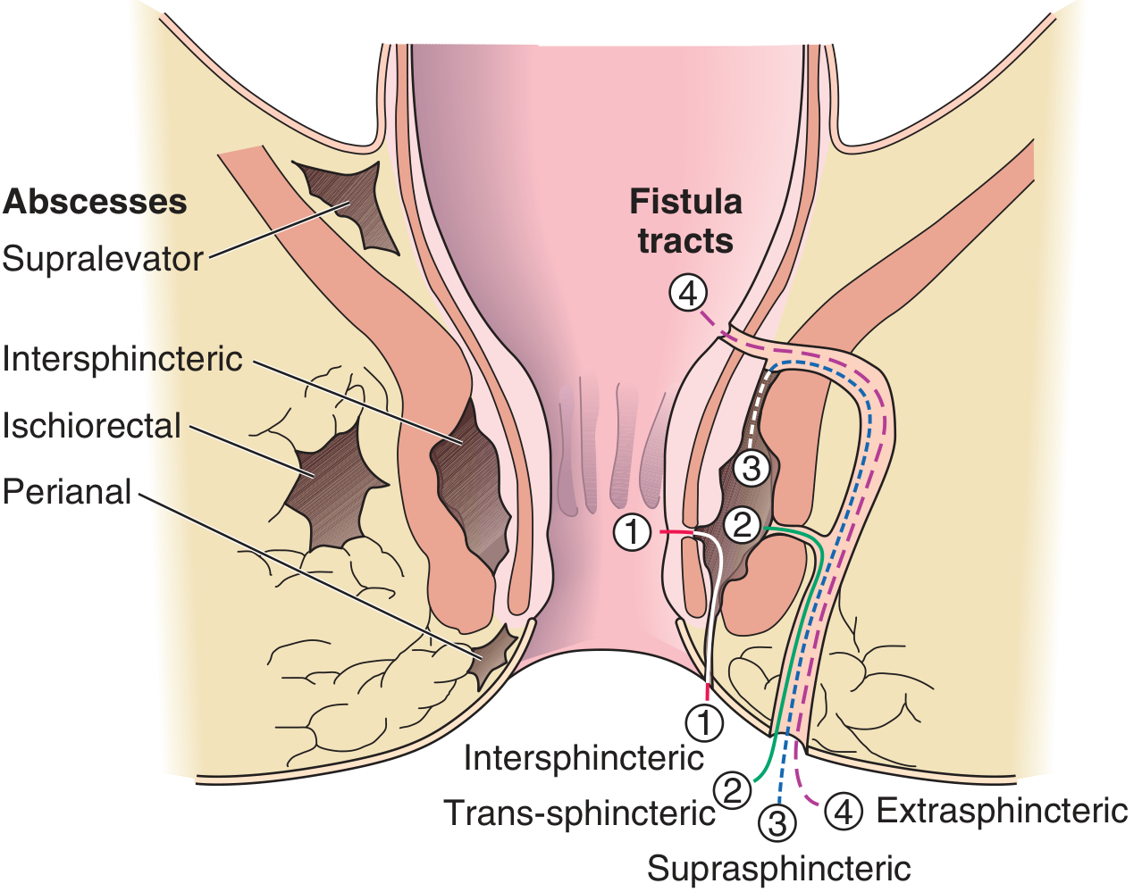
Types of Perianal Abscess (by location):
- Perianal - 40-50% (most common, superficial near the anal verge)
- Ischiorectal - 20-25%
- Intersphincteric - 2-5%
- Supralevator - 2.5% (deepest, most serious)
Fistula-in-Ano:
- 30-40% of abscesses eventually form a fistula - a tract connecting the abscess to the skin surface
- Ongoing pus/wound drainage = strongly suggests a fistula is already present
- Fistulas are classified as intersphincteric (70%), transsphincteric (23%), suprasphincteric (5%), extrasphincteric (2%)
What This Man Likely Has
- Pus draining from near the rectal opening - this is the external opening of a fistula
- Wound/sore around the rectal opening - the fistula tract
- WBC 14,600 with 77% neutrophils - active bacterial infection
Immediate Management Plan
Step 1 - Surgical Referral (URGENT)
- Antibiotics alone will NOT cure a perianal abscess/fistula
- The gold standard is surgical drainage - Harrison's states: "As with all abscesses, the gold standard is drainage"
Step 2 - Investigations Before Surgery
| Test | Purpose |
|---|---|
| Blood sugar (fasting + post-prandial) | Rule out diabetes - diabetics have higher risk of necrotizing fasciitis |
| HIV test | Immunocompromised patients have recurrent perianal infections |
| Stool culture / pus culture & sensitivity | Identify the causative organism and guide antibiotics |
| MRI pelvis (if complex fistula suspected) | Maps the fistula tract before surgery - 80% accuracy |
| Colonoscopy (if IBD suspected) | Rule out Crohn's disease (causes 10% of fistulas) |
| Serum B12 + Folate | Address the macrocytosis separately |
Step 3 - Surgical Treatment Options
| Procedure | Used for |
|---|---|
| Incision & Drainage (I&D) | Acute abscess - first priority |
| Fistulotomy | Simple, low intersphincteric/transsphincteric fistula |
| Seton placement | Complex/high fistula - staged approach to protect sphincter |
| LIFT procedure | Sphincter-sparing surgery for higher fistulas |
| Advancement flap | Complex fistulas, preserves continence |
Step 4 - Antibiotic Therapy (Adjunct only)
- There is obvious surrounding cellulitis
- Patient is diabetic or immunocompromised
- Common regimen: Metronidazole + Ciprofloxacin (covers anaerobes and gram-negatives common in perianal infections)
Red Flags - Go to ER Immediately If:
- High fever (>38.5°C) with chills
- Rapidly spreading redness/swelling around the area
- Inability to sit, severe pain
- Swelling spreading to scrotum/thighs
Also Address: Macrocytosis (MCV 103.45)
Bottom line: This man needs an urgent surgical consultation for the perianal fistula/abscess. The elevated WBC is the body's response to this active infection. He should not delay - untreated perianal abscesses can spread deeply and become life-threatening.
This analysis is for educational purposes. Please consult a surgeon for proper examination and treatment.
Can you provide me some clinical pictures original
perianal abscess clinical photo
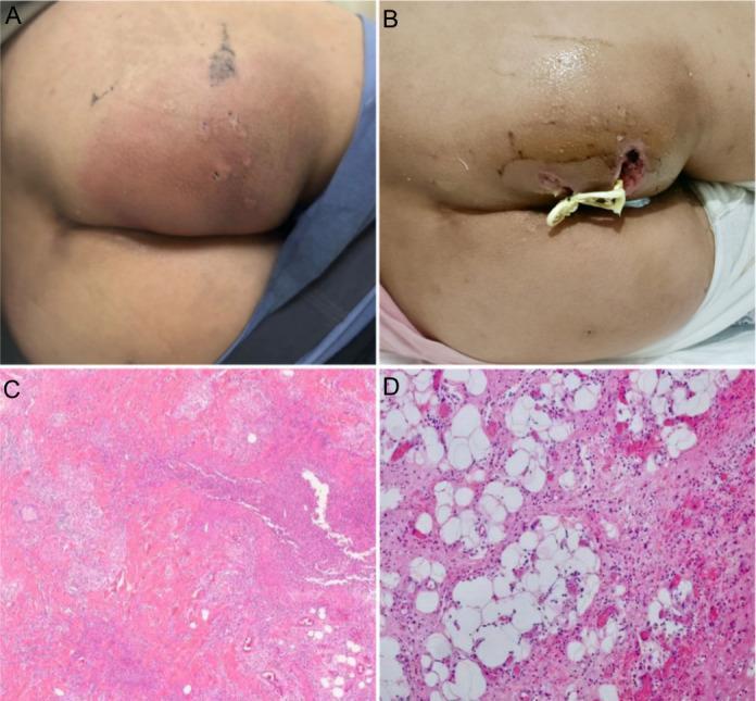
This composite image illustrates the clinical presentation, surgical management, and histopathology of a perianal abscess. (A) A clinical photograph shows a large, localized, erythematous swelling in the perianal region. The skin is notably red, tense, and bulging, indicating acute inflammation and an underlying fluctuant mass. (B) A subsequent photograph depicts the post-surgical status following incision and drainage. An open wound with irregular margins is visible, with a Penrose drain or surgical packing inserted into the cavity to facilitate the evacuation of purulent material and prevent premature closure. (C, D) Hematoxylin and eosin (H&E) stained histopathological sections at 40x and 200x magnification, respectively, show a dense inflammatory infiltrate. There is a prominent influx of neutrophils and lymphocytes into the dermal appendages and fibrous adipose tissue. The images demonstrate active abscess formation characterized by tissue necrosis and a heavy polymorphonuclear leukocyte presence, confirming the acute infective process within the perianal soft tissue.
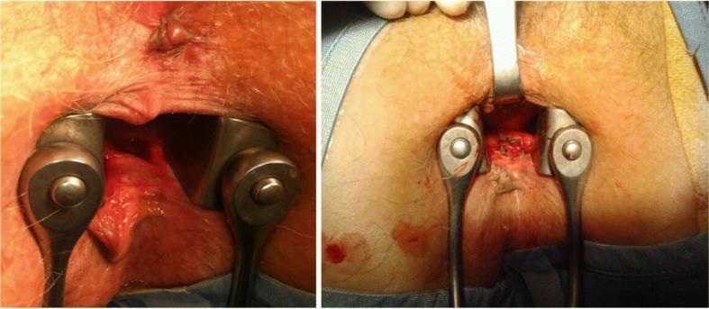
Two clinical photographs demonstrate the surgical management of a perianal abscess. The left image provides an intraoperative view of the anal verge and canal, stabilized with metal anal retractors. A deep, erythematous recess approximately 2-3 cm in diameter is visible on the right posterior-lateral anal wall, exhibiting signs of acute inflammation and tissue breakdown consistent with an infected abscess. The right image shows the post-operative state after surgical intervention. The procedure involved opening and deroofing the abscess cavity followed by curettage of the fundus. The surgical site appears as an open, raw-surfaced wound with irregular margins, facilitating drainage and secondary intention healing. The surrounding perianal skin remains erythematous, and surgical instruments remain in place to visualize the debrided cavity. This educational visual illustrates the fundamental surgical technique for treating deep-seated perianal infections that are non-responsive to conservative antibiotic management.
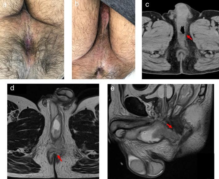
This composite educational image illustrates the clinical and radiological presentation of a perianal abscess with perineal involvement. Panels (a) and (b) are clinical photographs: (a) shows a normal appearing perianal region, while (b) demonstrates mild erythema across the perineum. Panel (c) is an axial non-contrast CT scan of the pelvis showing an air-containing, low-density collection in the perineum (red arrow) indicative of an anaerobic or gas-forming infection. Panels (d) and (e) are T2-weighted MRI scans in axial and sagittal planes, respectively. The axial MRI (d) highlights a high-intensity fluid signal at the 12 o'clock position relative to the anal canal, suggesting the primary track of the abscess. The sagittal MRI (e) confirms the continuity of the fluid collection with the anal canal and its anatomical position posterior to the bladder. This case highlights the importance of multi-modal diagnostic imaging (CT and MRI) for identifying deep-seated infections when external clinical signs, such as perianal redness, may be minimal or absent.
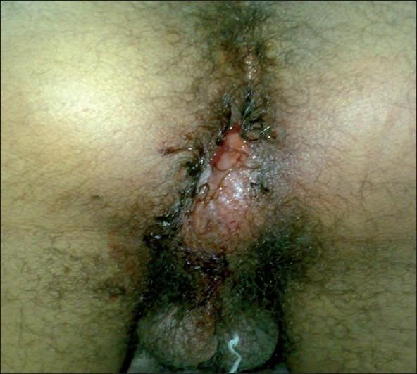
Clinical photograph of the perianal and gluteal region in a male patient, demonstrating a significant localized inflammatory process consistent with a perianal abscess. The image reveals a prominent, raised, and erythematous mass located at the 6 o'clock position relative to the anal opening. The skin over the lesion appears tense, shiny, and demonstrates marked hyperemia, indicating acute inflammation. There is evidence of skin breakdown or a small rupture point with associated serosanguinous discharge and crusting. The surrounding perianal skin shows secondary irritation and discoloration. This clinical presentation illustrates the typical morphology of a superficial perianal abscess, an acute anorectal condition characterized by localized infection and pus accumulation in the perianal space, often requiring surgical incision and drainage.
fistula in ano anal fistula clinical picture
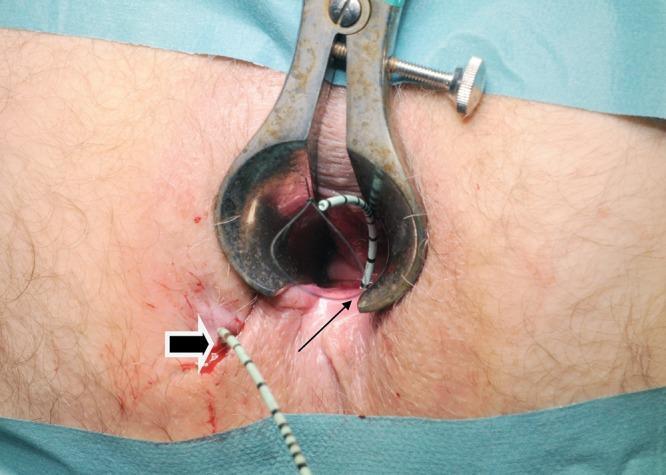
Clinical photograph of a surgical procedure for a complex fistula-in-ano. The image shows the perianal region with a metal anal speculum/retractor positioned to provide visualization of the anal canal. A 6Fr open-end ureteral catheter is seen threaded through the fistula track. A thick white-outlined arrow indicates the external cutaneous opening of the fistula, while a thin black arrow points to the internal opening within the anal canal. A 1/0 nylon suture is visible passing through the lumen of the catheter, forming a loop to facilitate the subsequent placement of a vascular sling or seton. The surrounding skin shows mild surgical inflammation and minor hemorrhagic spotting consistent with the procedure. This image illustrates a technique for seton placement in colorectal surgery, demonstrating the pathway from the internal to the external sphincter openings. Target audience: Colorectal surgeons and medical students studying proctology and surgical interventions for perianal disease.
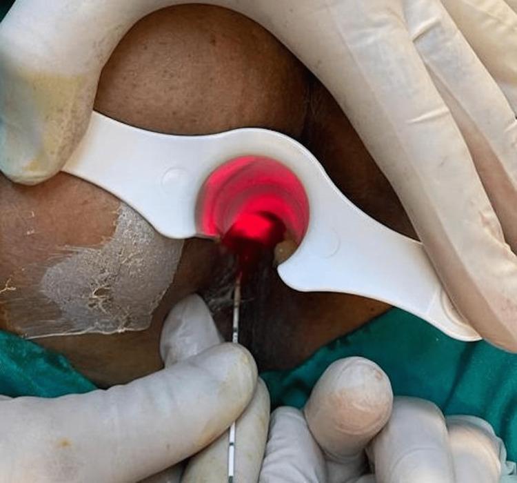
This clinical photograph provides an intraoperative view of a laser ablation procedure for a fistula-in-ano. The surgical field focuses on the perianal region, where a white, plastic, single-use anal retractor is being held by gloved hands to facilitate exposure. A thin, graduated laser probe from the neoV1470 Laser System is inserted into the fistula tract. A distinctive bright red laser light is visible within the anal canal, indicating the active ablation of the tract lumen. The image demonstrates the precise placement of the fiber-optic probe and the use of specialized retraction to maintain a clear visual field during minimally invasive colorectal surgery. The educational focus is on the technique of Laser-assisted Closure of Fistula (FiLaC), highlighting the equipment used, including the marked probe for depth measurement and the laser's thermal effect on tissue.
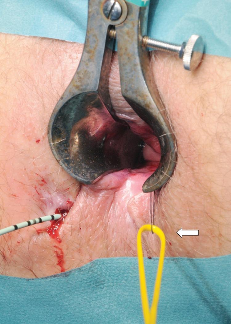
This clinical photograph captures an intraoperative view of a surgical procedure for a complex fistula-in-ano. A metallic Parks or similar anal retractor is positioned to provide exposure of the anal canal. The surgical field demonstrates the management of a fistula track using specialized instrumentation. On the left side of the image, a urological guidewire with black and white markings is seen entering the external fistula opening, which shows minor associated hemorrhage. A yellow vascular sling (indicated by a white arrow) is being threaded through the track using a 1/0 nylon suture loop. The educational focus of the image is the technique of passing a vascular sling from the internal opening to the external opening to serve as a loose seton. This procedure is critical in colorectal surgery for maintaining drainage of the fistulous track while preventing abscess formation and protecting the anal sphincter muscles. The image is intended for intermediate to advanced medical learners focusing on proctological surgical techniques.
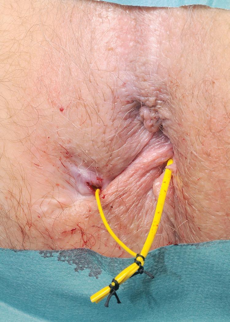
Clinical photograph of the perianal region demonstrating the management of a fistula-in-ano with a loose seton. The image shows a flexible, yellow vascular sling or vessel loop inserted through an external fistula opening located lateral to the anus and exiting through an internal opening within the anal canal. The two ends of the yellow tubing are secured together externally using black non-absorbable sutures to create a continuous, non-constricting loop (loose seton). The surrounding perianal skin shows mild erythema and minor blood spotting consistent with a recent surgical procedure. This clinical finding is characteristic of the surgical treatment for complex or high anal fistulas, where a seton is utilized to promote drainage, prevent recurrent abscess formation, and facilitate the gradual fibrous maturation of the fistula tract while minimizing the risk of fecal incontinence.
Clinical Pictures: Perianal Abscess
1. Acute Perianal Abscess - Classic Presentation
2. Perianal Abscess - Pre and Post Surgical Drainage
3. Intraoperative View - Abscess Drainage
4. Imaging (CT + MRI) of Deep Perianal Abscess
Clinical Pictures: Fistula-in-Ano (What Develops After Abscess)
5. Fistula-in-Ano - Seton Placement Surgery
6. Loose Seton in Place (Post-op Fistula Management)
7. Laser Treatment of Fistula (FiLaC - Modern Technique)
Summary: What This Patient's Condition Looks Like
These are real published clinical images from peer-reviewed medical literature for educational purposes.
In marathi this case called as
मराठीमध्ये या आजाराचे नाव
विविध नावे:
| मराठी / हिंदी नाव | वैद्यकीय नाव |
|---|---|
| भगंदर (Bhagandar) | Fistula-in-Ano |
| गुदद्वाराजवळील गळू | Perianal Abscess |
| नासूर (Nasoor) | Fistula / Chronic wound with pus |
भगंदर म्हणजे काय? (सोप्या भाषेत)
- गुदद्वाराच्या (शौचाच्या जागेच्या) आजूबाजूला एक बोगदा (tract) तयार होतो
- या बोगद्यातून सतत पू (pus) आणि घाण बाहेर येत राहते
- बाहेरच्या बाजूला एक जखम / छिद्र दिसते
- हा आजार स्वतःहून बरा होत नाही - शस्त्रक्रिया (operation) लागते
आयुर्वेदात भगंदर:
- शतपोनक - वातज
- उष्ट्रग्रीव - पित्तज
- परिक्षेपी - कफज
- शंबुकावर्त - त्रिदोषज
- उन्मार्गी - आगंतुज (बाहेरील कारणाने)
क्षारसूत्र चिकित्सा (Ayurvedic Treatment):
रुग्णाला सांगण्यासाठी (सोप्या मराठीत):
"तुम्हाला भगंदर झाला आहे. गुदद्वाराजवळ आतून एक बोगदा तयार झाला आहे ज्यातून पू येतो. हा आजार फक्त औषधांनी बरा होत नाही - शस्त्रक्रिया किंवा क्षारसूत्र उपचार लागतो. लवकरात लवकर सर्जनला दाखवा."