

Describe and tell me best medicine
ECU tenosynovitis treatment best medicine NSAIDs corticosteroid 2024
ECU extensor carpi ulnaris tenosynovitis wrist ultrasound anatomy
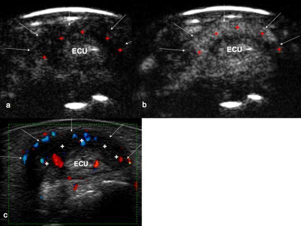
This diagnostic image series presents a transverse ultrasound comparison of the extensor carpi ulnaris (ECU) tendon at the wrist to assess for tenosynovitis. Panel (a) shows a Contrast-Enhanced Ultrasound (CEUS) image before contrast wash-in, displaying a hypoechoic peritendinous space. Panel (b) depicts the same region after contrast medium wash-in, showing significant hyperechoic enhancement within the peritendinous space and subtle intratendinous enhancement, indicating active synovial inflammation. Panel (c) utilizes Power Doppler Ultrasound (PDUS) to demonstrate increased vascularity (red and blue signals) within the thickened synovial sheath, marked with crosses. White arrows delineate the border of the tendon sheath. This side-by-side comparison illustrates the use of advanced ultrasound modalities to grade inflammatory activity and vascularization in musculoskeletal disorders. The images represent Grade 2 active tenosynovitis, characterized by significant synovial thickening and vascular signal occupying more than 50% of the proliferation area.
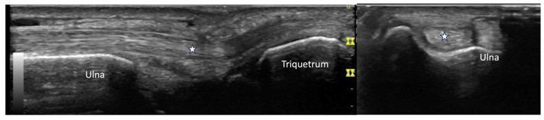
Two grayscale musculoskeletal ultrasound images display the extensor carpi ulnaris (ECU) tendon at the ulnar aspect of the wrist in two orthogonal planes. The left image shows a long-axis (longitudinal) view, where the ECU tendon (marked with a white star) appears as a hyperechoic, fibrillar structure extending superficially over the distal ulna and triquetrum. The cortical margins of both the ulna and triquetrum are well-defined, smooth, and continuous, showing no evidence of erosions or osteophytes. The right image provides a short-axis (transverse) view, depicting the ECU tendon (white star) as an oval, echogenic structure nestled within the ulnar groove of the distal ulna. There is no significant anechoic fluid or synovial thickening visible around the tendon in either view, suggesting a normal sonographic appearance. This imaging is commonly used to evaluate for tenosynovitis, tendon subluxation, or underlying cortical irregularities in rheumatological conditions like systemic sclerosis or rheumatoid arthritis.
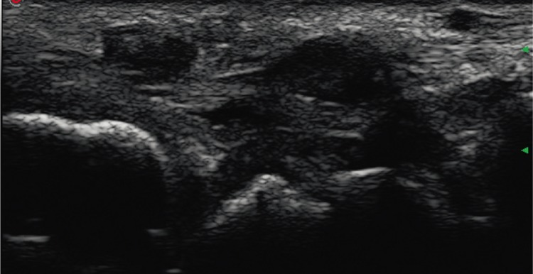
**Imaging Modality:** Musculoskeletal Ultrasound (grey-scale) **Anatomical Region:** Ulnar aspect of the wrist, transverse view. **Observed Pathology:** Tenosynovitis of the extensor carpi ulnaris (ECU). **Characteristic Visual Features:** The image demonstrates significant hypoechoic thickening of the synovial sheath surrounding the ECU tendon. The tendon itself is visualized within the sixth extensor compartment. There is evidence of synovial hypertrophy and anechoic to hypoechoic fluid accumulation (effusion) within the tendon sheath, indicative of an inflammatory process. The underlying bony cortex of the distal ulna is visible as a hyperechoic curvilinear interface with posterior acoustic shadowing. **Diagnostic Differentiating Features:** The circumferential distribution of hypoechoic material around the tendon within the fibro-osseous tunnel is characteristic of exudative and proliferative tenosynovitis. This presentation is distinct from simple tendinosis due to the primary involvement of the surrounding sheath rather than the internal fibrillar structure of the tendon itself.
Report Summary - Suman Lalta, 36F | Right Wrist Ultrasound (22/07/2026)
What the Report Says
- Mild soft-tissue swelling on the back-outer (dorsoulnar) side of the right wrist near the distal ulna bone
- Mild-to-moderate thickening of the ECU tendon sheath (synovial/peritendinous thickening) in the 6th extensor compartment
- Surrounding soft tissue inflammation (peritendinous edema)
- Good news: The ECU tendon itself is intact - no full-thickness tear, no abscess, no bone damage
- Orthopedic (Ortho) consultation
- MRI of the right wrist (for detailed soft tissue assessment)
- Blood tests: CBC, ESR, CRP (to rule out systemic inflammatory/rheumatological causes like rheumatoid arthritis)
What is ECU Tenosynovitis?
- Repetitive wrist movements (sports, typing, lifting)
- Sudden injury or overuse
- Systemic inflammatory conditions (rheumatoid arthritis, etc.)
ECU Tenosynovitis Illustration
Treatment - Best Medicines and Management
Important: This information is for general guidance only. The treating orthopedic doctor should be consulted before starting any medication. The radiologist has already recommended an ortho consultation.
Step 1 - Conservative / First-Line Treatment (Most Cases Respond to This)
| Treatment | Details |
|---|---|
| Rest & Activity Modification | Avoid repetitive wrist twisting, gripping, lifting for 4-6 weeks |
| Wrist Splint/Brace | Immobilize the wrist in slight extension to rest the ECU tendon. A forearm-based wrist brace is typically used |
| Ice/Cold Therapy | Apply ice pack wrapped in cloth for 15-20 min, 3-4 times a day for the first 48-72 hours |
Step 2 - Medicines (Pharmacological Treatment)
A. NSAIDs (Anti-inflammatory Painkillers) - First Choice
| Drug | Dose (Typical Adult) | Duration |
|---|---|---|
| Ibuprofen (e.g., Brufen, Combiflam) | 400-600 mg, 3 times a day with food | 5-7 days (short course) |
| Diclofenac (e.g., Voveran, Diclomol) | 50 mg, 2-3 times a day with food | 5-7 days |
| Naproxen (e.g., Naprosyn) | 500 mg, twice a day with food | 5-7 days |
| Aceclofenac + Paracetamol (e.g., Zerodol-P) | 1 tablet twice daily | 5-7 days |
Take NSAIDs with food to protect the stomach. Avoid if you have gastric ulcers, kidney problems, or are pregnant.
B. Topical (Local) Anti-inflammatories - Useful Alongside Oral NSAIDs
- Diclofenac gel (Voveran gel / Volini gel) - apply over the outer wrist 2-3 times daily
- Ketoprofen gel - alternative topical option
- These have minimal systemic side effects and are good for localised pain
C. Corticosteroid Injection - If NSAIDs + Splinting Do Not Work
- Triamcinolone acetonide or Methylprednisolone injection into or around the ECU tendon sheath
- This is a powerful anti-inflammatory given by the orthopedic doctor, usually guided by ultrasound
- Research shows a high success rate for tendon sheath steroid injections when other measures fail
- Usually given only 1-2 times (not repeated often, as it can weaken the tendon)
D. For Systemic Inflammatory Causes (if Blood Tests Show Raised ESR/CRP or Rheumatoid Arthritis)
- The orthopedic doctor may refer to a rheumatologist who might add:
- DMARDs (Disease-Modifying Anti-Rheumatic Drugs) like Methotrexate
- Hydroxychloroquine
- These are only if a systemic cause is found on blood workup
Step 3 - Physiotherapy (After Pain Reduces)
- Gentle wrist range-of-motion exercises
- Isometric ECU strengthening
- Eccentric exercises to promote tendon remodelling
- Research from PMC 2024 confirms that structured physiotherapy with isometric and eccentric exercises significantly helps ECU tendinopathy
Step 4 - MRI (As Recommended)
- Ultrasound shows the tendon sheath inflammation but MRI gives a clearer picture of the tendon fibres, any partial tear, and the bone
- This will guide whether surgery is needed
Step 5 - Surgery (Rare, Last Resort)
- Only if conservative treatment fails after 3-6 months
- ECU tenosynovectomy (removal of inflamed tendon sheath)
- As noted in Campbell's Operative Orthopaedics (2026), surgery (tenosynovectomy) is recommended only when nonoperative treatment has not resolved the condition
Summary - What to Do Next
- See an Orthopedic doctor immediately (as recommended in the report)
- Get the blood tests done: CBC, ESR, CRP
- Get the MRI of right wrist done
- Start rest + wrist splint/brace now
- Take Ibuprofen 400 mg 3x/day with food or Diclofenac 50 mg 2x/day for pain relief (only after confirming with your doctor that it is safe for you)
- Apply Diclofenac/Volini gel locally 2-3 times daily
- Avoid heavy wrist activity
Tell me the medicine to reduce edema
medicines reduce soft tissue edema tenosynovitis wrist anti-inflammatory 2024
Medicines to Reduce Edema (Swelling) in ECU Tenosynovitis
1. Oral NSAIDs - First Line (Best for Reducing Inflammatory Edema)
| Medicine | Brand Name (India) | Dose | How Often |
|---|---|---|---|
| Ibuprofen | Brufen, Combiflam | 400-600 mg | 3 times/day with food |
| Diclofenac | Voveran, Dicloran | 50 mg | 2-3 times/day with food |
| Aceclofenac + Paracetamol | Zerodol-P, Hifenac-P | 1 tablet | 2 times/day with food |
| Naproxen | Naprosyn | 500 mg | 2 times/day with food |
| Etoricoxib | Nucoxia 60/90 | 60-90 mg | 1 time/day with food |
Take ALL of these with food to avoid stomach irritation. Duration: 5-7 days (short course). Do not take on an empty stomach.
2. Topical Anti-inflammatory Gels - Apply Directly Over the Swollen Area
| Medicine | Brand Name (India) | How to Use |
|---|---|---|
| Diclofenac 1% gel | Voveran gel, Volini gel | Apply 2-3 times/day, gently massage into swollen area |
| Ketoprofen gel | Ketostal gel | Apply 2 times/day |
| Piroxicam gel | Dolonex gel | Apply 2 times/day |
These are safe to use along with oral tablets. Do not apply on broken skin.
3. Corticosteroid Injection - Strongest Anti-edema (Given by Doctor Only)
- Triamcinolone acetonide (Kenacort) - most commonly used
- Methylprednisolone (Depo-Medrol) - alternative
4. Serratiopeptidase / Trypsin-Chymotrypsin - Anti-edema Enzyme Tablets
| Medicine | Brand Name (India) | Dose |
|---|---|---|
| Serratiopeptidase | Serratiopeptidase (Sermion, Rofeta) | 10 mg, 2-3 times/day on empty stomach |
| Trypsin + Chymotrypsin | Chymoral Forte, Enzomac Forte | 1 tablet, 3 times/day on empty stomach |
Take enzyme tablets on an empty stomach (30 min before food or 2 hours after) for best absorption. They help dissolve inflammatory proteins causing the swelling.
5. Non-Medicine Measures That Actively Reduce Edema
- Ice pack - 15-20 min, 3-4 times/day for first 48-72 hrs - directly reduces capillary leakage and swelling
- Wrist elevation - Keep the wrist raised above heart level when resting - drains fluid by gravity
- Compression bandage - A light crepe bandage around the wrist reduces fluid accumulation
- Rest + Wrist splint - Stops mechanical irritation that keeps re-triggering inflammation
Quick Summary - Best Combination for This Patient
- Diclofenac 50 mg or Aceclofenac + Paracetamol (Zerodol-P) - 2 times/day after food x 5-7 days
- Serratiopeptidase (Chymoral Forte) - 3 times/day on empty stomach x 7-10 days
- Volini/Voveran gel - Apply locally 2-3 times/day
- Ice pack + wrist elevation + splint - daily
Always confirm with the orthopedic doctor first - especially before taking oral medicines, as individual health conditions (stomach, kidneys, blood pressure) affect which drug is safest for you.
- Miller's Review of Orthopaedics 9th Ed confirms rest, splinting, NSAIDs, and corticosteroid injections as the standard nonoperative approach for wrist extensor tenosynovitis.
- Mayo Clinic guidelines confirm ibuprofen and naproxen sodium as first-line anti-swelling medicines.
Tell me one thing which is best to use chymoral forte or etoricoxib or zerodol sp
Zerodol SP vs Etoricoxib vs Chymoral Forte tenosynovitis soft tissue edema best comparison
The Winner: Zerodol SP
What Each Drug Contains and Does
| Zerodol SP | Etoricoxib | Chymoral Forte | |
|---|---|---|---|
| Composition | Aceclofenac 100mg + Paracetamol 325mg + Serratiopeptidase 15mg | Etoricoxib 60/90mg | Trypsin 48mg + Chymotrypsin 60,000 AU |
| Type | NSAID + Painkiller + Enzyme (3-in-1) | Selective COX-2 inhibitor NSAID | Proteolytic enzyme only |
| Reduces inflammation | Yes (Aceclofenac) | Yes (Etoricoxib) | Mild |
| Reduces pain | Yes (Aceclofenac + Paracetamol) | Yes | No |
| Reduces edema/swelling | Yes (Serratiopeptidase) | Indirectly (via anti-inflammation) | Yes (primary role) |
| Speed of action | Fast | Fast | Slow |
| Stomach safety | Moderate | Better (COX-2 selective) | Very safe |
Why Zerodol SP Wins for This Case
- Aceclofenac - reduces the tendon sheath inflammation directly (COX inhibitor)
- Paracetamol - gives additional pain relief without extra stomach load
- Serratiopeptidase - specifically breaks down inflammatory proteins and fibrin that are causing the peritendinous edema (swelling) seen in the report
- Inflammatory soft-tissue edema → Aceclofenac handles this
- Peritendinous thickening/swelling → Serratiopeptidase specifically digests this fibrinous tissue
- Pain → Paracetamol + Aceclofenac together give better pain relief than either alone
Why Not the Other Two?
- It is an enzyme only - no direct anti-inflammatory or painkilling activity
- Works well for edema but cannot address the inflammation or pain from tenosynovitis on its own
- Good as an add-on, but not as a standalone for this condition
- Excellent anti-inflammatory and very stomach-friendly
- But it is a single-action drug - no enzyme for edema, no extra painkiller
- Better suited for chronic arthritis (osteoarthritis, rheumatoid arthritis) or gout
- Has a higher cardiovascular risk with long-term use
- Research comparing aceclofenac vs etoricoxib found both equally effective for pain, but etoricoxib had more cardiovascular concerns
Final Verdict
✅ Zerodol SP - 1 tablet, twice daily, after food