Few small intraparotid lymphnodes seen on left side
Reading File
search_medical_text
intraparotid lymph nodes normal findings significance
search_medical_image
intraparotid lymph nodes ultrasound parotid gland
GradeResponse
GradeResponse
Intraparotid Lymph Nodes — Left Side
Anatomy & Normal Findings
The parotid gland is unique among the major salivary glands in that it contains lymph nodes within its parenchyma (intraparotid lymph nodes). This occurs because the parotid gland encapsulates late in embryological development, after lymph node formation — meaning lymph nodes become enclosed within the gland tissue. This is a normal anatomical variant.
- The parotid gland typically harbors 2–10 intraparotid lymph nodes, predominantly in the superficial lobe
- On imaging, normal intraparotid lymph nodes appear as small, well-defined, oval/reniform hypoechoic structures with a smooth border and hilar vascularity on Doppler
Imaging Appearance of Normal Intraparotid Lymph Nodes
The image below illustrates the normal appearance across modalities:
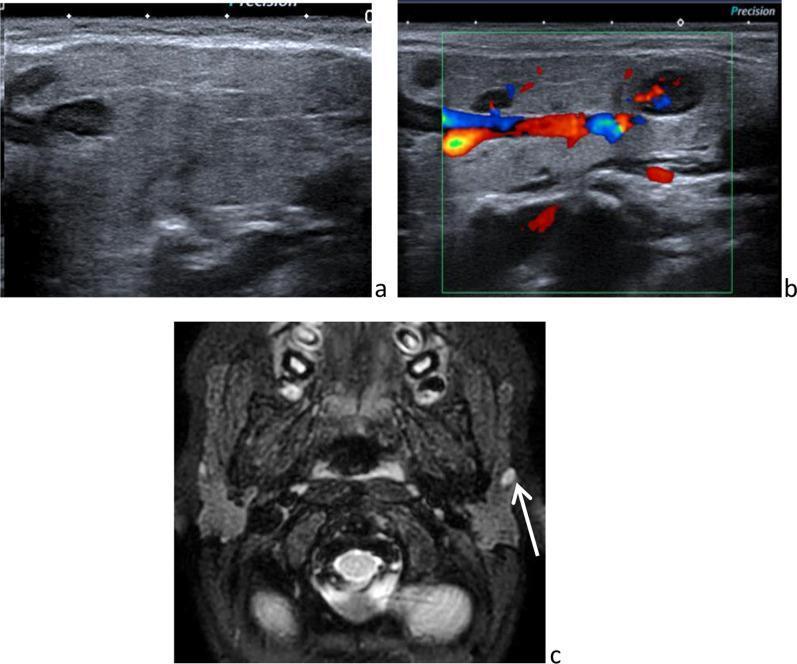
(a) B-mode ultrasound: hypoechoic, oval node with smooth border within echogenic parotid parenchyma
(b) Color Doppler: hilar vascularity pattern (benign feature)
(c) Fat-sat T2 MRI: well-defined hyperintense node within parotid
Clinical Interpretation of "Few Small Intraparotid Lymph Nodes"
This is a common incidental finding, often reported in imaging of the neck/parotid. The key assessment hinges on their size and morphology:
| Feature | Benign / Reactive | Suspicious / Pathological |
|---|---|---|
| Size | < 1 cm (short axis < 5 mm typical) | > 1 cm or rapidly enlarging |
| Shape | Oval/reniform | Round |
| Echogenicity | Homogeneous hypoechoic | Heterogeneous, cystic areas |
| Border | Well-defined, smooth | Irregular, poorly defined |
| Hilum | Preserved hilar vascularity | Absent hilum, peripheral flow |
| Number | Few, stable | Multiple, increasing |
Causes of Intraparotid Lymph Node Enlargement
If nodes are enlarged or morphologically abnormal, consider:
-
Reactive/Inflammatory (most common)
- Dental/oropharyngeal infection
- Viral illness (EBV, CMV)
- Upper respiratory tract infection
-
HIV-Associated
- Benign lymphoepithelial cysts (HIV-related reactive lymphoproliferation within intraparotid nodes; parotid glandular epithelium trapped in nodes → cystic enlargement; rare malignant conversion to lymphoma) (Bailey & Love's, p. 857)
-
Granulomatous disease
- Sarcoidosis, tuberculosis
-
Primary parotid lymphoma
- Rare; usually NHL (MALT type)
-
Metastatic disease
- From skin cancers of the face/scalp (melanoma, SCC), or oropharyngeal primaries
Clinical Significance of the Reported Finding
"Few small intraparotid lymph nodes on left side" — in the absence of abnormal morphological features — is most consistent with a normal or reactive finding requiring no immediate intervention.
Follow-up is recommended if:
- Nodes are > 1 cm in short axis
- Loss of fatty hilum
- Round shape or irregular borders
- Peripheral/chaotic vascularity on Doppler
- Associated parotid mass or skin lesion in the drainage area
- Systemic symptoms (fever, night sweats, weight loss)
- History of immunocompromise (HIV) or prior malignancy
In most routine imaging reports, this is a descriptive observation that can be correlated with clinical context and followed with serial imaging if there is any uncertainty.
This is a shared conversation. Sign in to Orris to start your own chat.