
Pnumonia notes imp points from robbins textbook of pathology onlyy
lobar pneumonia stages red hepatization gray hepatization histology
bronchopneumonia patchy consolidation lung histology
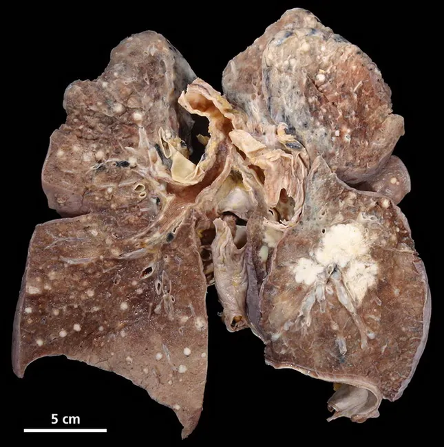
Imaging modality and specimen: Gross pathology photograph of a surgically resected lung specimen (likely right lower lobe lobectomy) illustrating invasive adenocarcinoma with extensive intrapulmonary metastases manifesting as lymphangitic carcinomatosis across both lungs and secondary bronchopneumonia of the left lower lobe. The specimen measures several inches in diameter; darker brown to tan parenchyma with multiple white nodules scattered through the lung margins and along the peribronchial/perivascular interstitium. The right lower lobe demonstrates focal tumor invasion at the hilum with contiguous soft-tissue mass; the left lower lobe shows patchy consolidation with bronchopneumonia. Microscopically, adenocarcinoma may display glandular differentiation, mucin production, and invasion into lung parenchyma, though histology is not visible in this gross image. Lymphangitic spread is indicated by nodular and band-like thickening along septa and peribronchial interstitium; this pattern correlates with rapid respiratory compromise and poor prognosis. Clinically relevant: This image supports diagnosis of primary lung adenocarcinoma as the most common non-small cell lung cancer; aligns with epidemiologic trends of increased adenocarcinoma incidence; highlights intrapulmonary metastases and associated infectious bronchopneumonia. Potential uses include medical education, radiology-pathology correlation, and gross-pathology reference in pulmonary oncology. Correlation with clinical history, imaging, and histopathology strengthens diagnostic confidence for staging and therapeutic planning in lung cancer.
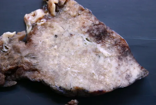
Gross pathology photograph of a lung tissue resection specimen illustrating an invasive mucinous adenocarcinoma with a prominent lepidic component. The tumor demonstrates ill-defined, infiltrative borders at the periphery, with a mucin-rich, gelatinous appearance and patchy grey-white parenchymal involvement. The gross pattern resembles infectious processes such as bronchopneumonia or even tuberculosis, posing a diagnostic pitfall on radiologic and clinical assessment prior to histology. The lesion is located in the peripheral lung (subpleural involvement), with predominant lepidic spread along alveolar septa contributing to the indistinct margins. On macroscopic inspection there is absence of discrete, well-circumscribed nodules; instead, broad-based infiltration disrupts normal parenchyma. Corresponding microscopic diagnosis would require correlation with histopathology to confirm adenocarcinoma with mucin production and lepidic growth, often associated with mucin pools and gland-forming cells. Clinically, recognizing this entity is essential for accurate staging (T modifiers reflecting lepidic invasion) and for planning surgical resection and adjuvant therapy. This image supports education on differential diagnosis for radiologic consolidation, emphasizes the mimicry of infection, and serves as a visual reference for pathologists and surgeons assessing suspicious lung lesions. The gross features underscore the need for tissue confirmation and multidisciplinary discussion to optimize prognosis and therapeutic strategy, correlating radiology, pathology, and clinical data together.
Pneumonia - Robbins Pathology (Important Notes)
1. Classification of Pneumonia Syndromes (Table 15.7)
| Syndrome | Key Pathogens |
|---|---|
| Community-Acquired Acute | S. pneumoniae, H. influenzae, M. catarrhalis, S. aureus, Legionella, Klebsiella, Mycoplasma, Chlamydia, Coxiella, Viruses |
| Healthcare-Associated | MRSA, MSSA, P. aeruginosa, S. pneumoniae |
| Hospital-Acquired (Nosocomial) | Gram-negative rods (Klebsiella, Serratia, E. coli, Pseudomonas), MRSA |
| Aspiration | Anaerobic oral flora (Bacteroides, Prevotella, Fusobacterium, Peptostreptococcus) ± aerobic bacteria |
| Chronic | Nocardia, Actinomyces, Granulomatous (TB, atypical mycobacteria, Histoplasma, Coccidioides, Blastomyces) |
| Necrotizing / Lung Abscess | Anaerobes (most common), S. aureus, Klebsiella, Strep pyogenes |
| Immunocompromised Host | CMV, Pneumocystis jirovecii, MAC, Aspergillus, Candida |
2. Community-Acquired Bacterial Pneumonia (CAP)
- Bacterial invasion of lung parenchyma → alveoli filled with inflammatory exudate → consolidation ("solidification")
- Often follows an upper respiratory viral infection
- Predisposing factors: extremes of age, CHF, COPD, diabetes, immune deficiency, absent splenic function (risk for encapsulated bacteria)
Morphological Patterns
- Entire lobe (or most of it) is homogeneously consolidated
- Typically caused by S. pneumoniae
- 4 classic stages:
| Stage | Gross | Microscopy |
|---|---|---|
| 1. Congestion | Lung heavy, red, boggy | Vascular engorgement, few neutrophils, many bacteria in alveoli |
| 2. Red Hepatization | Lung red, firm, liver-like | Alveoli packed with neutrophils, RBCs, fibrin - "hepatized" |
| 3. Gray Hepatization | Gray-brown, dry, firm | RBCs lysed, fibrin+neutrophils+macrophages remain |
| 4. Resolution | Normal | Enzymatic digestion of exudate; macrophages clear debris; architecture restored |
- Patchy consolidation centered on bronchioles/bronchi
- Multiple foci, often bilateral, more basal
- Any lobe may be involved
- Common in: hospital-acquired, aspiration, S. aureus, Klebsiella, H. influenzae
- Bacterial pneumonia: predominantly intraalveolar neutrophilic inflammation
- Viral pneumonia: interstitial lymphocytic inflammation (predominantly confined to alveolar septa); respiratory distress out of proportion to clinical/radiologic signs
3. Specific Organisms - Key Points
Streptococcus pneumoniae (Pneumococcus)
- Most common cause of CAP
- Gram-positive, lancet-shaped diplococci
- Distribution: usually lobar
- Gram stain of sputum: neutrophils with typical diplococci
- Important: S. pneumoniae is normal flora in 20% of adults → false-positives on sputum culture
- Blood culture: positive in only 20-30% early in illness (more specific, less sensitive)
- Vaccine: capsular polysaccharide-based
Haemophilus influenzae
- Common cause of acute exacerbations of COPD and CAP
- Also associated with M. catarrhalis for COPD exacerbations
Moraxella catarrhalis
- Associated with acute exacerbations of COPD
Staphylococcus aureus
- Important cause of secondary bacterial pneumonia after viral respiratory illness (measles in children, influenza in adults)
- High incidence of complications: empyema, abscess, pneumatocele
- Also a leading cause of hospital-acquired pneumonia (often MRSA)
Klebsiella pneumoniae
- Most frequent cause of gram-negative bacterial pneumonia
- Classic setting: chronic alcoholics and debilitated/malnourished patients
- Produces thick, mucoid (currant jelly) sputum due to capsule
- Associated with upper lobe involvement and cavitation
Pseudomonas aeruginosa
- Seen in: cystic fibrosis, burn victims, neutropenic patients
- A leading nosocomial pathogen
Legionella pneumophila
- Seen particularly in: individuals with comorbidities (heart/lung disease), organ transplant recipients
- Source: contaminated water systems (air conditioning, cooling towers)
- "Atypical" organism - does not grow on standard culture media
- Causes Legionnaires disease (pneumonia) and Pontiac fever (flu-like, no pneumonia)
Mycoplasma pneumoniae
- Most common atypical/walking pneumonia in young adults
- Interstitial pattern on CXR
- Cold agglutinins positive
4. Community-Acquired Viral Pneumonia
- Characterized by respiratory distress out of proportion to clinical and radiologic signs
- Inflammation predominantly confined to alveolar septa (interstitial)
- Important causes: SARS-CoV-2 (COVID-19), Influenza A & B, RSV, human metapneumovirus, parainfluenza, adenovirus
- Children: RSV, parainfluenza, human metapneumovirus
- Adults: Influenza A and B
- Military recruits: Adenovirus
Key Robbins point: "Flu condemns, and additional infection executes" - the most common cause of death in viral influenza epidemics is superimposed bacterial pneumonia (secondary bacterial pneumonia)
5. Aspiration Pneumonia
- Occurs in debilitated patients or those who aspirate gastric contents (unconscious, post-stroke, repeated vomiting)
- Abnormal gag and swallowing reflexes are the predisposing mechanism
- Pathology: partly chemical (irritating gastric acid) + partly bacterial (oral flora)
- Cultures: more than one organism (polymicrobial); aerobes more common than anaerobes
- Pattern: often necrotizing, fulminant clinical course
- Common complication: lung abscess
- Microaspiration: occurs commonly in almost all adults - usually cleared by mucociliary mechanism
6. Healthcare-Associated Pneumonia
- Risk factors: hospitalization ≥2 days in last 90 days; nursing home/long-term care facility; IV antibiotics/chemo/wound care recently; hemodialysis clinic
- Key organisms: MRSA and P. aeruginosa
- Higher risk of multidrug-resistant (MDR) pathogens → requires broad-spectrum empirical treatment
- Higher mortality than CAP
7. Hospital-Acquired (Nosocomial) Pneumonia
- Defined as pulmonary infections acquired during hospitalization
- High risk: severe underlying disease, immunosuppression, prolonged antibiotics, invasive devices, mechanical ventilation (ventilator-associated pneumonia - VAP)
- Organisms: Gram-positive cocci (mainly S. aureus) + gram-negative rods (Enterobacteriaceae + Pseudomonas)
- In VAP: gram-negative bacilli somewhat more common
8. Chronic Pneumonia
- Usually in immunocompetent patients
- Localized lesion in a single organ, most often lung
- Caused by:
- Nocardia, Actinomyces
- Granulomatous organisms: M. tuberculosis, atypical mycobacteria, Histoplasma capsulatum, Coccidioides immitis, Blastomyces dermatitidis
9. Pneumonia in the Immunocompromised Host
- May be caused by organisms considered non-pathogenic in healthy individuals
- Key organisms: CMV, Pneumocystis jirovecii (PCP), MAC (Mycobacterium avium-intracellulare), Aspergillus, Candida
- Often requires bronchoalveolar lavage (BAL) or biopsy for diagnosis
10. KEY CONCEPTS Summary (Robbins Key Concepts Box)
- S. pneumoniae is the most common cause of CAP; distribution usually lobar
- Lobar pneumonia has 4 stages: congestion → red hepatization → gray hepatization → resolution
- Other common bacterial CAP causes:
- H. influenzae / M. catarrhalis: COPD exacerbations
- S. aureus: secondary to viral infections
- K. pneumoniae: alcoholics
- P. aeruginosa: cystic fibrosis, neutropenia
- Legionella: comorbid conditions, transplant recipients
- Important viral CAP causes: COVID-19, influenza, metapneumovirus
- Bacterial pneumonia = intraalveolar neutrophilic inflammation
- Viral pneumonia = interstitial lymphocytic inflammation
- Aspiration pneumonia: necrotizing, polymicrobial, often leads to lung abscess
- Hospital-acquired: gram-negative rods + S. aureus dominate; VAP is most severe
- Immunocompromised: CMV, P. jirovecii, Aspergillus, Candida