
1.Child birth - stages , management and complications 2.muskuloskeletal problems during pregnancy
stages of labour childbirth diagram
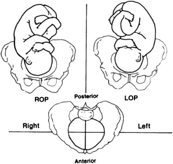
This medical anatomical diagram illustrates the various orientations of fetal occipitoposterior (OP) positions within the maternal pelvis during labour. The content is presented as three line-art diagrams from a superior perspective, focusing on the relationship between the fetal head and maternal pelvic landmarks (ilium, ischium, and sacrum). The top-left diagram shows the Right Occipito-Posterior (ROP) position, where the fetal occiput is directed toward the right posterior quadrant of the maternal pelvis. The top-right diagram depicts the Left Occipito-Posterior (LOP) position, with the occiput oriented toward the left posterior quadrant. The bottom center diagram illustrates the Occiput at Sacrum (OS) position, or Direct OP, where the occiput is positioned directly against the maternal sacrum. These diagrams demonstrate malpresentations where the fetal face is oriented anteriorly toward the maternal symphysis pubis. Educational objectives include the classification of fetal station and rotation, understanding the mechanisms of prolonged labour, and clinical management of malpositions in obstetrics.
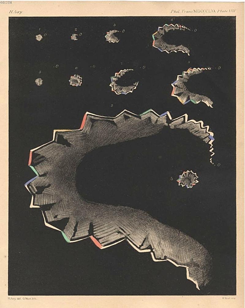
This historical medical illustration, titled 'Stages of Teichopsia' (1870), is a landmark clinical diagram representing the progression of a migraine aura across the visual field. The image depicts nine chronological stages (numbered 1-9) of a visual disturbance known as a scintillation scotoma or 'fortification spectrum'. The sequence begins with a small, central point of light and evolves into an expanding, crescentic arc. The figures are characterized by their jagged, crenellated edges, resembling the bastions of a fortified town, which Hubert Airy termed 'teichopsia'. These edges feature a prismatic or polychromatic effect, with alternating segments of red, green, blue, and yellow against a dark background, illustrating the 'shimmering' or 'boiling' nature of the aura. The diagram is an important record in the history of neurology and ophthalmology, providing a subjective yet scientifically detailed mapping of transient hemiopsia and the physiological manifestation of cortical spreading depression as perceived by a patient. This content is relevant for studying the clinical presentation of migraine with aura and the history of diagnostic visualization in neurology.
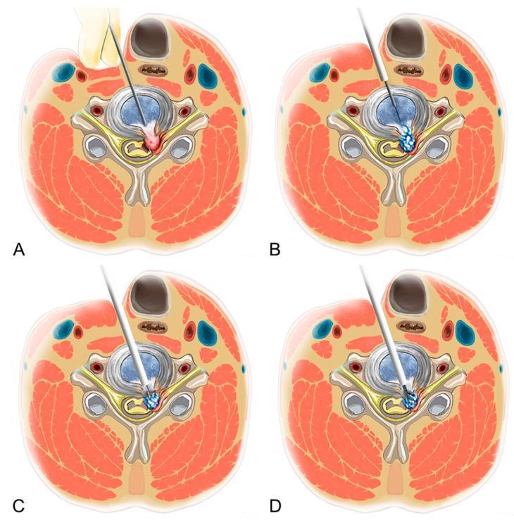
Anatomical diagram in axial cross-section illustrating the four key stages of a Percutaneous Endoscopic Cervical Discectomy (PECD). Stage A: Demonstrates the anterior percutaneous approach, showing manual displacement of the trachea medially and the carotid sheath laterally to establish a safe entry corridor for the spinal needle. Stage B: Illustrates sequential dilation over the initial guide needle, creating a path through the annulus fibrosus toward the herniated disc material. Stage C: Depicts the use of a side-firing laser or radiofrequency dissector to release the annular anchorage and fragment the herniated disc tissue while maintaining visualization. Stage D: Shows the selective removal of the herniated fragment using endoscopic forceps, achieving decompression of the neural structures while preserving the central nucleus pulposus. The illustrations highlight the relationship between the surgical instruments and key neck anatomy, including the vertebral body, spinal cord, nerve roots, and surrounding musculature.
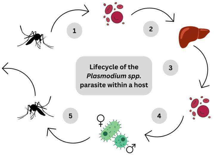
A pathophysiology diagram illustrating the human lifecycle of the Plasmodium spp. parasite, the causative agent of malaria. The diagram follows a numbered, clockwise circular flow consisting of five stages. Stage 1 depicts a mosquito vector, representing the inoculation of sporozoites into the host bloodstream. Stage 2 shows the transition of sporozoites through the bloodstream toward the liver, represented by a reddish-brown anatomical illustration of the organ. Stage 3 illustrates the intrahepatic phase where sporozoites mature into schizonts and merozoites within hepatocytes. Stage 4 depicts the erythrocytic cycle, where merozoites infect and lyse red blood cells, eventually developing into male and female gametocytes (marked with biological sex symbols). Stage 5 shows the uptake of these gametocytes by a second mosquito during a blood meal. The visual serves as an educational summary of the parasite's progression from the initial bite to the liver stage and subsequent blood-stage infection, highlighting key anatomical sites including the circulatory system, liver, and erythrocytes.
musculoskeletal changes pregnancy back pain
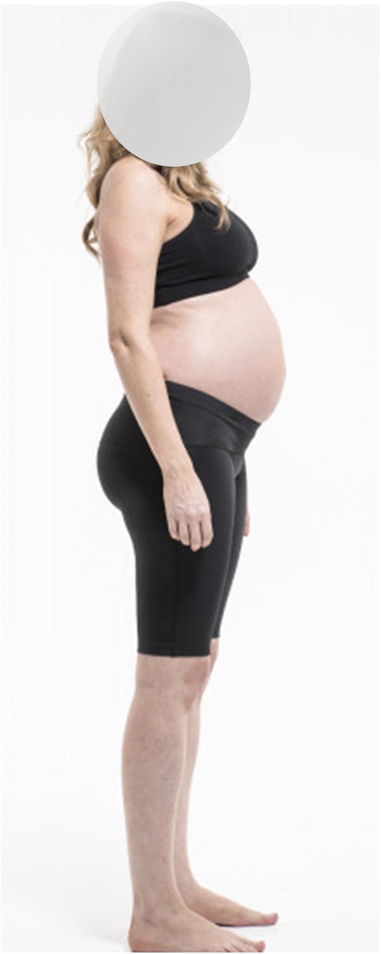
Clinical photograph of a pregnant individual in the third trimester, captured from a lateral profile view to demonstrate anatomical changes and the use of maternity support garments. The image highlights the physiological abdominal protrusion and the compensatory postural adjustments typical of late pregnancy, including an increased lumbar lordosis (swayback) and slightly posterior positioning of the shoulders. The individual is wearing black maternity support shorts (Dynamic Elastomeric Fabric Orthoses) designed with a high waistband that sits beneath the abdominal curve to provide pelvic and lower back stability. This visual serves to illustrate the mechanical management of pregnancy-related pelvic girdle pain (PGPP) and low back pain (LBP) through compression therapy. The educational focus is on maternal health, obstetrics, and physical therapy interventions for musculoskeletal changes during gestation.
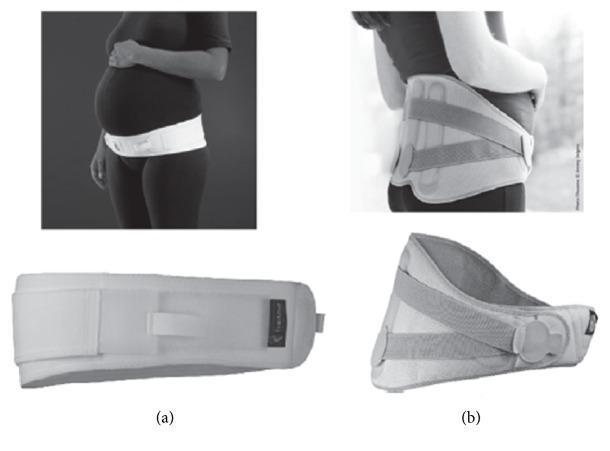
Comparison of two medical-grade maternity supports used for pregnancy-related pelvic and back pain. Panel (a) illustrates the Ortel-P pelvic maternity belt, a narrow, non-rigid, and adjustable orthopedic device. It is shown applied low on the torso, positioned at the symphysis pubis to provide targeted compression and stabilization to the pelvic girdle. Panel (b) depicts the LombaMum maternity lumbar brace, a wider, semi-rigid orthopedic garment. This brace features posterior metal reinforcements and a system of crisscrossed tension straps to provide broader structural support to the lumbar spine and sacroiliac joints. While the narrow pelvic belt (a) prioritizes mobility and pelvic stability, the lumbar brace (b) is designed for more significant postural support and alleviation of lower back pain. These devices are clinical interventions for managing pelvic girdle pain (PGP) and lower back pain (LBP) during pregnancy by distributing abdominal weight and stabilizing musculoskeletal structures.
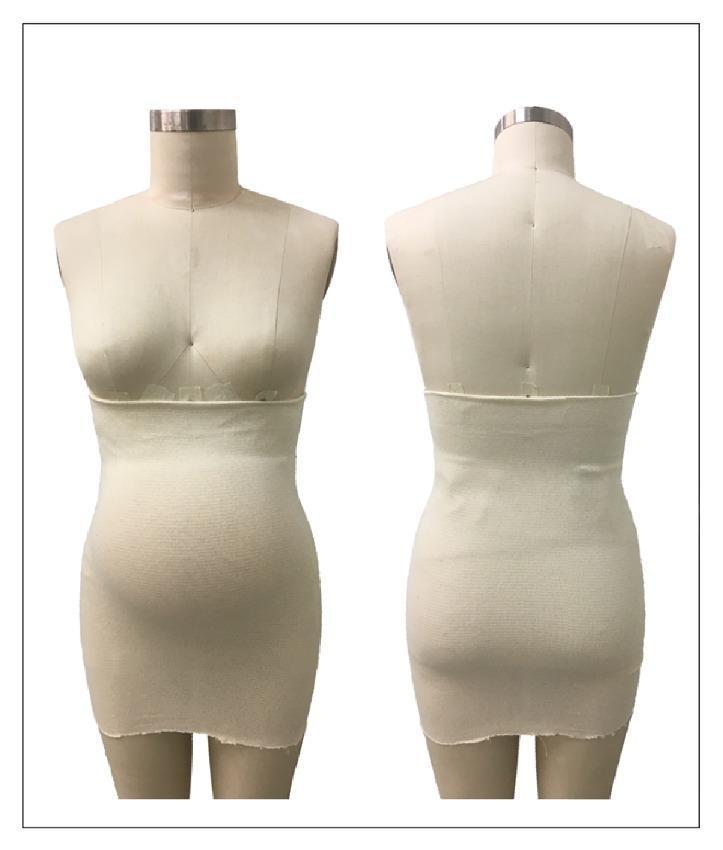
This clinical photograph displays a front and back view of a Tubigrip elastic support garment fitted on a female mannequin representing a pregnant torso. The garment is an elastic, one-piece tubular band with a ribbed texture, indicating a knit fabric construction. It is designed without fasteners or seams for a form-fitting, snug application. Anatomically, the band begins just below the inframammary fold and extends inferiorly to the mid-thigh level, covering the entire abdominal and pelvic regions. The visual demonstrates how the material conforms to the body's contours, specifically the protruding gravid abdomen and the lower back/lumbar region. In an obstetric and musculoskeletal context, such garments are used for the management of pregnancy-related low back pain (LBP) and pelvic girdle pain (PGP). The educational focus is on the distribution of circumferential compression to provide abdominal support, stabilize the pelvic area, and potentially reduce mechanical strain on the symphysis pubis and sacroiliac joints during pregnancy.
1. Childbirth - Stages, Management & Complications
Stages of Labour
Stage 1: Cervical Effacement and Dilatation
- Cervix effaces (thins out) and dilates from 0 to 6 cm
- Contractions irregular, becoming regular - every 5-10 min
- Duration: up to 20 hrs (nulliparous), up to 14 hrs (multiparous)
- Active management generally not indicated
- Cervix dilates from 6 cm to 10 cm
- Contractions regular, strong - every 2-3 min, lasting 60 sec
- Expected rate: ≥1 cm/hr (Friedman curve; modern evidence suggests ≥0.5-0.7 cm/hr is normal in active phase)
- Station of presenting part descends
- ROM (rupture of membranes) may occur here
- Continuous CTG (cardiotocography) for high-risk; intermittent auscultation acceptable for low-risk
- IV access, baseline bloods (FBC, group & save)
- Partogram monitoring (cervical dilatation, fetal heart rate, contractions, descent, maternal vitals)
- Pain relief: epidural analgesia, IV/IM opioids (pethidine, remifentanil), Entonox (N2O), TENS
- Encourage mobility, upright positioning
- Hydration - oral fluids in low-risk labour
- Amniotomy (ARM) only if progress is poor; not routine
- Oxytocin augmentation for established delay (after ARM if not already ruptured)
Stage 2: From Full Dilatation to Delivery of the Baby
- Duration: up to 3 hrs (nulliparous with epidural), up to 2 hrs (without epidural); up to 2 hrs (multiparous with epidural), 1 hr (without)
- Cardinal movements of labour (vertex presentation):
- Engagement
- Descent
- Flexion
- Internal rotation (occiput rotates anteriorly)
- Extension (head delivers under symphysis)
- Restitution and external rotation
- Expulsion of shoulders and body
- Maternal pushing with Valsalva or breathing-down technique
- Continuous fetal heart rate monitoring
- Perineal support to reduce tears
- Episiotomy - only if medically indicated (fetal distress, shoulder dystocia, instrumental delivery)
- Instrumental delivery (ventouse/forceps) for prolonged second stage or fetal compromise
- Emergency caesarean section (CS) if instrumental fails or contraindicated
Stage 3: Delivery of the Placenta (Birth to Placental Expulsion)
- Normal duration: up to 30 min with active management
- Signs of placental separation: gush of blood, uterus becomes globular, cord lengthens
- Oxytocin 10 IU IM immediately after baby delivery
- Controlled cord traction (CCT) after signs of separation
- Uterine massage after delivery (reduces PPH risk)
- AMTS reduces PPH risk by ~60% vs. expectant management
Stage 4: Immediate Postpartum Period (First 1-2 Hours)
- Monitoring for haemorrhage, uterine involution, perineal inspection
- Blood loss >500 mL vaginal delivery (>1000 mL CS) = PPH
- Breastfeeding initiation, skin-to-skin contact
- Vital signs monitoring q15 min
Complications of Labour and Delivery
Intrapartum Complications
| Complication | Key Features | Management |
|---|---|---|
| Fetal distress | CTG abnormalities (late decelerations, prolonged deceleration, reduced variability) | Repositioning, O2, stop oxytocin, urgent delivery |
| Prolonged labour | Failure to progress past Friedman norms | ARM, oxytocin augmentation, operative delivery |
| Shoulder dystocia | Head delivered, shoulders impacted at symphysis pubis | McRoberts manoeuvre, suprapubic pressure, Rubin/Woods screw, Zavanelli manoeuvre |
| Cord prolapse | Cord descends below presenting part after ROM | Knee-chest position, manual elevation, immediate CS |
| Uterine rupture | Scar dehiscence (prior CS) or spontaneous; fetal bradycardia, maternal shock | Emergency laparotomy, hysterectomy if needed |
| Amniotic fluid embolism | Collapse, DIC, respiratory failure | Supportive; CPR, FFP, cryoprecipitate |
| PPH (Primary) | Blood loss >500 mL within 24 hrs; 4 T's: Tone, Tissue, Trauma, Thrombin | Oxytocin, carboprost, misoprostol; uterine tamponade; surgical ligation; hysterectomy |
| Eclampsia | Seizures in pre-eclamptic patient | IV magnesium sulphate, antihypertensives, deliver |
| Placenta praevia | Placenta overlying OS; painless antepartum haemorrhage | Elective CS at 37-38 wks |
| Placental abruption | Premature separation; painful haemorrhage, tense uterus | Urgent delivery, manage shock |
Neonatal Complications
- Birth asphyxia: Apgar score <7 at 5 min; require resuscitation, therapeutic hypothermia if HIE
- Meconium aspiration: Thick meconium in liquor; paediatric team at delivery
- Instrumental injury: Cephalhaematoma, facial nerve palsy, fractured clavicle (shoulder dystocia)
2. Musculoskeletal Problems During Pregnancy
Pathophysiology - Why MSK Problems Arise
- Relaxin (peaks in 1st trimester, elevated throughout): loosens symphyseal and sacroiliac ligaments, increases joint laxity throughout the body
- Progesterone: further promotes ligament relaxation and smooth muscle changes
- Oestrogen: alters collagen structure
- Progressive anterior shift of centre of gravity with growing uterus
- Compensatory increased lumbar lordosis and thoracic kyphosis
- Anterior pelvic tilt increases
- Weight gain (average 11-16 kg) loads joints
- Widening of the stance
Common Musculoskeletal Problems
1. Low Back Pain (LBP)
- Most common MSK complaint in pregnancy; affects 50-80% of pregnant women
- Peaks in 3rd trimester
- Due to: lumbar hyperlordosis, disc loading, ligament laxity, piriformis tension
- Often worse with prolonged standing, walking, end-of-day
- Management:
- Physiotherapy: core strengthening, pelvic tilts, aquatic therapy
- Lumbar support/maternity belt
- Safe analgesia: paracetamol (1st line); avoid NSAIDs after 20 wks; short-course codeine with caution
- Sleeping with a pillow between knees
2. Pelvic Girdle Pain (PGP) / Symphysis Pubis Dysfunction (SPD)
- Pain in the pelvic region due to asymmetrical movement/dysfunction of SI joints and symphysis pubis
- Affects ~20% of pregnant women
- Hallmark: pain with single-leg activities (climbing stairs, turning in bed), waddling gait
- ASLR (Active Straight Leg Raise) test is positive
- Management:
- Pelvic support belt (worn low over symphysis pubis)
- Physiotherapy: stabilisation exercises, pelvic floor strengthening
- Crutches for severe cases
- Avoid wide leg movements, heavy lifting
- Delivery: consider supported lithotomy position; note maximum safe hip abduction

3. Carpal Tunnel Syndrome (CTS)
- Very common (affects ~35% of pregnant women)
- Due to fluid retention and oedema compressing the median nerve in the carpal tunnel
- Symptoms: tingling, numbness, burning pain in thumb, index, middle finger; worse at night
- Management:
- Wrist splints in neutral position (especially nocturnal)
- Elevation of arms
- Corticosteroid injection if severe
- Usually resolves postpartum; surgery rarely needed during pregnancy
4. De Quervain's Tenosynovitis
- Stenosing tenosynovitis of APL and EPB tendons at radial styloid
- Often postpartum (related to repetitive lifting of newborn) but can occur during 3rd trimester
- Finkelstein's test positive
- Management: splinting, corticosteroid injection; NSAIDs avoided in pregnancy
5. Sacroiliac Joint (SIJ) Pain
- Relaxin-mediated increased SIJ mobility causes pain at the SI joints, often radiating to buttocks/thighs
- Distinguished from sciatica: SIJ pain does not usually go below the knee
- Management: Physiotherapy, SIJ belt, aquatic therapy
6. Sciatica / Lumbar Radiculopathy
- Disc herniation (most common at L4-L5, L5-S1) aggravated by pregnancy weight
- Pain radiates down one or both legs, below the knee, with dermatomal distribution
- Straight leg raise (SLR) positive
- Management:
- Physiotherapy, relative rest
- Paracetamol; short-course opioids for severe cases
- Epidural steroid injection (limited data in pregnancy)
- Surgery rarely needed; most improve after delivery
7. Diastasis Recti (Abdominis)
- Separation of the two rectus abdominis muscles along the linea alba
- Common especially in 3rd trimester and postpartum
- Presents with: visible midline bulge with Valsalva, weak core, back pain
- Assessment: Finger-width test; >2 cm separation = clinically significant
- Management: Core rehabilitation (avoid crunches/sit-ups), physiotherapy, abdominal binder
8. Trochanteric (Greater Trochanter) Bursitis
- Lateral hip pain due to bursitis, aggravated by altered gait and pelvic tilt
- Management: Physiotherapy, ice, activity modification, corticosteroid injection
9. Thoracic Pain / Rib Pain
- Rib flaring and thoracic kyphosis from uterus pressing upward; intercostal neuralgia
- Common in 3rd trimester
- Management: Posture correction, physiotherapy, transcutaneous electrical nerve stimulation (TENS)
10. Osteoporosis of Pregnancy (Rare)
- Transient osteoporosis; vertebral fractures rarely occur
- Related to calcium mobilisation from maternal skeleton for fetal bone development
- Risk factors: low calcium intake, vitamin D deficiency, multiparity
- Management: Calcium + Vitamin D supplementation (routine in pregnancy), bisphosphonates avoided in pregnancy
11. Pre-existing Rheumatological Conditions
- Rheumatoid Arthritis (RA): often improves during pregnancy (immune tolerance), may flare postpartum
- SLE/Lupus: may worsen; associated with antiphospholipid syndrome, fetal loss risk
- Ankylosing Spondylitis: variable; NSAIDs contraindicated; anti-TNF biologics generally stopped by 3rd trimester
- Disease-modifying drugs reviewed carefully (methotrexate and leflunomide contraindicated; hydroxychloroquine and sulphasalazine generally safe)
Summary Table: MSK Problems in Pregnancy
| Condition | Timing | Key Mechanism | Key Management |
|---|---|---|---|
| Low back pain | 2nd-3rd trimester | Hyperlordosis, ligament laxity | Physio, support belt, paracetamol |
| Pelvic girdle pain | Any trimester | Relaxin, SIJ/symphysis laxity | Pelvic belt, physio, crutches |
| Carpal tunnel syndrome | 3rd trimester | Fluid retention/oedema | Wrist splints, elevation |
| Sciatica | 3rd trimester | Disc herniation, nerve compression | Rest, physio, analgesia |
| Diastasis recti | 3rd trimester | Linea alba stretching | Core rehab, abdominal binder |
| De Quervain's | Postpartum/3rd trimester | Repetitive tendon use | Splint, steroid injection |
| Trochanteric bursitis | 3rd trimester | Altered gait, pelvic tilt | Physio, ice, steroid injection |
- Park's Textbook of Preventive and Social Medicine (care during labour and childbirth)
- Rheumatology, 2-Volume Set (Elsevier, 2022) - pregnancy and musculoskeletal disease
- Creasy & Resnik's Maternal-Fetal Medicine: Principles and Practice
- Firestein & Kelley's Textbook of Rheumatology
Simplyfy
1. Childbirth - Stages, Management & Complications
Stages of Labour (4 Stages)
Stage 1 - Cervix Opens (0 → 10 cm)
- Latent phase: Slow dilatation (0-6 cm), irregular contractions
- Active phase: Fast dilatation (6-10 cm), regular strong contractions every 2-3 min
- Management: Pain relief (epidural, gas & air), monitor with partogram, CTG, IV access
Stage 2 - Baby is Born
- From full dilatation (10 cm) → delivery of baby
- Mother pushes with contractions
- Cardinal movements: Engagement → Descent → Flexion → Internal rotation → Extension → Restitution → Expulsion
- Management: Support perineum, episiotomy only if needed, forceps/ventouse if prolonged
Stage 3 - Placenta Delivered
- From birth of baby → delivery of placenta (within 30 min)
- Active management (standard): Oxytocin 10 IU IM + controlled cord traction
- Reduces postpartum haemorrhage by ~60%
Stage 4 - First 2 Hours Postpartum
- Watch for bleeding, check uterus is contracted, monitor vitals
- Encourage breastfeeding and skin-to-skin
Key Complications
| Complication | Simple Explanation | Management |
|---|---|---|
| Fetal distress | Baby's heart rate abnormal on CTG | Stop oxytocin, reposition, emergency delivery |
| Prolonged labour | Labour not progressing | ARM (break waters), oxytocin drip |
| Shoulder dystocia | Shoulders stuck after head delivers | McRoberts manoeuvre, suprapubic pressure |
| Cord prolapse | Cord comes out before baby | Knee-chest position, emergency CS |
| PPH | Bleeding >500 mL after delivery | Oxytocin, massage, surgery if needed |
| Uterine rupture | Uterus tears (esp. previous CS scar) | Emergency surgery |
| Eclampsia | Seizures in pre-eclampsia | IV magnesium sulphate, deliver baby |
2. Musculoskeletal Problems in Pregnancy
Common Problems (Simple)
🔹 Low Back Pain
- Most common (affects 50-80%)
- Cause: exaggerated lumbar lordosis (swayback posture)
- Rx: physiotherapy, maternity belt, paracetamol
🔹 Pelvic Girdle Pain (PGP)
- Pain around pelvis/symphysis, worse climbing stairs or turning in bed
- Cause: loose SI joints from relaxin
- Rx: pelvic support belt, crutches, avoid wide-leg movements
🔹 Carpal Tunnel Syndrome
- Tingling/numbness in fingers (thumb, index, middle), worse at night
- Cause: fluid retention compresses median nerve at wrist
- Rx: wrist splints at night, usually resolves after delivery
🔹 Sciatica
- Pain shooting down the leg
- Cause: disc pressing on nerve due to extra weight
- Rx: physiotherapy, rest, safe painkillers
🔹 Diastasis Recti
- Gap in abdominal muscles (midline bulge visible when straining)
- Cause: uterus stretches linea alba
- Rx: core rehab exercises, abdominal binder (avoid crunches)
🔹 Rib/Thoracic Pain
- Rib flaring and upper back pain in 3rd trimester
- Cause: uterus pushes diaphragm up
- Rx: posture correction, TENS, physiotherapy
🔹 Trochanteric Bursitis
- Outer hip pain from altered walking gait
- Rx: physiotherapy, ice, steroid injection
Quick Summary Table
| Problem | Timing | Main Cause | Treatment |
|---|---|---|---|
| Low back pain | 2nd-3rd trimester | Posture change | Physio, belt |
| Pelvic girdle pain | Any time | Relaxin, lax joints | Belt, physio |
| Carpal tunnel | 3rd trimester | Fluid/oedema | Wrist splint |
| Sciatica | 3rd trimester | Disc compression | Rest, physio |
| Diastasis recti | 3rd trimester | Muscle separation | Core exercises |
Safe analgesia in pregnancy: Paracetamol is 1st choice. Avoid NSAIDs after 20 weeks. Avoid aspirin in high doses.