I now have all the material needed. Let me compose the comprehensive 30-mark exam answer.
Carcinoma Cervix - 30 Marks Comprehensive Answer
1. INTRODUCTION & EPIDEMIOLOGY
Carcinoma cervix is the most common gynecological malignancy worldwide and the second most frequent cause of cancer death in women in developing countries. In India, it is the most common cancer among women. The mean age of presentation in the United States is 47 years, with a bimodal distribution peaking at 35-39 years and 60-64 years.
- Globally: ~500,000 new cases/year
- In the U.S., the lifetime probability of developing cervical cancer is 1:128
- 30% of cases occur in women who have never had a Pap smear; in developing countries this rises to ~60%
(Berek & Novak's Gynecology)
2. ETIOLOGY & RISK FACTORS
Human Papillomavirus (HPV) is the necessary causal agent. High-risk oncogenic HPV types 16, 18, 31, 33, 45 are responsible for most cases.
| Category | Risk Factors |
|---|
| Sexual behaviour | Early age at first intercourse (<16 years), multiple sexual partners |
| Infection | High-risk HPV types 16, 18 (together account for ~70% of cervical cancers) |
| Lifestyle | Cigarette smoking, low socioeconomic status |
| Reproductive | High parity |
| Immunological | Chronic immune suppression (e.g., HIV/AIDS) |
| Other | Race (higher in Black and Hispanic women in the U.S.) |
HPV infection is necessary but not sufficient - dysregulation of oncogenes at the site of viral DNA insertion, and accumulation of additional mutations, contribute to malignant transformation.
(Berek & Novak's Gynecology; Robbins Basic Pathology)
3. PATHOGENESIS
Transformation Zone is the key anatomical site. HPV infects basal cells of the squamocolumnar junction. The virus produces E6 and E7 oncoproteins that inactivate p53 and Rb tumour suppressors respectively, leading to uncontrolled cell proliferation.
Progression sequence:
- Normal epithelium → HPV infection → Squamous Intraepithelial Lesion (SIL)
- Low-grade SIL (LSIL = CIN 1) → High-grade SIL (HSIL = CIN 2/3) → Invasive carcinoma
- This progression typically takes 10-15 years (SIL peak precedes invasive carcinoma peak by this margin)
(Robbins Basic Pathology)
4. PATHOLOGY
Histological Types
| Type | Frequency |
|---|
| Squamous cell carcinoma | ~80% |
| Adenocarcinoma + mixed adenosquamous carcinoma | ~15% |
| Small cell neuroendocrine carcinoma | <5% |
Note: The proportion of adenocarcinoma is increasing in recent decades due to the decreasing incidence of squamous cell carcinoma and the limited ability of Pap test to detect glandular precancerous lesions.
Gross Appearance
- Microscopic foci of stromal invasion → grossly conspicuous exophytic tumors
- Tumors encircling the cervix produce a "barrel cervix"
- Extension into parametrial soft tissues can affix the uterus to surrounding pelvic structures
Microscopic Features
- Invasive tumors consist of tongues and nests of squamous cells with desmoplastic stromal response
- Grading based on degree of squamous differentiation
- Well-differentiated tumors: elaborate keratin pearls
- Rare neuroendocrine tumors: resemble small cell lung carcinoma morphologically
(Robbins Basic Pathology; Berek & Novak's Gynecology)
5. PATTERNS OF SPREAD
- Direct extension - to vagina (downward), parametrium, uterine corpus, bladder (anterior), rectum (posterior)
- Lymphatic spread - pelvic lymph nodes first, then para-aortic nodes (most common route of metastasis)
- Haematogenous - lungs, liver, bone (late, uncommon)
The likelihood of pelvic lymph node spread correlates with depth of invasion:
- <3 mm invasion: <1% risk of metastasis
-
3 mm invasion: >10% risk of metastasis
(Robbins Basic Pathology)
6. CLINICAL FEATURES
Early disease may be asymptomatic or detected on screening.
Symptoms of invasive disease:
- Abnormal vaginal bleeding - most common (postcoital, intermenstrual, or postmenopausal bleeding)
- Vaginal discharge (leukorrhea) - watery or purulent, malodorous
- Dyspareunia (painful coitus)
- Dysuria, frequency (bladder involvement)
- Haematuria or rectal bleeding (advanced disease)
- Leg oedema, deep pelvic pain (parametrial/pelvic wall involvement)
- Weight loss, anorexia, cachexia (advanced/metastatic disease)
Cervical cancer is most often diagnosed in patients who have never had a Pap test or who have not been screened for many years. (Robbins Basic Pathology)
7. DIAGNOSIS
Screening
- Pap (Papanicolaou) smear - exfoliative cytology from transformation zone
- HPV co-testing - more sensitive, used in women 30+ years
- VIA/VILI (Visual Inspection with Acetic acid/Lugol's Iodine) - used in low-resource settings
Diagnosis of invasive disease
- Pap smear → suspicious cytology → referral to colposcopy
- Colposcopy - acetic acid applied; abnormal areas show acetowhite changes, punctation, mosaic patterns, atypical vessels
- Biopsy (directed/punch biopsy, or cone biopsy) - histological confirmation is essential
- EUA (Examination Under Anaesthesia) - for clinical staging
Workup for staging
- Chest X-ray (lung metastases)
- IVP/renal function (hydronephrosis - stage IIIB)
- Cystoscopy + proctoscopy (bladder/rectal involvement - stage IVA)
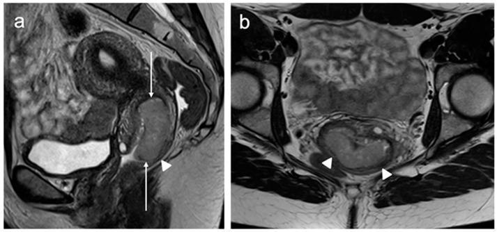
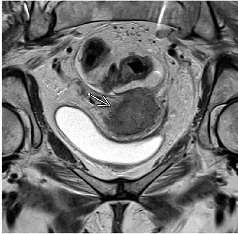
- MRI pelvis - best for local staging; parametrial assessment
- CT abdomen/pelvis - lymph node assessment
- PET-CT - most sensitive for nodal and distant metastases
- FNA/biopsy of suspicious lymph nodes
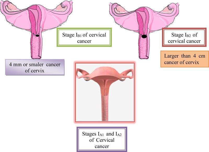
8. FIGO STAGING (2018 REVISED)
(Source: Berek & Novak's Gynecology)
| Stage | Description |
|---|
| I | Confined strictly to the cervix |
| IA | Microscopic only; depth of invasion <5 mm |
| IA1 | Stromal invasion <3 mm |
| IA2 | Stromal invasion ≥3 mm and <5 mm |
| IB | Invasion ≥5 mm, limited to cervix |
| IB1 | Invasion ≥5 mm depth, <2 cm greatest dimension |
| IB2 | ≥2 cm and <4 cm |
| IB3 | ≥4 cm in greatest dimension |
| II | Beyond uterus, not to lower 1/3 vagina or pelvic wall |
| IIA | Upper 2/3 vagina, no parametrial involvement |
| IIA1 | <4 cm |
| IIA2 | ≥4 cm |
| IIB | Parametrial involvement (not up to pelvic wall) |
| III | Lower 1/3 vagina, or pelvic wall, or hydronephrosis, or lymph nodes |
| IIIA | Lower 1/3 vagina involved, no pelvic wall extension |
| IIIB | Pelvic wall and/or hydronephrosis/non-functioning kidney |
| IIIC | Pelvic (IIIC1) or para-aortic (IIIC2) lymph node metastasis |
| IV | Beyond true pelvis or bladder/rectal mucosa involvement |
| IVA | Adjacent organ involvement (bladder/rectum) |
| IVB | Distant metastases |
Key 2018 changes:
- Horizontal spread no longer considered in stage IA - only depth of invasion
- Stage IB now has 3 subdivisions (IB1, IB2, IB3); IB1 <2 cm reflects fertility-sparing potential
- New Stage IIIC added for lymph node metastasis (worsens prognosis)
- Imaging and pathology can now inform staging (previously clinical only)
9. MANAGEMENT (BY STAGE)
Principles
- Early stage (I to IIA): Either radical surgery OR radiation therapy (equivalent survival)
- Advanced stage (IIB to IV): Concurrent chemoradiation + brachytherapy
Surgery
Cone biopsy:
- Stage IA1 with no lymphovascular space invasion (LVSI): cone biopsy alone (fertility-preserving)
- Must have clear margins; both endocervical and ectocervical margins free
Simple (extrafascial) hysterectomy:
- Stage IA1 with no LVSI (when fertility not desired)
Radical trachelectomy:
- Stage IA1/IA2/IB1 - fertility-sparing option; removes cervix but preserves uterus
- Allows future pregnancy (cervical cerclage required)
Radical (Wertheim's) hysterectomy + bilateral pelvic lymphadenectomy:
- Standard for Stages IA2, IB1, IB2, IIA1
- Removes uterus, upper vagina, parametria, and pelvic lymph nodes
- 5-year survival for Stage I: ~85%
- Advantages over radiotherapy: ovarian conservation, avoids radiation-related complications
- Urinary fistula rate: <2%; operative mortality: <1%
- Contraindicated for tumors >4 cm (will require postoperative radiation anyway)
Advantages of surgery over radiotherapy:
- Ovarian conservation (important in premenopausal women)
- Avoids vaginal fibrosis, atrophy, radiation cystitis/proctitis
- Surgical injuries more repairable than radiation injuries
Radiotherapy
- Can be used for all stages
- External beam radiotherapy (EBRT) to whole pelvis
- Brachytherapy (intracavitary) - delivers high local dose to cervix and parametria
- Equivalent to radical surgery for Stage I: 5-year survival ~85%
Concurrent Chemoradiation (CRT) - Standard for Advanced Disease
- Cisplatin-based chemotherapy concurrent with radiotherapy
- Significantly improves survival compared to radiotherapy alone
- Used for: IIB-IVA, post-operative high-risk features (positive nodes, positive margins, parametrial involvement)
Management by Stage Summary
| Stage | Treatment |
|---|
| IA1 (no LVSI) | Cone biopsy (fertility desired) or simple hysterectomy |
| IA1 (with LVSI) / IA2 | Modified radical hysterectomy + pelvic lymphadenectomy, OR RT |
| IB1, IB2, IIA1 | Radical hysterectomy + lymphadenectomy OR RT/brachytherapy |
| IB3, IIA2 | Chemoradiation (preferred) OR radical hysterectomy + adjuvant CRT |
| IIB - IVA | Concurrent chemoradiation + brachytherapy |
| IVB | Palliative chemotherapy/radiotherapy |
(Berek & Novak's Gynecology)
10. POSTOPERATIVE HIGH-RISK FEATURES (Indications for Adjuvant CRT)
"High-risk" (Sedlis criteria) - Adjuvant CRT recommended after radical hysterectomy:
- Positive lymph nodes
- Positive surgical margins
- Parametrial involvement
"Intermediate-risk" - Adjuvant RT may be considered:
- Large tumour size
- Deep stromal invasion
- Lymphovascular space invasion
11. SPECIAL SITUATIONS
Cervical cancer in pregnancy:
- Managed based on gestational age, stage, and patient's wishes
- Early stage + first trimester: treatment may be offered or deferred
- Definitive treatment generally delayed until fetal viability
Barrel-shaped cervix:
- Tumour >4 cm encircling cervix
- Worse prognosis, often managed with neoadjuvant chemoradiation followed by surgery
Recurrent disease:
- Pelvic exenteration: anterior (bladder removed), posterior (rectum removed), or total - for central pelvic recurrence in previously irradiated field
- Chemotherapy (cisplatin-based combinations) for systemic recurrence
- Immunotherapy (pembrolizumab/bevacizumab) - newer options
12. PROGNOSIS (5-Year Survival by Stage)
| Stage | 5-Year Survival |
|---|
| IA | ~95-99% |
| IB | ~80-90% |
| IIA | ~70-80% |
| IIB | ~65-70% |
| IIIA/B | ~40-50% |
| IIIC | ~30-40% |
| IV | <15-20% |
Poor prognostic factors: advanced stage, lymph node metastasis, parametrial involvement, large tumour size, neuroendocrine histology (worst prognosis)
13. PREVENTION
Primary Prevention - HPV Vaccination
- Bivalent vaccine (Cervarix): HPV types 16, 18
- Quadrivalent vaccine (Gardasil): HPV types 6, 11, 16, 18
- Nonavalent vaccine (Gardasil-9): HPV types 6, 11, 16, 18, 31, 33, 45, 52, 58 - covers ~90% of cervical cancers
- Recommended for girls 9-26 years (ideally before sexual debut)
- Administered in 2-dose (age <15) or 3-dose schedule
Secondary Prevention - Screening
- Cytology alone (Pap smear): Annually × 3, then every 3 years if normal (ages 21-65)
- Co-testing (Pap + HPV DNA): Every 5 years from age 30
- VIA: Cost-effective in resource-limited settings - "screen and treat" approach
- Colposcopy + biopsy: For abnormal screening results
QUICK REFERENCE MNEMONICS
Risk factors - "SMILCH":
- Sexually active young age / multiple partners
- Smoking
- Multiple sexual partners
- Immunocompromised
- Low socioeconomic status
- Chronic HPV infection (16, 18)
- High parity
Stages I → IV:
- I - confined to Cervix
- II - beyond cervix, not pelvic Wall or lower 1/3 vagina
- III - pelvic Wall / lower vagina / Hydronephrosis / lymph Nodes
- IV - bladder/rectum or distant metastasis
SUMMARY TABLE
| Feature | Key Point |
|---|
| Causative agent | HPV 16, 18 (high-risk oncogenic types) |
| Most common type | Squamous cell carcinoma (80%) |
| Site of origin | Transformation zone (squamocolumnar junction) |
| Most common symptom | Postcoital bleeding |
| Staging | FIGO 2018 (clinical + imaging + pathology) |
| Early stage treatment | Radical hysterectomy OR radiotherapy |
| Advanced stage | Concurrent cisplatin-based chemoradiation + brachytherapy |
| Best fertility-sparing option | Radical trachelectomy (IB1) or cone biopsy (IA1) |
| Screening | Pap smear / HPV co-testing / VIA |
| Prevention | HPV vaccination (nonavalent Gardasil-9) |
| Best prognosis | Stage IA1 (>95%) |
| Worst histology | Neuroendocrine/small cell carcinoma |
Sources: Berek & Novak's Gynecology; Robbins & Kumar Basic Pathology (10th ed.)