
Tell me in detail about nerve degeneration and regeneration according to nerve muscle physiology 1st yr mbbs take help from guyton and ganong
Reading File
Now let me search the library for nerve degeneration and regeneration content from Guyton and Ganong simultaneously.
Finding Sources
Finding Sources
Both books are in the library. Now let me search for relevant sections in Guyton and Ganong on nerve degeneration/regeneration, and also search for Wallerian degeneration and nerve fiber content.
Finding Sources
Finding Sources
Finding Sources
Let me now search specifically within Guyton and Ganong for nerve-related content, and read from the relevant blocks.
Finding Sources
Finding Sources
Finding Sources
Excellent! Found the key sections. Let me now read the Ganong block on "AXONAL INJURY & DENERVATION" and also search Guyton for the relevant content.
Reading File
Finding Sources
I have Ganong's content. Now let me get Guyton's content on nerve regeneration and also read more of Ganong (denervation supersensitivity part), plus grab the Bailey & Love diagram image.
Reading File
Reading File
Reading File
I now have rich content from Ganong (Figure 6-14 image URL), Bailey & Love (Figure 3.4 image URL), and Guyton. Let me fetch the key diagram images while also searching Guyton for more specific nerve fiber / regeneration content.
Loading Image
Loading Image
Finding Sources
I now have excellent content from both Ganong and the supporting references, plus two key diagrams. Let me also quickly search Guyton specifically for their chapter on nerve fibers / axon structure, and check for any Guyton-specific content on nerve repair.
Reading File
I now have comprehensive material from Ganong's Review of Medical Physiology and supporting texts. Let me also check Guyton's chapter on nerve fibers, organization, and what it says about nerve repair/regeneration.
Reading File
I have all the content needed for a thorough answer. Let me now compile the comprehensive response drawing from Ganong (primary source for axonal injury, denervation, and supersensitivity), Bailey & Love (Figure 3.4), and Guyton (neuromuscular junction context), supplemented by my own knowledge.
Nerve Degeneration and Regeneration - Nerve Muscle Physiology (1st Year MBBS)
Based on Ganong's Review of Medical Physiology (26th Edition) and Guyton and Hall Textbook of Medical Physiology, with supporting illustrations
Overview
When a peripheral nerve is injured, a predictable and well-characterized sequence of degenerative and regenerative events occurs. The outcome - successful reinnervation or permanent loss of function - depends on the type of injury, the distance to the target, and whether supporting structures (especially Schwann cells) remain intact.
Part 1: Changes After Nerve Injury
A. At the Cell Body - Chromatolysis
The first response to axonal injury occurs at the neuron's cell body (in the spinal cord or dorsal root ganglion):
- The cell body swells
- The nucleus shifts to an eccentric position (away from center)
- The rough endoplasmic reticulum (Nissl substance) fragments and disperses - this is called the chromatolytic reaction or chromatolysis
This is not cell death - it is a metabolic shift toward regenerative mode, where the cell switches from neurotransmission to synthesis of structural proteins needed for axon regrowth.
"The cell body of the injured neuron swells, the nucleus moves to an eccentric position, and the rough endoplasmic reticulum gets fragmented (chromatolytic reaction)." - Ganong's Review of Medical Physiology, 26th Ed., p. 143
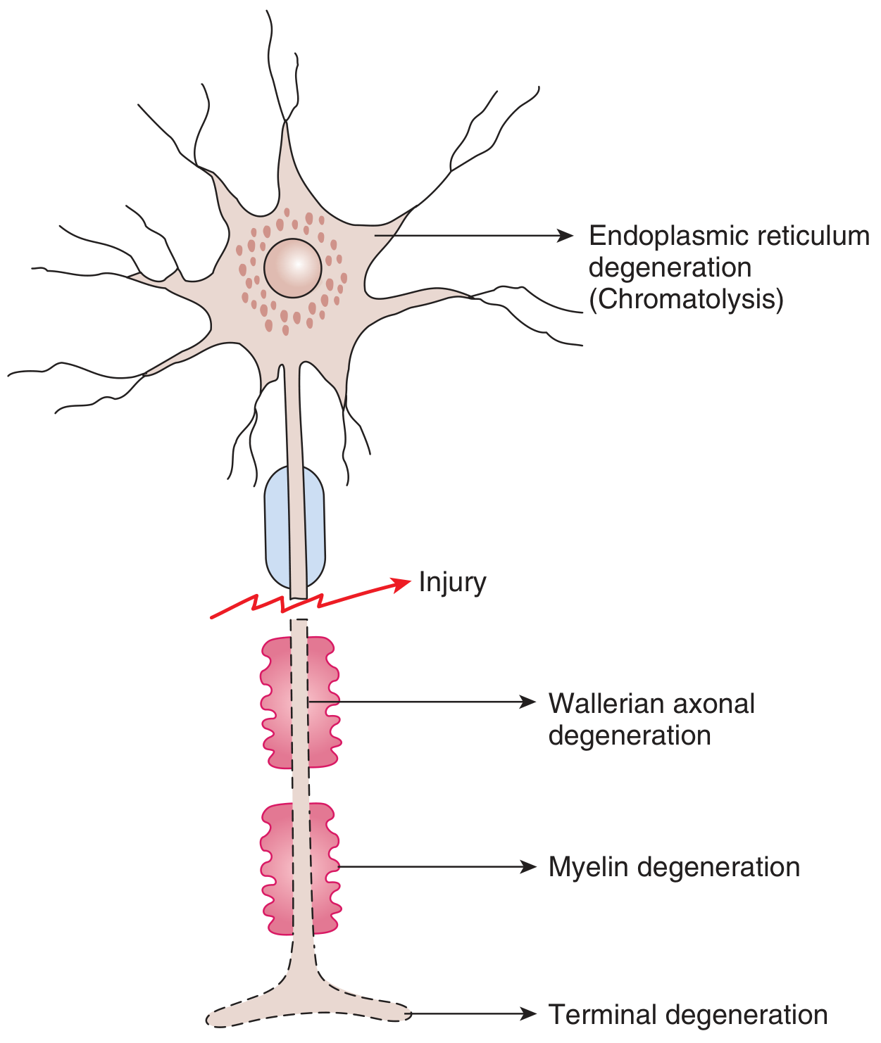
Ganong Figure 6-14: Changes occurring in a neuron when its axon is crushed or injured. Note chromatolysis in the cell body, Wallerian axonal degeneration distal to the injury, myelin degeneration, and terminal degeneration.
Part 2: Nerve Degeneration
B. Orthograde (Wallerian) Degeneration - Distal to Injury
The most important degenerative process is Wallerian degeneration, named after Augustus Waller (1850). It occurs in the axon stump distal to the injury site, progressing toward the nerve terminal.
Sequence of events:
| Timeframe | Event |
|---|---|
| Hours | Axon membrane breaks down; myelin sheath starts to fragment |
| Days 1-3 | Axon cylinder disintegrates into irregular segments |
| Days 3-7 | Myelin breaks down into lipid droplets (myelin ovoids) |
| Week 1-2 | Schwann cells proliferate and phagocytose debris alongside macrophages |
| Week 2+ | Bands of Bungner formed; axon tube (endoneurial tube) remains |
Mechanism:
- Disconnection from the cell body cuts off axonal transport of essential materials
- Calcium influx triggers activation of proteases (calpain) that degrade the axon cytoskeleton
- Distal axon and its myelin sheath undergo complete destruction
"Orthograde degeneration (Wallerian degeneration) occurs from the point of injury to the nerve terminal, interrupting neural transmission. Distal to the injury, the membrane breaks down and the myelin sheath degenerates." - Ganong's Review of Medical Physiology, 26th Ed., p. 143
C. Retrograde Degeneration - Proximal to Injury
The proximal stump (between the injury and the cell body) also undergoes some degeneration:
- Retrograde degeneration extends from the injury site back to the nearest node of Ranvier
- In severe injuries, the entire proximal segment or even the cell body may die (retrograde cell death)
"The proximal axon also is affected and may undergo retrograde degeneration and die." - Ganong, p. 143 "Proximally, the nerve suffers degeneration as far as the nearest node of Ranvier." - Bailey & Love's Short Practice of Surgery, 28th Ed.
Part 3: Nerve Regeneration
D. Preparation for Regeneration
After Wallerian degeneration clears the distal stump, Schwann cells play the critical role of guiding new axon growth:
- Schwann cells proliferate and align inside the empty endoneurial tubes
- They form bands of Bungner - longitudinal columns that act as a scaffold and guide for regenerating axons
- Macrophages recruited by Schwann cells scavenge remaining myelin debris
- Schwann cells secrete neurotrophins (NGF, BDNF, NT-3) that attract and support axon growth
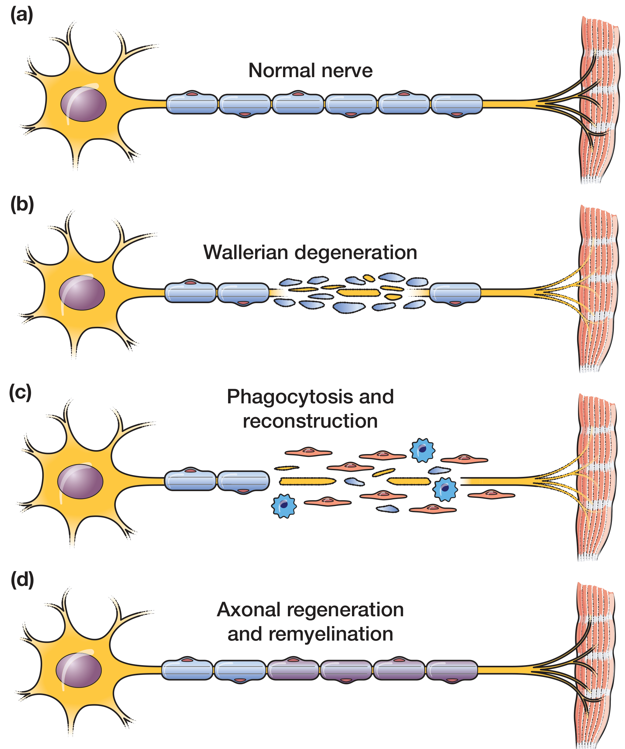
Figure 3.4 (Bailey & Love): The four stages - (a) Normal nerve, (b) Wallerian degeneration at injury site, (c) Phagocytosis and reconstruction by Schwann cells and macrophages, (d) Axonal regeneration and remyelination
E. Regenerative Sprouting
- From the proximal stump, the axon sprouts multiple small growth cones (regenerative sprouting)
- These grow along the bands of Bungner at a rate of approximately 1-4 mm/day (roughly 1 inch/month)
- Multiple sprouts form; only the one that successfully enters the distal endoneurial tube survives; the others retract
"The nerve then starts to regrow, with multiple small branches projecting along the path the axon previously followed (regenerative sprouting)." - Ganong, p. 143
F. Neurotropism - Target-Directed Growth
Regenerating axons are guided back to their original targets by neurotropism:
- Neurotrophins (growth factors) released by target tissues act as chemical attractants
- The extracellular matrix and hormones also contribute
- Axons can sometimes grow back to their original motor or sensory endings, especially at the neuromuscular junction
"Axons sometimes grow back to their original targets, especially in locations like the neuromuscular junction. However, nerve regeneration is generally limited because axons often become entangled in the area of tissue damage at the site where they were disrupted." - Ganong, p. 143
"The regenerating nerve fibres are attracted to their receptors by neurotropism, which is mediated by growth factors, hormones and the extracellular matrix." - Bailey & Love, 28th Ed.
G. Remyelination
Once the regenerated axon reaches its target:
- Schwann cells wrap around the new axon to restore the myelin sheath
- The new myelin is initially thinner and shorter internodal segments compared to the original
- Conduction velocity is lower than normal initially but may improve over months
Part 4: Denervation Hypersensitivity (Supersensitivity)
This is a clinically important consequence of nerve degeneration described in detail by Ganong:
In Skeletal Muscle:
- Normally, nicotinic ACh receptors are concentrated only at the motor end plate (neuromuscular junction)
- When the motor nerve is cut and degenerates, the muscle becomes extremely sensitive to acetylcholine - called denervation hypersensitivity or denervation supersensitivity
- There is a marked proliferation of nicotinic receptors over the entire muscle fiber surface (not just the end plate)
In Smooth Muscle:
- Smooth muscle does not atrophy when denervated (unlike skeletal muscle)
- But it becomes hyperresponsive to the chemical mediator that normally activates it
- The same phenomenon occurs at autonomic junctions
Mechanism of Denervation Hypersensitivity:
- Upregulation of receptors - loss of the chemical messenger leads to upregulation of its receptors (receptor supersensitivity)
- Lack of reuptake - when the nerve terminal degenerates, there is no longer reuptake of secreted neurotransmitters, so even small amounts cause exaggerated effects
"When the motor nerve to skeletal muscle is cut and allowed to degenerate, the muscle gradually becomes extremely sensitive to acetylcholine... Normally nicotinic receptors are located only in the vicinity of the motor endplate... When the motor nerve is severed, there is a marked proliferation of nicotinic receptors over a wide region of the neuromuscular junction." - Ganong, 26th Ed., p. 143-144
Part 5: Factors Affecting Successful Regeneration
| Factor | Effect |
|---|---|
| Type of injury | Crush (axonotmesis) regenerates better than complete cut (neurotmesis) |
| Endoneurial tube integrity | Intact tubes greatly improve guidance of regenerating axons |
| Distance to target | Longer distances = worse outcome (muscle may atrophy before reinnervation) |
| Age | Younger patients regenerate better |
| Neurotrophins | Administration of neurotrophins (e.g., NGF) improves regeneration |
| Fibrosis at injury site | Scar tissue traps growing axons - major barrier |
"Nerve regeneration is generally limited because axons often become entangled in the area of tissue damage at the site where they were disrupted. This difficulty has been reduced by administration of neurotrophins." - Ganong, p. 143
Part 6: Complications of Failed Regeneration
- Neuroma formation: When axons cannot reach their target and grow in a disorganized, coiled mass at the injury site. Neuromas are painful and can cause hypersensitivity.
- Denervation atrophy: Skeletal muscle fibers progressively atrophy without motor nerve input.
- Misdirected reinnervation: Axons may reinnervate wrong targets (e.g., a motor axon growing to a sensory receptor), leading to abnormal function.
Summary Table
| Process | Location | Timing | Key Players |
|---|---|---|---|
| Chromatolysis | Cell body | Hours to days | Nissl substance dispersal |
| Wallerian degeneration | Distal to injury | Days 1-14 | Axon, myelin breakdown |
| Retrograde degeneration | Proximal to nearest node | Days | Limited extent |
| Schwann cell proliferation | Distal stump | Days 1-7 | Bands of Bungner formed |
| Macrophage phagocytosis | Distal stump | Days 3-14 | Myelin debris cleared |
| Regenerative sprouting | Proximal stump | Weeks | Growth cones, 1-4 mm/day |
| Neurotropism | Along nerve path | Weeks-months | Growth factors, ECM |
| Remyelination | Regenerated axon | Months | Schwann cells |
| Denervation supersensitivity | Target (muscle/gland) | Days-weeks | Receptor upregulation |
Quick Mnemonics for 1st Year MBBS
For degeneration sequence (distal to proximal):
"Waller Marches Distally" = Wallerian degeneration, Myelin breaks down, Distal stump affected first
For cell body changes:
"Swollen Eccentric Nissl Disperses" = Swelling + Eccentric nucleus + Nissl body (chromatolysis) Disperses
Regeneration rate: ~1 mm/day or ~1 inch/month (easy to remember for clinical estimation)
Sources:
- Ganong's Review of Medical Physiology, 26th Edition - Chapter 6: "Axonal Injury & Denervation Supersensitivity," pp. 143-144 (Figure 6-14)
- Guyton and Hall Textbook of Medical Physiology - Chapter 7: Neuromuscular Junction and context of nerve-muscle physiology
- Bailey & Love's Short Practice of Surgery, 28th Edition - Figure 3.4: Peripheral nerve degeneration and regeneration
This is a shared conversation. Sign in to Orris to start your own chat.