
Tell me about iris in antomy
The Iris
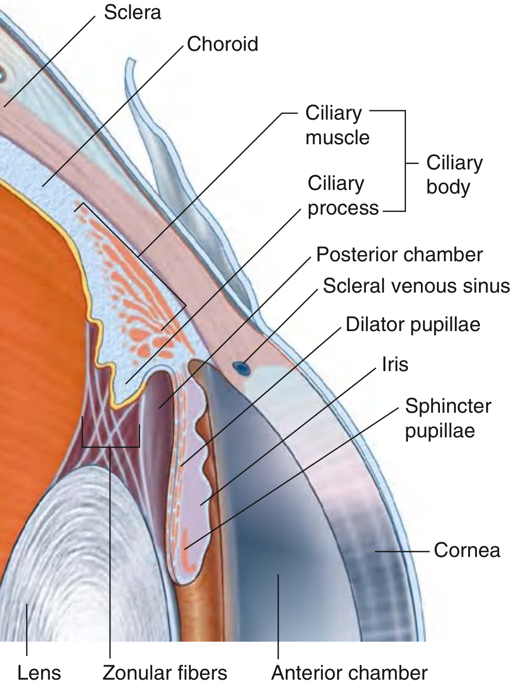
Position and Overview
- Anterior chamber - between the cornea and the iris
- Posterior chamber - between the iris and the lens
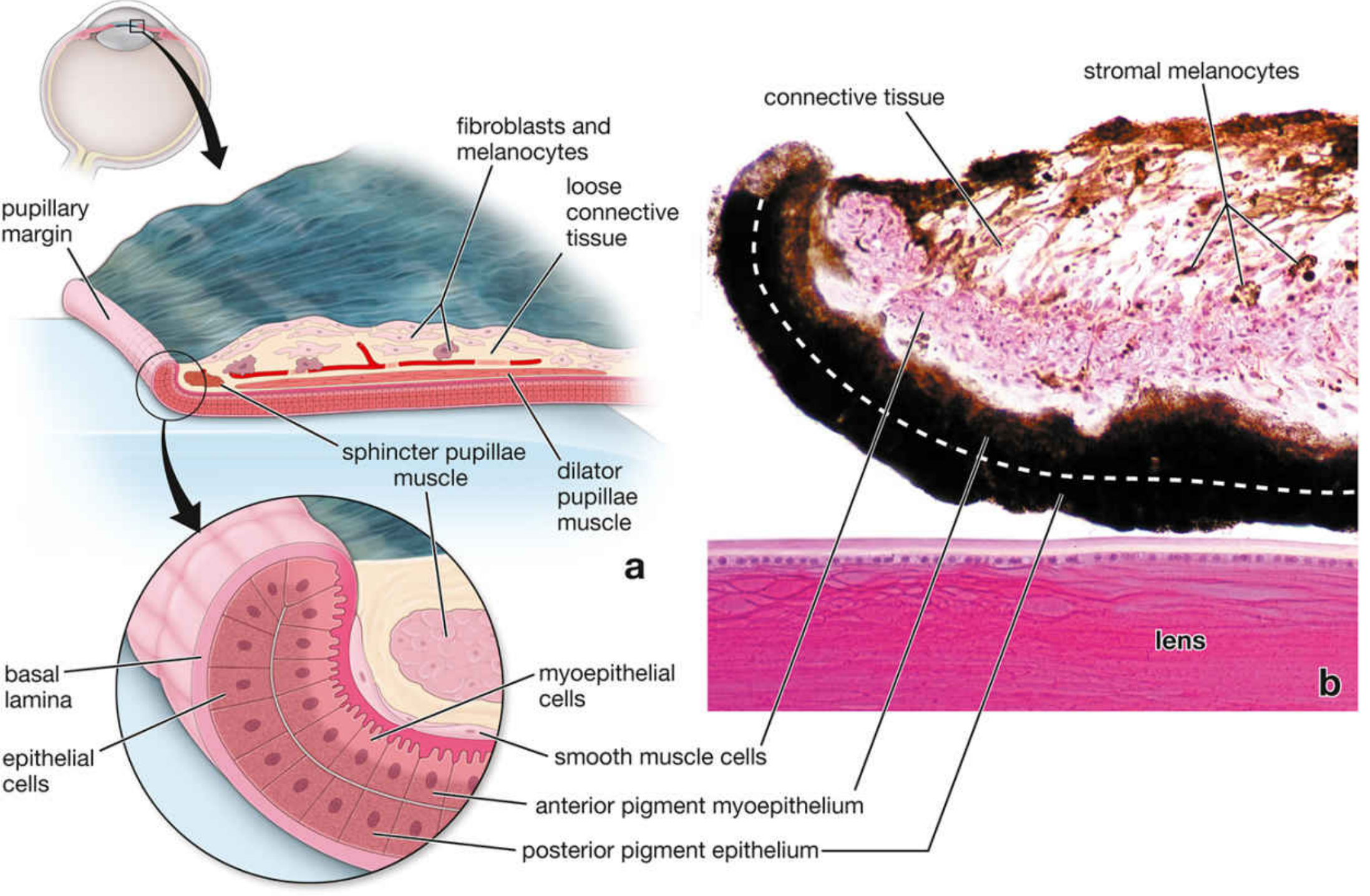
Layers of the Iris (from anterior to posterior)
-
Anterior surface - a discontinuous layer of fibroblasts and melanocytes (not a true epithelium). It shows numerous ridges and grooves visible on clinical examination.
-
Stroma - highly vascularized loose connective tissue containing:
- Blood vessels
- Stromal melanocytes (whose number determines eye color)
- The sphincter pupillae muscle near the pupillary margin
-
Anterior pigment myoepithelium - a layer of myoepithelial cells with:
- Apical (posterior) portions laden with melanin granules
- Basal (anterior) portions with contractile processes forming the dilator pupillae muscle
-
Posterior pigment epithelium - intensely pigmented cells; so heavily pigmented that neither nuclei nor cytoplasmic detail is visible under light microscopy. Basal lamina faces the posterior chamber.
Muscles of the Iris
| Muscle | Arrangement | Innervation | Action |
|---|---|---|---|
| Sphincter pupillae | Circular fibers near pupillary margin (in stroma) | Parasympathetic (CN III) | Constricts pupil (miosis) |
| Dilator pupillae | Radial myoepithelial cell processes | Sympathetic | Dilates pupil (mydriasis) |
Eye Color
- Few stromal melanocytes → light scatter from the posterior pigment epithelium produces a blue appearance
- More melanocytes → progressively green, hazel, or brown eyes
- The posterior pigment epithelium is uniformly dark in all individuals; it is only the stromal melanocyte density that varies
Clinical Relevance
- The iridocorneal angle (where iris meets cornea) contains the trabecular meshwork through which aqueous humor drains into the scleral venous sinus (canal of Schlemm). Obstruction here causes glaucoma.
- Laser iridotomy creates a small opening at the base of the iris to widen the iridocorneal angle in angle-closure glaucoma.
- Mydriatic drugs (atropine, phenylephrine) dilate the pupil by blocking parasympathetics or stimulating sympathetics.
- Miotic drugs (pilocarpine) constrict the pupil via parasympathomimetic action.
Five elements of accupuntre
five elements acupuncture TCM Wood Fire Earth Metal Water theory
five elements acupuncture TCM Wood Fire Earth Metal Water diagram
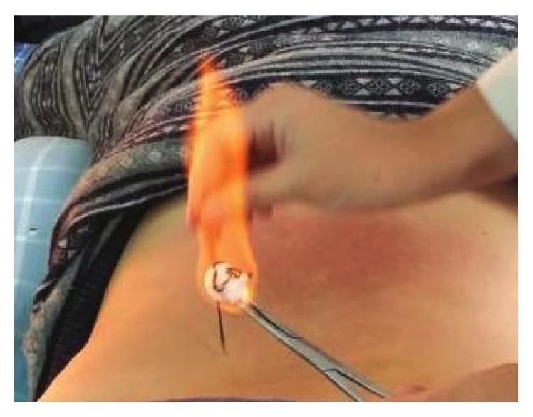
This clinical procedural photograph demonstrates fire-needle moxibustion, a technique in Traditional Chinese Medicine (TCM). The image shows a practitioner's hand using a metallic surgical clamp or forceps to hold a thin, dark acupuncture needle that has been inserted perpendicularly into the patient's skin, likely at a localized acupoint on the torso. A cotton ball soaked in a flammable substance is ignited, creating a bright orange flame that engulfs the distal end of the needle to heat it during the procedure. The surrounding skin displays a localized patch of mild erythema (redness), representing an inflammatory response to the thermal stimulation or the needling itself. This image serves as an educational example of integrative medicine, specifically showing the application of heat through a high-temperature-resistant needle to treat conditions like osteoarthritis or chronic pain. The procedural focus highlights the interaction between clinical tools, thermal elements, and the physiological skin response in external therapies.
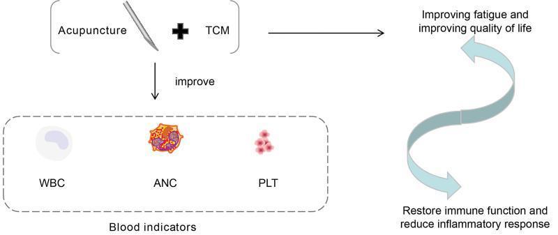
A medical conceptual diagram illustrating the integrative oncological approach for lung cancer management using Acupuncture and Traditional Chinese Medicine (TCM). The left side of the flowchart depicts the intervention—acupuncture (symbolized by a needle) and TCM—leading to the improvement of 'Blood indicators,' specifically identifying White Blood Cells (WBC), Absolute Neutrophil Count (ANC), and Platelets (PLT) through clinical illustrations of these hematologic components. The right side of the diagram outlines the physiological and symptomatic outcomes, connected by a bidirectional curved arrow suggesting a synergistic cycle: 'Improving fatigue and improving quality of life' and 'Restore immune function and reduce inflammatory response.' The diagram serves as a visual summary of how integrative therapies target cancer-related fatigue and hematological health, ultimately aiming to restore homeostatic immune function in cancer patients.
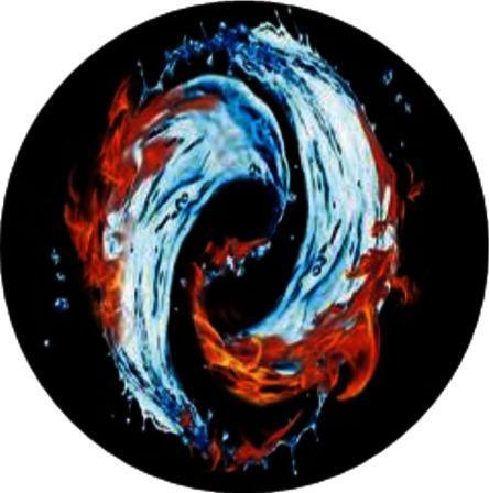
This medical illustration represents the concept of 'intermixed rooting of yin and yang' within the context of Traditional Chinese Medicine (TCM) philosophy applied to oncology. The diagram features two swirling, interconnected elements forming a circular Taiji-style composition against a black background. One element is depicted as flowing blue and white water, symbolizing 'Yin,' while the opposing element is shown as red and orange flames, symbolizing 'Yang.' The fluid and flickering textures illustrate a dynamic, cyclical relationship where the elements merge and interact at their boundaries. In a clinical context, this visual serves as a conceptual model for the balance between chemotherapeutic efficacy and toxicity, or the synergistic interaction between conventional oncology treatments (e.g., chemotherapy for NSCLC) and natural medicines like the Shenyi capsule. It illustrates the pharmacological principles of 'attenuation, sensitization, and synergistic effects' through the framework of maintaining homeostatic balance between opposing biological forces.
Five Elements in Acupuncture (Wu Xing - 五行)
1. 🌿 Wood (木 - Mù)
| Attribute | Correspondence |
|---|---|
| Yin organ | Liver |
| Yang organ | Gallbladder |
| Season | Spring |
| Emotion | Anger / frustration |
| Color | Green |
| Taste | Sour |
| Body tissue | Tendons & sinews |
| Sense organ | Eyes |
2. 🔥 Fire (火 - Huǒ)
| Attribute | Correspondence |
|---|---|
| Yin organ | Heart (+ Pericardium) |
| Yang organ | Small Intestine (+ Triple Warmer) |
| Season | Summer |
| Emotion | Joy / over-excitement |
| Color | Red |
| Taste | Bitter |
| Body tissue | Blood vessels |
| Sense organ | Tongue |
3. 🌍 Earth (土 - Tǔ)
| Attribute | Correspondence |
|---|---|
| Yin organ | Spleen |
| Yang organ | Stomach |
| Season | Late summer / transition |
| Emotion | Worry / overthinking |
| Color | Yellow |
| Taste | Sweet |
| Body tissue | Muscles & flesh |
| Sense organ | Mouth / lips |
4. 🪨 Metal (金 - Jīn)
| Attribute | Correspondence |
|---|---|
| Yin organ | Lungs |
| Yang organ | Large Intestine |
| Season | Autumn |
| Emotion | Grief / sadness |
| Color | White |
| Taste | Pungent / spicy |
| Body tissue | Skin & body hair |
| Sense organ | Nose |
5. 💧 Water (水 - Shuǐ)
| Attribute | Correspondence |
|---|---|
| Yin organ | Kidneys |
| Yang organ | Bladder |
| Season | Winter |
| Emotion | Fear / anxiety |
| Color | Black / dark blue |
| Taste | Salty |
| Body tissue | Bones & marrow |
| Sense organ | Ears |
The Two Governing Cycles
Sheng Cycle (Generating / Nourishing)
Wood → Fire → Earth → Metal → Water → Wood
- Wood fuels Fire
- Fire creates Earth (ash)
- Earth produces Metal (minerals)
- Metal enriches Water (condensation)
- Water nurtures Wood (growth)
Ke Cycle (Controlling / Restraining)
Wood controls Earth | Earth controls Water | Water controls Fire | Fire controls Metal | Metal controls Wood
How Acupuncturists Use This System
- Pulse qualities at three positions on each wrist
- Tongue appearance (color, coating, shape)
- Emotional tendencies and presenting symptoms
- Seasonal aggravation of symptoms