
What is
What is atrophic rhinitis
atrophic rhinitis
atrophic rhinitis nasal cavity crusting ozena
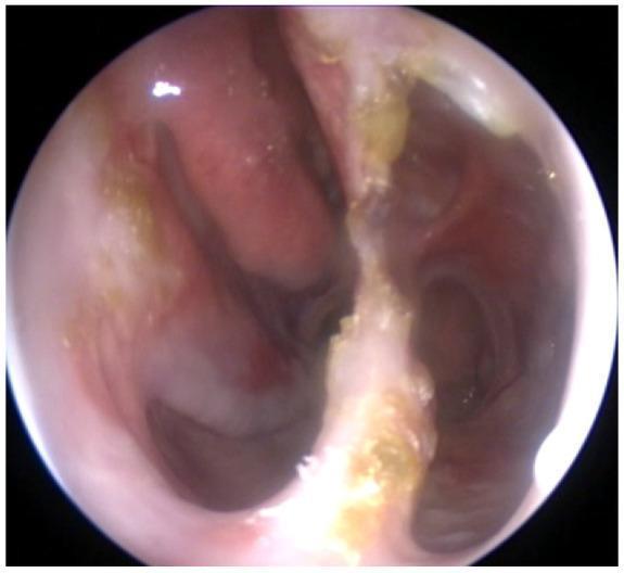
This clinical photograph is an endoscopic view of the internal nasal cavity, demonstrating significant structural and mucosal pathology. The image reveals a wide, single nasal cavity resulting from a large septal perforation and the subsequent loss of septal cartilage. The nasal mucosa appears markedly atrophic, thin, and friable. There is evidence of significant turbinate atrophy, with the inferior and middle turbinates being nearly absent. Extensive crusting is visible throughout the cavity; gray-colored crusts are distributed along the posterior regions toward the rhinopharynx, while yellowish-gold crusts are adherent to the remnants of the septal cartilage. The visual findings are consistent with chronic inflammatory or granulomatous processes, such as granulomatosis with polyangiitis (GPA), illustrating secondary atrophic rhinitis and structural collapse. This image serves as an educational tool for identifying end-stage nasal manifestations of systemic autoimmune diseases.
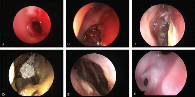
A composite clinical diagnostic image containing six nasal endoscopy frames (A–F) documenting postoperative complications following endonasal transsphenoidal surgery. (A) Active nasal hemorrhage characterized by bright red, fluid blood obscuring the field. (B) Evidence of previous hemorrhage with dark, solidified blood clots within the nasal passage. (C) Sphenoid sinusitis presenting with yellowish purulent discharge and polypoid mucosal changes. (D) Atrophic rhinitis featuring characteristic crusting and a greyish-white fungal ball/infection. (E) Nasal septum perforation showing a clear structural defect allowing visualization of the contralateral nasal cavity. (F) Right nasal synechia (adhesion) consisting of a fibrotic tissue bridge connecting the nasal septum to the lateral wall. This clinical photograph series serves as an educational reference for Otorhinolaryngology and Neurosurgery trainees to identify common and rare sequelae of endoscopic skull base procedures, emphasizing tissue healing, infection, and structural integrity of the sinonasal mucosa.
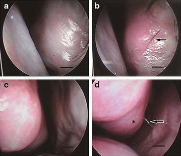
This set of four clinical endoscopic images (a-d) demonstrates the nasal cavity and turbinate morphology in patients with Empty Nose Syndrome (ENS) before and after regenerative treatment with fat tissue and adipose-derived stem cells (ADSCs). Panel (a) and (c) represent the preoperative state, showing a pale, atrophic nasal mucosa with diminished turbinate volume and limited secretions. Panel (b) highlights the emergence of rhinothrix (nasal hair, indicated by an arrow) on the regenerated mucosal surface. Panel (d) illustrates significant post-treatment recovery, characterized by more erythematous and healthy-appearing mucosa with increased mucus production (arrow) and the restoration of nasal turbinate morphology (asterisk). These images serve as visual evidence of mucosal regeneration and physiological recovery in the context of otorhinolaryngology and regenerative medicine, specifically for managing atrophic rhinitis and ENS. The clinical focus is on the structural and functional improvement of the nasal airway lining and turbinate bulk.
Atrophic Rhinitis
Definition
Types
1. Primary Atrophic Rhinitis
- More prevalent in developing countries in subtropical and temperate climate zones
- Predominantly affects women
- Etiology is not fully clear, but bacterial infection is strongly implicated
- Causative organisms: Klebsiella ozaenae, Staphylococcus aureus, Proteus mirabilis, Escherichia coli
- K. ozaenae is the organism most classically associated
2. Secondary Atrophic Rhinitis
-
More prevalent in developed countries; generally less severe and less progressive
-
Caused by identifiable local or systemic factors:
- Excessive sinonasal surgery (including aggressive turbinate removal - "empty nose syndrome")
- Granulomatous diseases (e.g., granulomatosis with polyangiitis, sarcoidosis, tuberculosis)
- Radiation therapy to the head and neck
- Trauma
- Chronic rhinosinusitis
- Rhinitis medicamentosa (rebound rhinitis from prolonged decongestant overuse)
-
Cummings Otolaryngology; Scott-Brown's Otorhinolaryngology, Vol. 1
Pathology & Histology
- Squamous metaplasia of the respiratory (ciliated columnar) epithelium
- Glandular atrophy - loss of mucous and serous glands
- Diffuse endarteritis obliterans - obliteration of small vessels leading to ischemia and further atrophy
- Loss of mucociliary function
Clinical Features
| Feature | Description |
|---|---|
| Crusting | Thick yellow/green nasal crusts |
| Ozena | Persistent foul smell (from bacterial colonization of crusts) |
| Anosmia | Loss of smell, from mucosal destruction |
| Nasal obstruction | Paradoxical - cavity is wide but patient feels congested |
| Epistaxis | From friable atrophic mucosa |
| Wide nasal cavity | Turbinate atrophy leads to spacious but dysfunctional nasal airway |
Diagnosis
- History of the characteristic symptoms
- Nasal endoscopy - shows wide, crusted nasal cavity with atrophied turbinates
- Microbiological cultures (to identify K. ozaenae and other organisms)
- CT scan of the sinuses (to assess degree of bony atrophy and rule out other pathology)
Management
- Nasal saline irrigation - regular, high-volume saline washes to clear crusts and moisturize mucosa (mainstay of treatment)
- Antibiotics - systemic or topical, targeting K. ozaenae (e.g., ciprofloxacin); used for acute bacterial colonization
- Nasal moisturizers and lubricants - glucose-glycerin drops, sesame oil, or other emollients
- Surgical reduction of nasal cavity size - procedures to narrow the abnormally wide nasal cavity, reducing the air-drying effect on the mucosa (e.g., submucosal injections, implants, or Young's procedure - closure of one or both nostrils to rest the mucosa)
- A 2025 systematic review (PMID: 40820344) explored platelet-rich plasma (PRP) injections as a regenerative treatment, representing an emerging approach to mucosal restoration
Distinction from Empty Nose Syndrome
Clinical Image
Do you know me
I m final year mbbs Student in india
- 🏥 Clinical subjects - Medicine, Surgery, OBG, Pediatrics, Orthopedics, ENT, Ophthalmology, Psychiatry
- 💊 Pharmacology & Pathology - Drug mechanisms, side effects, lab findings, histology
- 🔬 Preclinical revision - Anatomy, Physiology, Biochemistry
- 📋 Case discussions - Patient workups, differentials, management plans
- 📝 Exam prep - High-yield topics, MCQs, short/long answer questions (university exam style)
- 🧬 Recent guidelines - Updated treatment protocols from PubMed and clinical sources
Create a quick reference sheet on ENT common disorders
What format do you want the quick reference sheet in? · Which ENT areas should it cover?
🩺 NOSE - Quick Reference Sheet
Final Year MBBS | ENT | High-Yield Topics
1. EPISTAXIS (Nosebleed)
| Feature | Details |
|---|---|
| Most common site | Little's area / Kiesselbach's plexus (anterior septum) |
| Blood supply to Kiesselbach's | Ant. ethmoidal + Post. ethmoidal + Sphenopalatine + Greater palatine + Superior labial arteries |
| Anterior epistaxis | More common, less severe, easier to control |
| Posterior epistaxis | From posterior 2/3 of nasal cavity - severe, harder to control |
- First aid: Pinch soft part of nose, lean forward, 10-15 min
- Anterior bleed: Chemical cautery (silver nitrate) > electrical cautery > anterior nasal packing (BIPP/Merocel)
- Posterior bleed: Posterior nasal packing / Foley catheter balloon / surgical ligation / embolization
- Correct coagulopathy if present; control hypertension
2. RHINITIS
Allergic Rhinitis
| Feature | Details |
|---|---|
| Type | Type I hypersensitivity (IgE-mediated) |
| Symptoms | Sneezing, watery rhinorrhoea, nasal itching, bilateral obstruction, postnasal drip |
| Signs | Pale/bluish boggy turbinates, allergic salute, allergic shiners |
| Diagnosis | Skin prick test, serum IgE, nasal provocation test |
| Treatment | Allergen avoidance, intranasal corticosteroids (1st line), antihistamines, LTRA, immunotherapy |
Atrophic Rhinitis (Ozena)
| Feature | Details |
|---|---|
| Types | Primary (idiopathic, K. ozaenae) / Secondary (post-surgery, radiation, granulomatous) |
| Symptoms | Paradoxical nasal obstruction, foul smell (ozena), anosmia, thick yellow/green crusts |
| Histology | Squamous metaplasia, glandular atrophy, endarteritis obliterans |
| Treatment | Nasal saline irrigation, antibiotics (ciprofloxacin), Young's procedure (surgical cavity narrowing) |
Vasomotor (Non-Allergic) Rhinitis
- Triggered by temperature change, smoke, strong smells, alcohol
- IgE negative, no eosinophilia
- Treatment: Ipratropium bromide nasal spray, intranasal steroids
3. SINUSITIS
| Acute | Chronic | |
|---|---|---|
| Duration | < 4 weeks | > 12 weeks |
| Organisms | S. pneumoniae, H. influenzae, M. catarrhalis | Polymicrobial + anaerobes; fungi (in immunocompromised) |
| Symptoms | Facial pain/pressure, purulent nasal discharge, fever, nasal obstruction | Dull facial pressure, nasal obstruction, postnasal drip, anosmia |
| Most common sinus | Maxillary (largest, low ostium) | Maxillary |
| Frontal sinus complication | Pott's puffy tumour (osteomyelitis) | - |
| Orbit complication | Orbital cellulitis / abscess (Chandler's stages) | - |
| Intracranial complication | Meningitis, cavernous sinus thrombosis, brain abscess | - |
| Investigation | Clinical; CT scan (gold standard for chronic/complicated) | CT sinuses |
| Treatment | Saline irrigation, decongestants, analgesics; antibiotics if bacterial (amoxicillin ± clavulanate) | Intranasal steroids; FESS (Functional Endoscopic Sinus Surgery) |
4. NASAL POLYPS
| Feature | Details |
|---|---|
| Character | Pale, grey, glistening, insensitive to touch (unlike turbinates), mobile, bilateral in inflammatory type |
| Associations | Chronic rhinosinusitis, allergic rhinitis, aspirin sensitivity (Samter's triad), cystic fibrosis, Kartagener's |
| Samter's Triad | Nasal polyps + Bronchial asthma + Aspirin sensitivity |
| Unilateral polyp | Always send for histology - may be malignant |
| Bleeding polyp | Suspect malignancy |
| Polyp in child | Exclude meningocele/encephalocele before removal |
| Histology | Oedematous stroma, eosinophil infiltration, respiratory epithelium |
| Treatment | Intranasal/systemic corticosteroids; surgical removal with powered microdebrider (FESS) |
5. DEVIATED NASAL SEPTUM (DNS)
| Feature | Details |
|---|---|
| Symptoms | Unilateral/alternating nasal obstruction, headache, snoring, recurrent sinusitis |
| Types | C-shaped, S-shaped, spur |
| Investigation | Anterior rhinoscopy, nasal endoscopy, CT |
| Treatment | Septoplasty (after 17-18 years - when growth complete) |
| Note | Compensatory hypertrophy of inferior turbinate on opposite side |
6. NASAL FOREIGN BODY
- Common in children; object usually in floor of nasal cavity (below inferior turbinate)
- Presents with: unilateral foul-smelling purulent discharge in a child = foreign body until proved otherwise
- Long-standing = rhinolith (calcified foreign body)
- Removal: Positive pressure technique (mother's kiss), hook or forceps under direct vision; GA if uncooperative child
7. JUVENILE NASOPHARYNGEAL ANGIOFIBROMA (JNA)
| Feature | Details |
|---|---|
| Who | Adolescent males (15-25 years) |
| Origin | Sphenopalatine foramen |
| Nature | Benign but locally aggressive, highly vascular |
| Presentation | Recurrent profuse epistaxis + progressive unilateral nasal obstruction |
| Investigation | CT/MRI (do NOT biopsy - causes torrential bleeding) |
| Treatment | Pre-op embolization + surgical excision (endoscopic or open) |
8. KEY "ONE-LINERS" FOR EXAMS
| Condition | High-Yield Fact |
|---|---|
| Most common sinus involved in sinusitis | Maxillary |
| Most common site of epistaxis | Kiesselbach's plexus (Little's area) |
| Fungal sinusitis in diabetic/immunocompromised | Mucormycosis - emergency, invades orbit/brain |
| Ozena caused by | Klebsiella ozaenae |
| Samter's triad | Polyps + Asthma + Aspirin sensitivity |
| Angiofibroma biopsy | CONTRAINDICATED |
| Unilateral nasal polyp | Always exclude malignancy |
| Polyp in child | Exclude meningocele/encephalocele |
| Pott's puffy tumour | Frontal sinusitis + osteomyelitis of frontal bone |
| Cavernous sinus thrombosis | Dangerous complication of sphenoid/ethmoid sinusitis |
| Kartagener's syndrome triad | Sinusitis + Bronchiectasis + Situs inversus |
| Young's procedure | Surgical closure of nostril for atrophic rhinitis |