
Give me an info graphic on Clostridium difficile for MD Microbiology exam
Medical exam infographic on Clostridium difficile (C. diff) for MD Microbiology. Clean, professional, dark navy blue background with color-coded sections. Include the following clearly labeled sections: 1. ORGANISM PROFILE (top): Gram-positive, large anaerobic rod (0.5-1.9 x 3.0-17 µm), spore-forming, obligate anaerobe, produces volatile fatty acids ("barnyard" odor), name means "difficult to isolate". Gram stain illustration showing purple rods with terminal spores. 2. VIRULENCE FACTORS (left panel, orange): Toxin A (Enterotoxin) - chemotactic for neutrophils, disrupts tight junctions, increases intestinal permeability, causes diarrhea. Toxin B (Cytotoxin) - depolymerizes actin, destroys cytoskeleton in vivo and in vitro. Both act SYNERGISTICALLY. Binary toxin CDT in hypervirulent NAP1/BI/027 strain. 3. PATHOGENESIS FLOWCHART (center): Antibiotic exposure (clindamycin, cephalosporins, fluoroquinolones, quinolones) → disrupts normal colonic flora → C. difficile overgrowth → toxin A+B production → neutrophil infiltration + cytokine release → colonic inflammation → pseudomembrane formation. 4. EPIDEMIOLOGY (right panel, teal): #1 cause of nosocomial diarrhea in adults. >300,000 cases/year in USA. ~25% of antibiotic-associated diarrhea. 50-75% of antibiotic-associated colitis. Colonizes <5% healthy individuals (endogenous). Spores persist on hospital surfaces. Community-acquired cases rising since 2000s. 5. CLINICAL SPECTRUM (bottom left, red gradient): Mild: watery diarrhea. Moderate: lower abdominal cramping + systemic fever/malaise. Severe: PSEUDOMEMBRANOUS COLITIS - profuse diarrhea, whitish plaques on colonic mucosa, abdominal cramping, fever. Fulminant: toxic megacolon → colonic perforation → peritonitis → mortality up to 38%. 6. DIAGNOSIS (bottom center, yellow): Gold standard = Tissue culture cytotoxicity assay (sensitivity 80-90%, specificity ~99%, takes 48h). EIA for toxin A or B (sensitivity 65-85%, specificity 95-100%, rapid). NAAT/PCR for toxin genes (high sensitivity/specificity, similar to cytotoxin assay). GDH (glutamate dehydrogenase) screening antigen assay. Stool culture (takes up to 96h). Endoscopy for pseudomembranous colitis (avoid in fulminant colitis - perforation risk). 7. TREATMENT (bottom right, green): 1. STOP offending antibiotic. 2. Mild-moderate: Metronidazole (oral). 3. Severe: Vancomycin (oral). 4. Recurrent/refractory: Fecal Microbiota Transplant (FMT). NOTE: Relapse common - antibiotics don't kill spores! Second course usually successful. Multiple courses may be needed. 8. PREVENTION (bottom, purple): Contact precautions, hand washing with SOAP (alcohol gel INEFFECTIVE against spores), gloves + gown, thorough room decontamination with BLEACH after discharge. EXAM MNEMONICS in corner: "ABCDE" - Antibiotic associated, Broad-spectrum antibiotics trigger it, C. diff Colitis/pseudomembranes, Diarrhea + Death, Endoscopy for diagnosis. Use icons, arrows, small illustrations. High information density, exam-ready format.
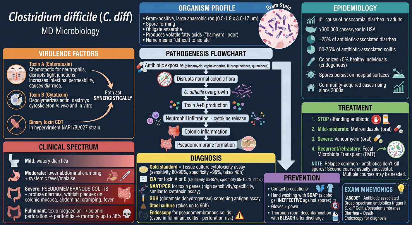
High-Yield C. difficile Summary
- Toxin A (Enterotoxin) - attracts neutrophils, disrupts tight junctions, increases intestinal permeability → diarrhea
- Toxin B (Cytotoxin) - depolymerizes actin, destroys cytoskeleton in vivo and in vitro
- Mild: self-limited watery diarrhea
- Severe: Pseudomembranous colitis - whitish plaques on colonic mucosa, profuse diarrhea, fever, cramping
- Fulminant: toxic megacolon → perforation → mortality up to 38%
| Test | Sensitivity | Specificity | Time |
|---|---|---|---|
| Tissue culture cytotoxin assay (gold std) | 80-90% | ~99% | 48h |
| EIA (toxin A/B) | 65-85% | 95-100% | Rapid |
| NAAT/PCR | High (similar to gold std) | High | Rapid |
| GDH antigen screen | Screening only | - | Rapid |
- Stop the offending antibiotic
- Mild-moderate: Metronidazole (oral)
- Severe: Vancomycin (oral)
- Recurrent/refractory: Fecal Microbiota Transplant (FMT)
- Relapse is common - spores are NOT killed by antibiotics!
Microbiology diagnosis
What do you mean by 'Microbiology diagnosis'?
Detailed medical exam infographic: "Microbiology Diagnosis of Clostridium difficile" for MD students. Clean, professional layout on a deep dark blue/charcoal background. Organized into clearly labeled color-coded sections with headers, icons, and arrows. High information density, exam-ready. TITLE at top: "C. difficile: Microbiology Diagnosis" with subtitle "MD Microbiology Exam Reference" SECTION 1 - SPECIMEN (bright teal header): - Specimen: DIARRHEAL STOOL only (do NOT test formed stool) - One stool sample usually sufficient - Must be diarrheal (>3 loose stools/24h) - NAATs/EIAs cannot distinguish asymptomatic carriage from active infection SECTION 2 - DIAGNOSTIC ALGORITHM FLOWCHART (central, white/yellow): Step 1: GDH (Glutamate Dehydrogenase) Antigen Immunoassay (rapid screening) - Positive GDH → Step 2: EIA for Toxins A and B - Toxin positive → CONFIRM C. difficile infection - Toxin negative (discordant) → Step 3: NAAT/PCR for toxin genes → confirm or rule out - Negative GDH → Low probability, consider other diagnosis Draw clear arrows connecting these steps. SECTION 3 - INDIVIDUAL TESTS TABLE (left panel, orange): 5 rows with test name, sensitivity, specificity, turnaround time, key notes: 1. Tissue Culture Cytotoxicity Assay Sens: 80-90% | Spec: ~99% Time: 48 hours | GOLD STANDARD Detects cytotoxin B | Detects as little as 10 pg toxin Requires tissue culture facility 2. GDH Antigen EIA High negative predictive value Does NOT distinguish toxigenic vs nontoxigenic strains Used as SCREENING ONLY Rapid 3. EIA (Toxin A and/or B) Sens: 65-85% | Spec: 95-100% Time: 4 hours | Detects >1000 pg toxin Simple, rapid, preferred in >6 stools/day inpatients Lower sensitivity than gold standard 4. Stool Culture + Cytotoxin Assay Sens: >90% | Spec: >98% Time: 72-96 hours | Labor intensive Best for OUTBREAK INVESTIGATION + strain typing 5. NAAT/PCR (Nucleic Acid Amplification) Sens: 93-100% | Spec: High (similar to gold standard) Rapid result | Detects toxin genes (tcdA, tcdB) LIMITATION: Cannot distinguish carriage vs active disease Use for DISCORDANT GDH/Toxin results SECTION 4 - ENDOSCOPY (right panel, red): Pseudomembranous Colitis diagnosis: - Endoscopic visualization required - Shows: WHITISH/YELLOW PLAQUES (pseudomembranes) on intact colonic mucosa - Classic "volcano lesion" appearance - WARNING: AVOID endoscopy in fulminant colitis → risk of PERFORATION - CT scan alternative: "accordion sign" on contrast CT (mucosal thickening with oral contrast trapped in folds) SECTION 5 - KEY EXAM POINTS (bottom, purple banner): ★ Gold standard = Tissue Culture Cytotoxin Assay (but slow - 48h) ★ Fastest practical test = EIA Toxin A/B (4h) ★ Best sensitivity = NAAT/PCR (93-100%) ★ Best for outbreaks = Stool culture (strain typing) ★ GDH alone NEVER confirms infection - needs toxin confirmation ★ Test ONLY diarrheal stools ★ NAAT cannot distinguish carriage vs disease ★ Endoscopy: pseudomembranes = pathognomonic (but avoid in fulminant disease) Include small icons: microscope, test tube, DNA helix, stool sample, endoscope. Use color consistently. Bold the key numbers and percentages.
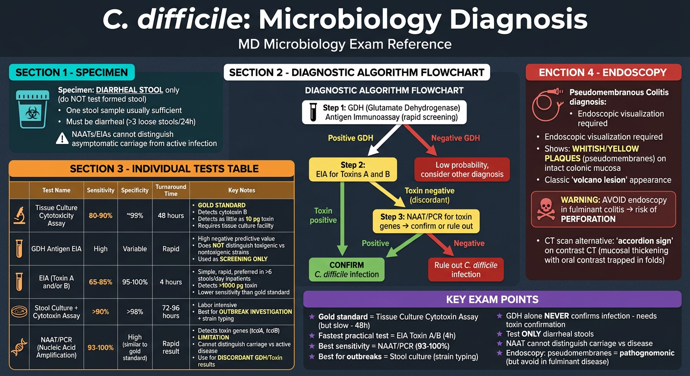
C. difficile Diagnosis - High-Yield Summary
Diagnostic Tests at a Glance
| Test | Sensitivity | Specificity | Time | Role |
|---|---|---|---|---|
| Tissue culture cytotoxin assay | 80-90% | ~99% | 48h | Gold standard |
| EIA (Toxin A/B) | 65-85% | 95-100% | 4h | Rapid, practical |
| NAAT/PCR (toxin genes) | 93-100% | High | Rapid | Confirmatory |
| GDH antigen EIA | High NPV | - | Rapid | Screening only |
| Stool culture | >90% | >98% | 72-96h | Outbreak investigation |
Recommended Algorithm (3-step)
- Screen with GDH antigen EIA
- If positive GDH → confirm with Toxin A/B EIA
- If discordant → resolve with NAAT/PCR
Critical Limitations to Know
- NAAT/PCR - cannot distinguish asymptomatic carriage vs active disease
- GDH - cannot distinguish toxigenic vs non-toxigenic strains (always needs toxin confirmation)
- EIA - detects >1,000 pg of toxin; less sensitive than cytotoxin assay (detects as little as 10 pg)
Endoscopy
- Required for diagnosing pseudomembranous colitis directly
- Appearance: whitish/yellow plaques (pseudomembranes) on intact colonic mucosa
- AVOID in fulminant colitis - risk of perforation
- CT alternative: "accordion sign"
In same way explain Clostridium botulinum
Comprehensive medical exam infographic on Clostridium botulinum for MD Microbiology. Dark navy blue background, clean professional layout, color-coded sections with icons, arrows, bold key facts. High information density, exam-ready format. TITLE: "Clostridium botulinum" with subtitle "MD Microbiology Exam Reference" SECTION 1 - ORGANISM PROFILE (top teal band): - Gram-positive, large anaerobic rod, spore-forming obligate anaerobe - Spores: HIGHLY heat resistant - survive 100°C for HOURS (killed only at 121°C autoclave) - Spore resistance reduced at: acid pH or high salt concentration - Name: "botulus" = sausage (first outbreak from insufficiently smoked sausage) - Toxin: heat LABILE - destroyed by heating 20 min at 100°C (or 10 min at 60-100°C) - Lethal dose in humans: ~1-2 µg/kg (one of the MOST TOXIC substances known) - Found in: soil worldwide, animal feces SECTION 2 - TOXIN TYPES (left panel, orange): 7 serotypes A-G: - Types A, B, E, F → Human disease - Type A, B → most common, associated with variety of foods - Type E → mainly fish products - Type C → limberneck in birds - Type D → botulism in cattle/mammals - Type G → NOT associated with disease Structure: 150 kDa protein - Heavy chain (H) = 100 kDa → binds presynaptic membrane - Light chain (L) = 50 kDa → cleaves SNARE proteins - Linked by disulfide bond SNARE protein targets: - Types A, C, E → cleave SNAP-25 - Type C also → cleaves Syntaxin - Types B, D, F, G → cleave Synaptobrevin (VAMP) SECTION 3 - MECHANISM OF ACTION (center, illustrated flowchart): Toxin absorbed from gut → enters bloodstream → binds presynaptic membrane of peripheral motor neurons and cranial nerve endings → L chain cleaves SNARE proteins → BLOCKS ACETYLCHOLINE RELEASE → FLACCID PARALYSIS Key: Does NOT cross blood-brain barrier. Does NOT affect CNS. Icon: nerve terminal with vesicles and blocked ACh release. SECTION 4 - CLINICAL FORMS (right panel, red-orange gradient): 4 forms with descending incubation: 1. FOODBORNE BOTULISM Onset: 18-24 hours after ingestion Classic "4 Ds + 2 Cs": Diplopia, Dysarthria, Dysphagia, Descending paralysis + Constipation, dry mouth (Cholinergic blockade) Also: blurred vision, abdominal pain, NO fever, patient remains CONSCIOUS until near death Progression: bilateral descending flaccid paralysis → respiratory failure → death Mortality: HIGH if untreated. No fever, No GI symptoms typically. 2. INFANT BOTULISM (most common form in USA) Vehicle: HONEY (most common), contaminated soil/dust Pathogenesis DIFFERENT: spores ingested → germinate in gut → vegetative cells produce toxin in situ → absorbed Signs: Poor feeding, weak cry, constipation, failure to thrive → "FLOPPY BABY" → respiratory arrest Possible cause of SUDDEN INFANT DEATH SYNDROME (SIDS) Note: Toxin in FECES not serum (unlike foodborne) 3. WOUND BOTULISM Route: Skin-popping contaminated "black tar" heroin (IV drug users) Same clinical as foodborne BUT longer incubation, FEWER GI symptoms Spores in wound → germinate → produce toxin locally 4. INHALATION BOTULISM Bioterrorism concern Rapid onset flaccid paralysis + pulmonary failure HIGH mortality SECTION 5 - DIAGNOSIS (bottom left, yellow): GOLD STANDARD: Mouse Bioassay - Specimens: Serum, stool, gastric secretions, implicated food - Mice injected IP → die rapidly if toxin present - Toxin type identified by neutralization with specific antitoxin in mice Infant botulism: C. botulinum + toxin in STOOL (not serum) Wound botulism: toxin in serum OR wound culture Other methods: ELISA, PCR (PCR may detect gene without toxin expression) Contact PUBLIC HEALTH AUTHORITIES before submitting specimens! SECTION 6 - TREATMENT (bottom center, green): 1. Supportive care (MOST IMPORTANT) - ICU, mechanical ventilation up to 8 weeks 2. Trivalent antitoxin (A, B, E) - give PROMPTLY IV; does NOT reverse paralysis, prevents progression 3. Penicillin or Metronidazole (antibiotic) 4. Infant botulism: Human Botulinum Immune Globulin (BIG) - "BabyBIG" NOTE: Antitoxin is from HORSES - check for hypersensitivity Mortality: Reduced from 65% to <25% with modern ICU care SECTION 7 - PREVENTION (bottom right, purple): Foods: Canning must reach 121°C (destroys spores) OR boil food 20 min before eating - Spore germination prevented by: acid pH, high sugar content, storage at ≤4°C - Toxin destroyed by: heating 20 min at 100°C NEVER give honey to infants under 12 months Commercial canning strictly regulated Foods implicated: home-canned alkaline vegetables, smoked/vacuum packed fish, spiced meats EXAM MNEMONICS box (bottom, gold): "ABCDE of Botulism": A = ACh blocked (flaccid paralysis) B = Bilateral descending paralysis C = Canned/home-preserved foods, Cranial nerves first D = Diplopia/Dysphagia/Dysarthria (bulbar symptoms first) E = Emergency antitoxin + ventilator support "SNARE" proteins = Synaptobrevin, SNAP-25, Syntaxin Sources: Medical Microbiology 9e | Jawetz Melnick & Adelberg Medical Microbiology 28e
Focused medical exam infographic: "Microbiology Diagnosis of Clostridium botulinum" for MD students. Dark charcoal background, clean professional layout, color-coded sections, icons. Exam-optimized. TITLE: "C. botulinum: Microbiology Diagnosis" with subtitle "MD Microbiology Exam Reference" SECTION 1 - SPECIMENS BY DISEASE TYPE (top, teal): Table layout: FOODBORNE: Serum + Stool + Gastric secretions + Implicated food INFANT: Stool/Feces (toxin AND organism) - NOT serum WOUND: Serum + Wound swab/culture INHALATION: Serum + Nasal secretions Key rule: In INFANT botulism - toxin in FECES not serum (germination occurs in gut) Contact PUBLIC HEALTH AUTHORITIES first before submitting specimens! SECTION 2 - MOUSE BIOASSAY (center-left, orange - GOLD STANDARD): Step-by-step flowchart: 1. Collect specimen (serum/stool/food) 2. Inject intraperitoneally into mice 3. Mice die rapidly → POSITIVE for botulinum toxin 4. To TYPE the toxin: Neutralize specimen with specific antitoxin (A, B, C...) → if mouse survives = that type confirmed Why gold standard: most sensitive, identifies toxin type, detects all serotypes Limitation: requires animal facility, slow, not widely available SECTION 3 - OTHER LAB TESTS (center-right, yellow): 1. ELISA (Enzyme-Linked Immunosorbent Assay) - Detects toxin in food/serum - Faster than mouse bioassay - Used in food safety labs 2. PCR - Detects toxin-encoding genes - IMPORTANT LIMITATION: May detect gene without toxin EXPRESSION - Useful for C. botulinum in food/wound cultures - Confirms organism presence, not always active toxin 3. Culture of C. botulinum - From food remnants - Rarely done, questionable clinical significance - Anaerobic culture required 4. EMG (Electromyography) - clinical confirmation - Shows characteristic pattern: incremental response on repetitive stimulation (opposite of myasthenia gravis) SECTION 4 - CLINICAL DIAGNOSIS CLUES (left panel, red): Classic presentation that should trigger testing: - Acute DESCENDING FLACCID paralysis (starts bulbar - eyes, mouth, throat → arms → legs → diaphragm) - BILATERAL cranial nerve palsies (diplopia, dysphagia, dysarthria) - NO fever - FULLY CONSCIOUS patient - Onset 18-24h after eating suspicious food - Multiple cases from SAME meal/source = foodborne outbreak Differential diagnosis to rule out: - Guillain-Barré (ascending, not descending) - Myasthenia gravis (fatiguable, edrophonium positive) - Miller Fisher syndrome SECTION 5 - DIAGNOSTIC ALGORITHM FLOWCHART (center, white): Clinical suspicion of botulism ↓ CONTACT Public Health Lab / CDC IMMEDIATELY ↓ Collect specimens (serum + stool + food samples) ↓ Mouse Bioassay (Gold Standard) Positive → Type with specific antitoxin Negative but strong clinical suspicion → Repeat / ELISA / PCR ↓ For INFANT botulism: stool culture for C. botulinum + toxin in stool ↓ For WOUND botulism: wound culture (anaerobic) + serum toxin SECTION 6 - KEY EXAM COMPARISONS (bottom, purple): Comparison table: Foodborne vs Infant vs Wound botulism Headers: Source | Toxin location | Pathogenesis | Age group | Key specimen Foodborne: Preformed toxin in food | Serum + stool | Ingestion of toxin | Adults | Serum/food Infant: Spores in honey/environment | Feces (not serum) | In-gut germination + toxin | <12 months | Stool Wound: Spores in wound | Serum/wound | Local germination + toxin | IV drug users | Wound swab/serum SECTION 7 - HIGH-YIELD FACTS BOX (bottom right, gold): ★ Gold standard = Mouse bioassay (IP injection → death → neutralization typing) ★ NEVER give honey to infants <1 year (infant botulism risk) ★ Infant botulism: toxin in STOOL not serum ★ PCR limitation: detects gene, not necessarily active toxin ★ Toxin heat labile: 100°C for 20 min destroys toxin (spores resistant!) ★ No antitoxin available for infant botulism → use BIG (BabyBIG - human-derived) ★ Trivalent antitoxin (A, B, E) used for adult foodborne/wound botulism Sources: Jawetz Melnick & Adelberg Medical Microbiology 28e | Medical Microbiology 9e
Pathogenesis flowchart for polio virus
Detailed MD Microbiology exam infographic: "Poliovirus - Pathogenesis Flowchart". Dark navy background, professional medical illustration style with clear color-coded stages and labeled arrows. Vertical flowchart in the center with side annotations. High information density, exam-optimized. TITLE at top: "POLIOVIRUS PATHOGENESIS" bold white text, subtitle: "Picornaviridae | Enterovirus | ssRNA (+sense)" --- ORGANISM BOX (top, teal) --- - Family: Picornaviridae | Genus: Enterovirus - Non-enveloped, icosahedral, +ssRNA - Receptor: CD155 / PVR (Poliovirus Receptor) - immunoglobulin superfamily - 3 serotypes: Type 1 (most common cause of paralysis), Type 2, Type 3 - Resistant to: stomach acid, bile, proteases - Transmission: FECAL-ORAL route --- MAIN FLOWCHART (center, vertical with stages) --- STAGE 1 - ENTRY (blue box) ↓ Fecal-oral ingestion of virus Oropharynx → PRIMARY REPLICATION - Tonsils + pharyngeal mucosa - Shed in oral secretions (up to 3 weeks) - Swallowed → intestinal tract STAGE 2 - GUT REPLICATION (green box) ↓ Virus survives stomach acid Intestinal mucosa → Peyer's patches (M cells) → lymphocytes + enterocytes - Shed in STOOL (up to 12 weeks; >20 years in hypogammaglobulinemic patients) - Attenuated OPV can mutate here → revert to neurovirulent phenotype Side note: >90% of infections STOP HERE → subclinical STAGE 3 - PRIMARY VIREMIA (orange box) ↓ Virus enters bloodstream (day 3-5 post-infection) Primary viremia → spreads to: - Reticuloendothelial cells (lymph nodes, spleen, liver) ← ANTIBODY BLOCKADE HERE = STOPS DISEASE (IgG + secretory IgA) ↓ If immunity fails → continues STAGE 4 - SECONDARY VIREMIA (red box) ↓ Second phase of replication → secondary viremia → SYMPTOMS begin Non-specific febrile illness (abortive polio): fever, sore throat, headache, nausea [80-90% recover at this stage] STAGE 5 - CNS INVASION (dark red box) ↓ Two pathways to CNS (show both): PATHWAY A: Blood → crosses blood-CNS barrier (via reflex dilation of capillaries supplying anterior horn) PATHWAY B: Viremia → infects skeletal muscle at neuromuscular junction → travels retrograde UP axons → anterior horn cells (like rabies) Note: Poliovirus DOES NOT cross blood-brain barrier directly; uses capillary dilation mechanism OR axonal transport Virus binds CD155 on MOTOR NEURONS of: - Anterior horn of spinal cord - Brainstem motor nuclei (bulbar polio) STAGE 6 - NEURONAL DESTRUCTION (deep red/black box) ↓ Cytolytic replication in motor neurons: - Viral protease cleaves eIF4G → shuts down HOST protein synthesis - Viral mRNA outcompetes host mRNA - Perivenous mononuclear cell inflammatory infiltrate - MOTOR NEURONS destroyed (anterior horn cells) - Inflammatory infiltrate: lymphocytes + macrophages --- CLINICAL OUTCOMES BRANCHES (right side, fanning out) --- Show 4 outcome boxes connected by arrows from Stage 4/5: 1. SUBCLINICAL (90%) - yellow: No symptoms / unrecognized infection → lifelong immunity 2. ABORTIVE POLIO (4-8%) - light orange: Minor illness 2-3 days: fever, sore throat, malaise. NO CNS involvement. Full recovery. 3. NON-PARALYTIC / ASEPTIC MENINGITIS (1-2%) - orange: Meningeal signs: stiff neck, back pain/stiffness. Rapid complete recovery within days. 4. PARALYTIC POLIO (<2%) - bright red: HALLMARK: ASYMMETRIC FLACCID PARALYSIS (lower motor neuron) - NO sensory loss Types: a) SPINAL: Limb paralysis (asymmetric, proximal > distal, legs > arms) b) BULBAR: Cranial nerve paralysis + respiratory muscle paralysis → RESPIRATORY FAILURE c) BULBOSPINAL: Both combined (worst) Maximum paralysis within 2-3 days Recovery: may continue up to 6 MONTHS; after 6 months → PERMANENT POST-POLIO SYNDROME: Progressive weakness 15-40 years later --- LEFT SIDE ANNOTATIONS --- IMMUNITY BOX (green): - IgM: peaks early, <6 months duration - IgG: lifelong protection (VP1 capsid = main target) - Secretory IgA: reduces gut replication + shedding - Breast milk IgA: protects infants - Humoral immunity CRITICAL (agammaglobulinemia → severe disease) - Cellular immunity: NOT critical INCUBATION PERIOD BOX: - Range: 4-35 days - Usual: 7-14 days - Viremia detectable: day 3-5 RISK FACTORS FOR PARALYSIS: - Older age at infection - Pregnancy - Tonsillectomy (removes barrier) - IM injections during viremia (provocation poliomyelitis) - Immunodeficiency --- BOTTOM BANNER (purple) - KEY EXAM POINTS --- ★ Receptor = CD155/PVR (immunoglobulin superfamily) - only primates express it ★ Mechanism of paralysis = Cytolysis of ANTERIOR HORN MOTOR NEURONS → LMN flaccid paralysis (NO sensory loss) ★ Two CNS entry routes: Viremia → capillary → BBB | OR muscle → retrograde axonal transport ★ OPV can revert to neurovirulent strain in gut (vaccine-derived poliovirus / VDPV) ★ Hypogammaglobulinemia → shed virus for >20 years ★ Flaccid paralysis = ASYMMETRIC; DTRs absent; NO sensory loss (distinguishes from Guillain-Barré) ★ Post-polio syndrome: new weakness 15-40 years after initial infection SOURCES: Medical Microbiology 9e | Harrison's Principles of Internal Medicine 22e | Sherris & Ryan's Medical Microbiology 8e
400, message: Got more than 4194304 bytes when reading: b'data: {"candidates": [{"content": {"role": "model","parts": [{"inlineData": {"mimeType": "image/png"...'.
We encountered an error while processing your request. Please try again or reach out to us for help.