
Full details about whipples procedure including detailed steps of procedure
Reading File
The Whipple procedure (pancreaticoduodenectomy) is a major surgery for pancreatic head cancers and periampullary tumors. It involves resection of the pancreatic head, duodenum, proximal jejunum, gallbladder, and common bile duct, followed by reconstruction.
Searching sources...
Finding Sources
Searching Images
Whipple procedure pancreaticoduodenectomy surgical steps diagram
Finding Sources
Reading File
Reading File
Reading File
Reading File
Reading File
Reading File
Reading File
Finding Sources
Reading File
Finding Sources
Reading File
Reading File
Loading Image
Loading Image
Loading Image
I now have comprehensive information from multiple surgical textbooks. Let me compile the full, detailed answer.
Whipple Procedure (Pancreaticoduodenectomy) - Full Details
Overview
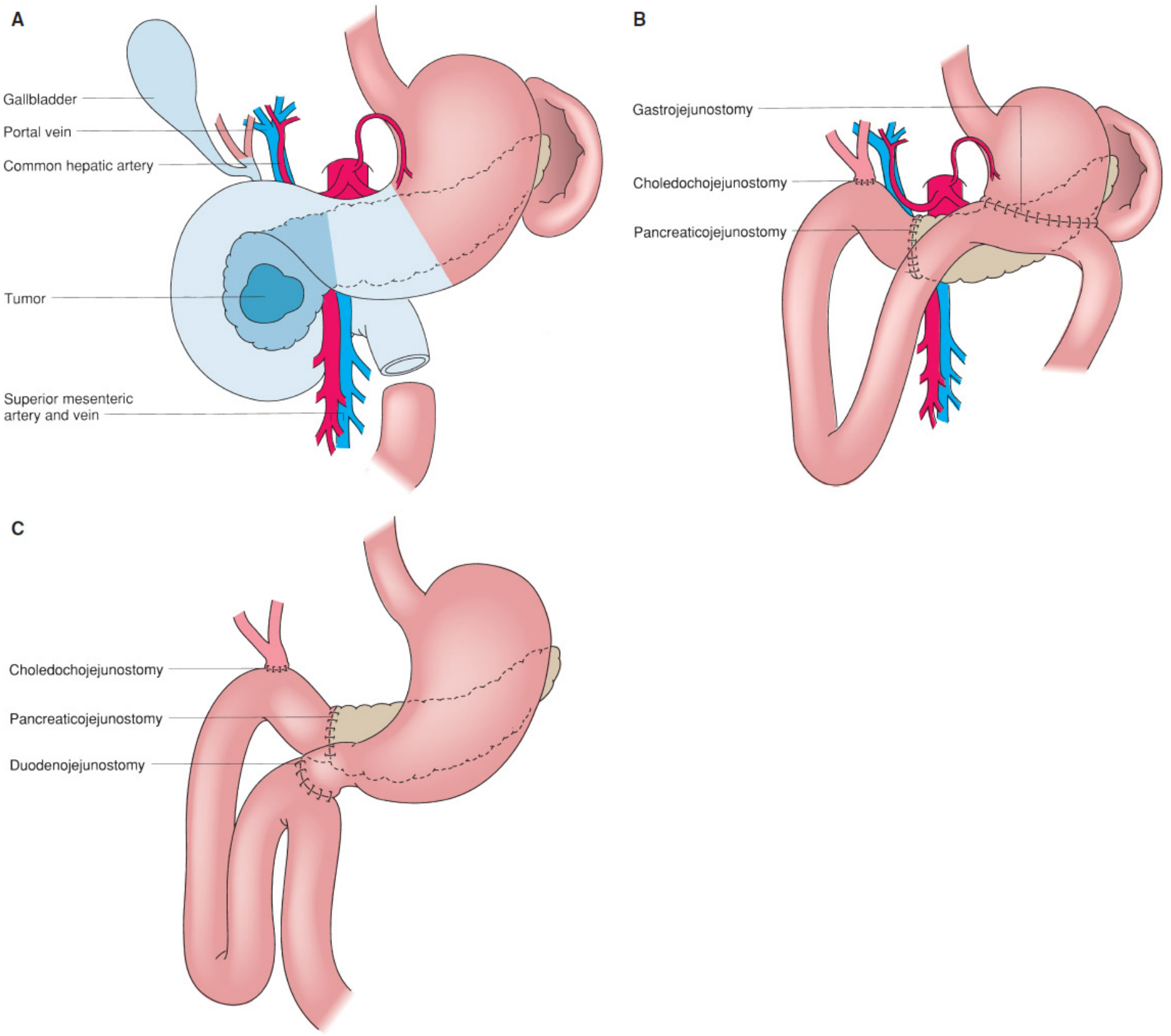
The Whipple procedure (pancreaticoduodenectomy, PD) is the most common operation for tumors of the periampullary region. It involves en-bloc removal of the pancreatic head, duodenum, gallbladder, distal bile duct, and (in the classic variant) the gastric antrum. Two variants exist:
- Classic Whipple: includes distal gastrectomy (antrectomy)
- Pylorus-preserving pancreaticoduodenectomy (PPPD): preserves the entire stomach and pylorus with division of the duodenum 2 cm distal to the pylorus
The diagrams below (from Mulholland and Greenfield's Surgery, 7e) illustrate resection extent and reconstruction:
Indications
- Adenocarcinoma of the pancreatic head (most common)
- Periampullary tumors (ampullary carcinoma, distal cholangiocarcinoma, duodenal carcinoma)
- Cystic neoplasms of the pancreatic head (IPMN, mucinous cystic neoplasm)
- Neuroendocrine tumors of the pancreatic head
- Chronic pancreatitis (head-dominant disease refractory to other measures)
- Benign disease: ampullary adenoma, duodenal polyposis
Preoperative Assessment
- Staging CT (arterial and venous phase): evaluates for distant metastases and local vascular involvement
- Involvement of SMA/celiac/hepatic artery = unresectable
- SMV/portal vein involvement can be addressed with vascular resection and reconstruction in select cases
- Look for "bird's beak" or teardrop deformity of the SMV indicating venous involvement
- CA 19-9, bilirubin, LFTs, nutritional status
- Biliary drainage: endoscopic stenting for obstructive jaundice before surgery in selected cases
- Neoadjuvant therapy: increasingly used for borderline-resectable disease; operations after neoadjuvant therapy are more complex due to fibrosis/adhesions
Patient Preparation & Positioning
- General endotracheal anesthesia
- Supine position with arms extended
- Nasogastric tube, Foley catheter
- Preoperative broad-spectrum antibiotics (gram-negative and anaerobic coverage)
- VTE prophylaxis
Incision
Either:
- Upper midline laparotomy (most common) - from xiphoid to umbilicus
- Bilateral subcostal (chevron) incision - for broader exposure in obese patients
Diagnostic Laparoscopy (Before Laparotomy)
- Recommended before the main incision: ~10% of patients will have small-volume metastatic disease (liver surface, peritoneum) not detected on preoperative imaging
- If metastases are found, proceed to palliation rather than resection
- Inspect all visceral and parietal peritoneal surfaces and the liver
RESECTION PHASE: Six Operative Steps
Step 1 - Define the Infrapancreatic Superior Mesenteric Vein (SMV)
- Elevate the greater omentum from the transverse colon
- Mobilize the hepatic flexure of the colon to the level of the duodenum
- Enter the lesser sac
- Incise the visceral peritoneum along the inferior border of the pancreas (medial to lateral) to expose the infrapancreatic SMV
- Identify the junction of the middle colic vein and SMV (may enter directly or share a common trunk with the gastroepiploic vein - the gastrocolic trunk)
- If shared trunk: either preserve the trunk (dividing only the gastroepiploic vein) or divide the entire trunk
- Purpose: confirm the SMV is free from tumor before committing to resection; establish the lower boundary of the pancreatic tunnel
- Note: If inflammatory scarring prevents early SMV identification, the vein will be exposed in Step 6 after pancreatic neck division
Step 2 - Extended Kocher Maneuver (Mobilize Duodenum and Pancreatic Head)
- Begin the Kocher maneuver at the third (transverse) portion of the duodenum by identifying the inferior vena cava
- Mobilize the duodenum and pancreatic head medially to the level of the left renal vein
- Divide the posterior leaf of peritoneum behind the mesenteric vessels
- Rotating the head of pancreas medially exposes the posterior aspect and facilitates subsequent SMA dissection
- Purpose: Allows palpation of the SMA to assess if a tumor-free plane exists between mass and artery; confirms resectability
Step 3 - Portal Dissection and Cholecystectomy
- Identify the common hepatic artery
- Remove the hepatic artery lymph node to improve visualization
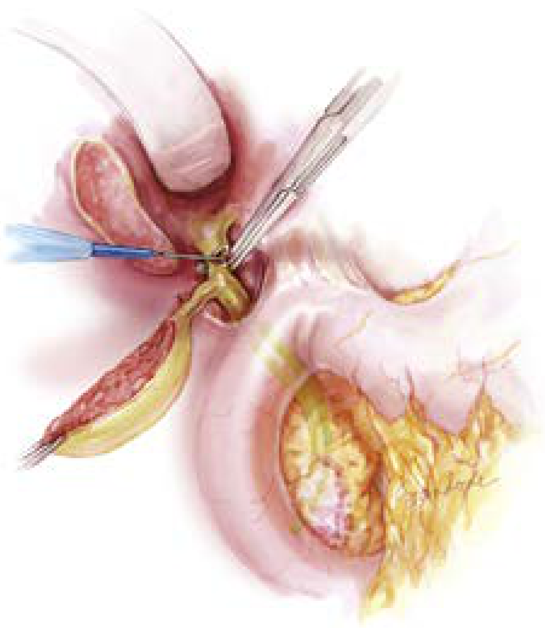
- Perform retrograde cholecystectomy (top-down), freeing the gallbladder from its bed
- Transect the common hepatic duct at the level of the cystic duct junction; send bile duct margin for frozen section
- CRITICAL: Before division, confirm no replaced/accessory right hepatic artery (arises from SMA, courses posterolateral to PV - present in ~15-20% of patients and easily injured)
- Place a bulldog clamp on the hepatic duct stump to prevent bile spillage; send biliary cultures
- Division of the common hepatic duct exposes the anterior surface of the portal vein
- Identify the gastroduodenal artery (GDA) - confirm proper hepatic artery pulsation before ligation
- Divide the GDA: exposes the superior border of the pancreas and the portal vein
- The superior pancreaticoduodenal vein (Pancreas Belcher vein) is a constant tributary at the superolateral aspect of the PV - handle with care to avoid traction hemorrhage
Step 4 - Transect the Stomach or Duodenum
Classic Whipple (antrectomy):
- Transect the stomach with a GI stapler at the level of the 3rd/4th transverse vein on the lesser curvature and at the confluence of the gastroepiploic veins on the greater curvature
- This completes a standard ~30-40% distal gastrectomy (antrectomy)
- Note: When opening the pars flaccida (hepatogastric ligament), specifically look for and preserve any accessory or replaced left hepatic artery arising from the left gastric artery
Pylorus-preserving (PPPD):
- Divide the duodenum approximately 2 cm distal to the pylorus to preserve adequate blood supply to the duodenojejunostomy
- Pylorus preservation is appropriate for small periampullary neoplasms; avoid in bulky pancreatic head tumors or duodenal tumors involving the 1st/2nd portion
- In patients who received preoperative radiation: antrectomy is recommended as the antrum lies within the radiation field
Step 5 - Divide the Jejunum and Mobilize the Ligament of Treitz
- Carefully take down the ligament of Treitz (loose attachments) - avoid injury to the inferior mesenteric vein (IMV)
- Transect the jejunum ~10-15 cm distal to the ligament of Treitz with a GI stapler
- Divide the mesentery to the level of the 3rd and 4th portions of the duodenum
- Reflect the devascularized duodenum/jejunum segment under the superior mesenteric vessels to the right side of the abdomen - this exposes the uncinate process for the final dissection
Step 6 - Remove the Pancreatic Head from the SMV/PV and SMA
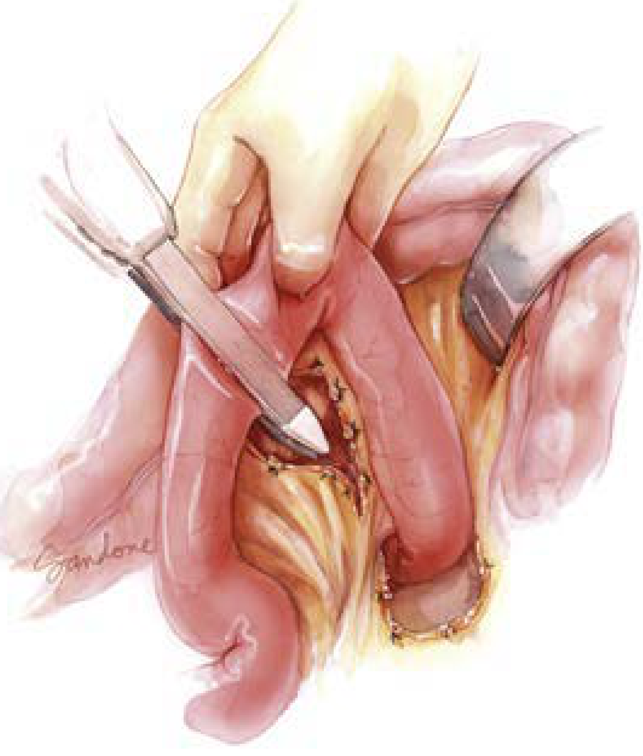
- Confirm the SMV-portal vein tunnel is patent (finger or blunt clamp passed posterior to pancreatic neck, anterior to PV/SMV)
- Place hemostatic sutures along superior and inferior borders of the planned transection line
- Transect the pancreatic neck with electrocautery at the level of the portal vein
- The pancreatic neck margin is sent for frozen section
- Pass vessel loops around the distal SMV, PV, and splenic vein for vascular control
- Retract the SMV-PV confluence medially to expose the SMA posteriorly
- Separate the pancreatic head and uncinate process from the SMA (the "SMA dissection" or "uncinate dissection"):
- Ligate and divide all branches from the uncinate/pancreatic head into the right lateral aspect of the PV/SMV: superior and inferior pancreaticoduodenal veins, right gastroepiploic vein, and any anomalous branches
- Ligate inferior pancreaticoduodenal arteries directly off the SMA - failure to control these results in post-resection hemorrhage
- Clear all tissue along the right lateral border of the SMA (the "retroperitoneal margin" or "SMA margin" - most common positive margin in pancreatic cancer)
- The specimen (pancreatic head, duodenum, gallbladder, bile duct, with or without gastric antrum) is now removed en bloc
RECONSTRUCTION PHASE: Three Anastomoses
Reconstruction uses a single jejunal limb brought up in the following order (proximal to distal along the jejunum):
1. Pancreaticojejunostomy (PJ) - most proximal anastomosis
- The most technically demanding anastomosis and the primary source of morbidity (pancreatic fistula)
- Three main techniques:
- End-to-end (terminoterminal) dunking: the pancreatic stump is invaginated (dunked) into the end of the jejunum
- End-to-side (terminolateral) dunking: pancreas invaginated into a side opening in the jejunum
- Mucosa-to-mucosa (duct-to-mucosa) pancreaticojejunostomy: the pancreatic duct is anastomosed directly to a corresponding enterotomy in the jejunum - preferred when the pancreatic duct is dilated (>3 mm)
- A pancreatic duct stent (internal or external) is often placed across the anastomosis
- Risk factors for pancreatic fistula: soft gland texture, small duct diameter (<3 mm), non-pancreatic cancer pathology, high intraoperative blood loss (these four factors constitute the Fistula Risk Score, FRS)
2. Hepaticojejunostomy (HJ) - biliary anastomosis
- Performed approximately 10 cm distal to the PJ
- End-to-side, single-layer technique with absorbable monofilament sutures
- A biliary stent may be placed if the bile duct is narrow
- The round ligament (preserved at the start of the case) is used as a pedicled flap to cover the GDA stump and serve as a physical barrier
3. Gastrojejunostomy (classic) or Duodenojejunostomy (pylorus-preserving)
- Performed approximately 50-60 cm distal to the HJ (antecolic or retrocolic position)
- Classic: side-to-side or end-to-side gastrojejunostomy (isoperistaltic, partial orallis partialis orientation)
- PPPD: end-to-side or end-to-end duodenojejunostomy ~2 cm distal to the pylorus
Variant: Pylorus-Preserving Whipple (PPPD)
The distinction from the classic procedure:
- The entire stomach is preserved
- Duodenum is divided 2 cm from the pylorus
- Reconstruction uses a duodenojejunostomy rather than gastrojejunostomy
- Advantages: avoids dumping syndrome, better nutritional outcomes, equivalent oncologic results
- Disadvantage: higher rates of delayed gastric emptying (DGE) compared to classic
Vascular Resection and Reconstruction
When the SMV or portal vein is involved by tumor:
- Tangential resection with primary suture repair (for partial involvement)
- Segmental resection with end-to-end anastomosis or interposition graft (saphenous vein, internal jugular vein) for circumferential involvement
- Arterial resection (SMA, hepatic artery) is controversial and performed at specialized centers only
- Pre-op CT signs: bird-beak deformity, teardrop deformity of the SMV indicate likely need for vascular reconstruction
Procedure-Specific Complications
Complication rates after PD remain high (40-60%), despite near-zero mortality at high-volume centers. The ISGPS (International Study Group of Pancreatic Surgery) has standardized definitions:
| Complication | Incidence | Key Features |
|---|---|---|
| Clinically Relevant Postoperative Pancreatic Fistula (CR-POPF) | ~15% | Dehiscence of PJ anastomosis; single largest contributor to mortality (36% of PD deaths); classified by Fistula Risk Score |
| Delayed Gastric Emptying (DGE) | 20-30% | Inability to tolerate oral diet by POD7; more common with PPPD; graded A/B/C by ISGPS |
| Postpancreatectomy Hemorrhage (PPH) | 5-10% | Early (within 24h, usually from surgical field) or late (pseudoaneurysm from pancreatic fistula eroding GDA stump) |
| Chyle leak | 5-10% | Lymphatic disruption during SMA dissection |
| Post-pancreatectomy acute pancreatitis | Uncommon | Remnant gland inflammation |
| Wound infection / SSI | Common | Particularly in patients with preoperative biliary stents |
| Bile leak | ~3% | Disruption of hepaticojejunostomy |
The Postoperative Morbidity Index (PMI) provides a quantitative severity-weighted measure; average PMI at high-volume centers is 0.18 (scale 0-1.0). Patients who do sustain complications average a PMI of 0.346, indicating these complications tend to be severe when they occur.
Outcomes
- Operative mortality: <3% at high-volume centers (>20 PD/year), up to 15% at low-volume centers
- 5-year survival: pancreatic adenocarcinoma ~25-30% with R0 resection (adjuvant chemotherapy with modified FOLFIRINOX or gemcitabine/capecitabine); ampullary/distal bile duct tumors ~40-60%
- Margin status (R0 vs R1): the SMA/retroperitoneal margin is most commonly positive; R0 resection is the primary determinant of long-term survival
- Extended lymphadenectomy: four randomized trials have failed to demonstrate survival benefit and show increased morbidity (higher DGE and fistula rates)
Sources:
- Current Surgical Therapy, 14e - Pancreaticoduodenectomy chapter (Cameron et al., with Sandone illustrations)
- Mulholland and Greenfield's Surgery, 7e, p. 2697-2700
- Fischer's Mastery of Surgery, 8e, Chapter on Pancreaticoduodenectomy
- Maingot's Abdominal Operations - Pancreaticoduodenectomy Technique
- Clinical Gastrointestinal Endoscopy, 3e - Whipple Procedure (endoscopic anatomy)
This is a shared conversation. Sign in to Orris to start your own chat.