
List of important emergency medicine emergencies like necrotising fasciitis
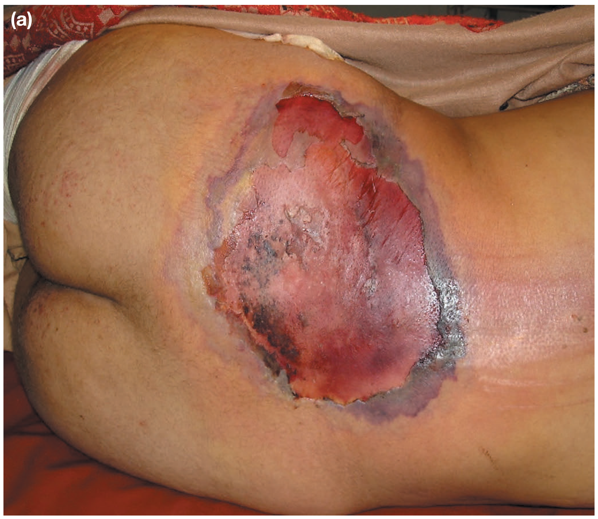
Surgical / Soft-Tissue Emergencies
1. Necrotising Fasciitis
- Rapidly spreading infection causing necrosis of subcutaneous tissues and overlying skin; caused by beta-haemolytic streptococci, S. aureus, or polymicrobial (Bacteroides, Clostridium, Pseudomonas, Klebsiella)
- Presentations: dusky blue/black skin, severe pain out of proportion, bullae, fever, septic shock; mortality approaches 70% without aggressive treatment
- Management: ICU admission, IV high-dose penicillin G + broad-spectrum antibiotics (cephalosporins + metronidazole), emergency surgical debridement - the cornerstone; daily wound revision; hyperbaric oxygen as adjunct
- Special forms: Fournier's gangrene (perineum), Meleney's gangrene (abdominal wall)
- Bailey and Love's Short Practice of Surgery 28th Edition
Cardiovascular Emergencies
2. Acute Myocardial Infarction (STEMI)
- Full-thickness myocardial ischemia from coronary artery occlusion; ST elevation on ECG
- Time is myocardium: door-to-balloon time target <90 minutes
- Management: aspirin + P2Y12 inhibitor, anticoagulation, primary PCI (preferred) or thrombolysis
3. Aortic Dissection
- Tear in the intima of the aorta; Stanford Type A (ascending) requires emergency surgery; Type B may be managed medically
- Classic presentation: tearing/ripping chest/back pain, unequal BP/pulses, widened mediastinum on CXR
- Immediate BP control with IV beta-blockers (target SBP 100-120 mmHg)
4. Cardiac Tamponade
- Fluid accumulation in pericardial sac compresses cardiac chambers; Beck's triad: hypotension, JVD, muffled heart sounds
- ECG: electrical alternans; Echo is diagnostic
- Treatment: immediate pericardiocentesis (needle drainage)
5. Hypertensive Emergency
- Severe hypertension (usually >180/120) with acute end-organ damage (encephalopathy, AKI, ACLE, aortic dissection)
- Controlled BP reduction by 20-25% in first hour with IV labetalol or nicardipine; not a rapid drop
- ROSEN's Emergency medicine
Respiratory Emergencies
6. Tension Pneumothorax
- Lung collapse with mediastinal shift; air trapped under pressure compresses great vessels
- Presents with hypoxia, hypotension, absent breath sounds, tracheal deviation (late sign), raised JVP
- Do not wait for CXR - immediate needle decompression (2nd ICS, mid-clavicular line) followed by chest drain
- Bailey and Love's; Roberts and Hedges' Clinical Procedures in Emergency Medicine
7. Pulmonary Embolism (Massive)
- Haemodynamically significant PE causes right heart failure and circulatory collapse
- Presentations: hypoxia, pleuritic chest pain, tachycardia, syncope; ECG shows S1Q3T3 (classic but non-specific)
- CT pulmonary angiography is diagnostic gold standard; treatment: systemic thrombolysis or catheter-directed therapy for massive PE
8. Acute Severe Asthma (Near-Fatal)
- Status asthmaticus: bronchospasm unresponsive to initial bronchodilators
- Silent chest is an ominous sign (no air movement)
- Management: continuous nebulized salbutamol, IV magnesium sulfate, IV corticosteroids; intubation as last resort (high risk)
9. Acute Respiratory Distress Syndrome (ARDS)
- Diffuse alveolar damage from direct/indirect lung injury; bilateral infiltrates, hypoxia (PaO2/FiO2 <200)
- Management: lung-protective ventilation (low tidal volume 6 mL/kg, PEEP titration), prone positioning
Neurological Emergencies
10. Status Epilepticus
- Continuous seizure activity >5 minutes or recurrent seizures without recovery of consciousness
- Management: benzodiazepines first-line (lorazepam/diazepam), then second-line agents (levetiracetam, valproate, phenytoin); refractory SE requires anaesthetic agents (propofol, midazolam, thiopental)
- Tintinalli's Emergency Medicine; ROSEN's Emergency Medicine
11. Acute Ischaemic Stroke
- "Time is brain" - 1.9 million neurons die per minute during occlusion
- IV alteplase (tPA) within 4.5 hours of symptom onset; mechanical thrombectomy for large vessel occlusion up to 24 hours in selected patients
- Rule out haemorrhage with CT before giving thrombolytics
12. Subarachnoid Haemorrhage (SAH)
- "Worst headache of my life" (thunderclap headache); caused by ruptured aneurysm in ~85% of cases
- CT head non-contrast first; if negative, LP for xanthochromia
- Neurosurgical consultation urgently; nimodipine for vasospasm prevention
13. Raised Intracranial Pressure (ICP) / Brain Herniation
- Cushing's triad: bradycardia, hypertension, irregular breathing
- Immediate: head elevation 30°, hyperventilation (transient), IV mannitol or hypertonic saline, neurosurgery
Abdominal / GI Emergencies
14. Ruptured Abdominal Aortic Aneurysm (rAAA)
- Classic triad: hypotension, pulsatile abdominal mass, back/flank pain
- Mortality >80% without surgery; bedside FAST ultrasound confirms; immediate theatre
- No time for CT if haemodynamically unstable
15. Acute Mesenteric Ischaemia
- Occlusion of superior mesenteric artery causes bowel infarction; "pain out of proportion to examination"
- Causes: arterial embolism (50%), arterial thrombosis, venous thrombosis, non-occlusive
- CT angiography diagnostic; surgical/endovascular revascularization; bowel resection if infarcted
- Schwartz's Principles of Surgery
16. Perforated Viscus (Perforated Peptic Ulcer)
- Free air under diaphragm on erect CXR; peritonitis on examination
- Surgical emergency: IV resuscitation, broad-spectrum antibiotics, urgent laparotomy/laparoscopy
17. Bowel Obstruction with Strangulation / Sigmoid Volvulus
- Closed-loop obstruction causes rapid bowel ischemia and gangrene
- CT abdomen is diagnostic; emergency surgery when strangulation suspected
Infectious / Sepsis Emergencies
18. Septic Shock
- Life-threatening organ dysfunction from infection; MAP <65 mmHg despite fluid resuscitation
- Hour-1 bundle: blood cultures, broad-spectrum antibiotics, IV fluid bolus (30 mL/kg crystalloid), vasopressors (norepinephrine), measure lactate
- ROSEN's Emergency Medicine
19. Toxic Shock Syndrome (TSS)
- Staphylococcal or streptococcal superantigen-mediated multi-organ failure
- Diffuse macular erythroderma ("sunburn rash"), desquamation, fever, hypotension
- Management: source control, clindamycin (toxin suppression), IV fluids, IVIG in severe cases
20. Bacterial Meningitis
- Triad: fever, neck stiffness, altered consciousness; do not delay antibiotics for CT
- Immediate: IV dexamethasone + ceftriaxone + vancomycin; add ampicillin if Listeria risk
- Lumbar puncture after antibiotics if CT required
21. Ludwig's Angina
- Rapidly spreading cellulitis of the floor of the mouth causing airway compromise
- Immediate airway management (awake fibreoptic intubation preferred - avoid blind intubation); surgical drainage; IV antibiotics
Endocrine / Metabolic Emergencies
22. Diabetic Ketoacidosis (DKA) / Hyperosmolar Hyperglycaemic State (HHS)
- DKA: anion gap metabolic acidosis, ketones, hyperglycaemia; HHS: extreme hyperglycaemia (>600 mg/dL), hyperosmolarity, no ketosis
- Management: IV fluids, insulin infusion, electrolyte replacement (especially K+); HHS needs very slow correction
23. Adrenal Crisis (Addisonian Crisis)
- Profound hyponatraemia, hyperkalaemia, hypoglycaemia, hypotension in adrenal insufficiency
- Immediate: IV hydrocortisone 100 mg bolus, aggressive IV saline, glucose; do not wait for cortisol results
24. Thyroid Storm
- Extreme hyperthyroidism; fever, tachycardia, heart failure, altered consciousness
- Management: propylthiouracil (blocks synthesis), Lugol's iodine (blocks release), beta-blockers, dexamethasone, cooling
Obstetric Emergencies
25. Ruptured Ectopic Pregnancy
- Intraperitoneal haemorrhage from fallopian tube rupture; hypovolaemic shock; positive urine hCG
- FAST ultrasound shows free fluid; resuscitate and take to theatre immediately - surgical emergency
26. Eclampsia
- Seizures in pregnancy/postpartum + hypertension; can cause stroke, placental abruption, multi-organ failure
- Immediate: IV magnesium sulfate (4 g loading dose), antihypertensives (hydralazine/labetalol), urgent delivery
27. Amniotic Fluid Embolism
- Catastrophic obstetric collapse: hypoxia, cardiac arrest, DIC
- Resuscitation per ACLS protocols; manage DIC aggressively
Toxicological Emergencies
28. Organophosphate / Nerve Agent Poisoning
- SLUDGE syndrome (salivation, lacrimation, urination, defecation, GI distress, emesis) + bronchospasm, miosis, seizures
- Atropine (in large doses), pralidoxime (oxime reactivator), benzodiazepines for seizures
29. Tricyclic Antidepressant (TCA) Overdose
- QRS widening (>100 ms), arrhythmia, seizures, hypotension
- IV sodium bicarbonate is the treatment (alkalinizes plasma to reduce drug binding to Na channels)
30. Carbon Monoxide Poisoning
- Cherry-red skin (unreliable sign); headache, confusion, coma; SpO2 is falsely normal
- 100% O2 via non-rebreather mask; hyperbaric oxygen for severe cases (COHb >25%, neurological signs, cardiac involvement)
ENT / Head and Neck Emergencies
31. Epiglottitis
- Rapidly progressive supraglottic inflammation; "tripod position," drooling, stridor, muffled voice
- Do not examine the throat in children - can precipitate complete obstruction
- Immediate anaesthetic/ENT team; controlled intubation in theatre; IV ceftriaxone
32. Retropharyngeal Abscess
- Deep neck space infection extending to mediastinum (descending mediastinitis); high mortality
- CT neck + chest; surgical drainage + IV antibiotics
Orthopaedic / Vascular Emergencies
33. Compartment Syndrome
- Raised pressure in a closed fascial compartment causing tissue ischaemia; the 6 P's: Pain (especially with passive stretch), Pressure, Paresthesia, Pallor, Paralysis, Pulselessness (late)
- Emergency fasciotomy - do not delay
34. Acute Limb Ischaemia
- Sudden arterial occlusion; the 6 P's again: Pain, Pallor, Pulselessness, Paresthesia, Paralysis, Poikilothermia (cold)
- Embolectomy or bypass within 6 hours to prevent irreversible muscle/nerve damage
Key Principle: "Time-Critical" Emergencies
| Emergency | Time Window |
|---|---|
| STEMI | PCI within 90 min |
| Ischaemic stroke | Thrombolysis within 4.5 h |
| Acute limb ischaemia | Revascularization within 6 h |
| Septic shock | Antibiotics within 1 h |
| Necrotising fasciitis | Debridement within hours |
| Tension pneumothorax | Needle decompression immediately |
| Ruptured AAA | Theatre without delay |
| Eclampsia | Magnesium + delivery |
Explain me a normal 12 lead ECG with normal axis and where would you normally find inverted t waves
normal 12 lead ECG with axis and waveform labels
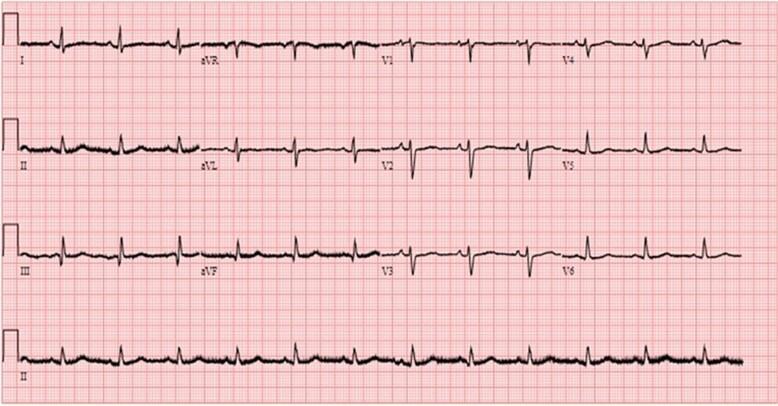
A standard 12-lead electrocardiogram (ECG) recording displayed on a red-lined grid paper. The image represents diagnostic cardiology imaging from a post-operative follow-up. The tracing demonstrates normal sinus rhythm characterized by consistent P waves preceding every QRS complex, normal PR intervals, and regular R-R intervals. The 12 leads (I, II, III, aVR, aVL, aVF, V1-V6) are organized into four columns, with a long rhythm strip of Lead II at the bottom. Waveform morphologies show appropriate axis and progression, including positive P waves in leads I and II, an inverted aVR, and normal R-wave progression across the precordial leads (V1-V6). QRS complexes and T waves appear within normal limits for duration and amplitude, with no evidence of acute ischemia, significant ST-segment deviation, or conduction abnormalities. This clinical photograph serves as an educational example of baseline cardiac electrical activity and post-surgical recovery monitoring.
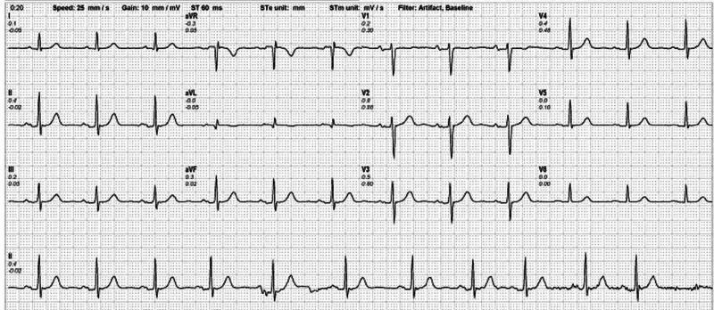
A standard 12-lead diagnostic resting electrocardiogram (ECG) displayed on a grid. The tracing includes the standard limb leads (I, II, III), augmented limb leads (aVR, aVL, aVF), and precordial leads (V1-V6), with a long rhythm strip of lead II at the bottom. The visual demonstrates a normal sinus rhythm characterized by consistent P-wave morphology preceding every QRS complex, regular R-R intervals, and a heart rate within normal physiological limits (approximately 80-85 bpm). Waveform analysis shows normal axis, absence of ST-segment elevation or depression, and normal T-wave morphology across all leads, indicating no acute ischemic changes at rest. Technical parameters visible at the top specify a paper speed of 25 mm/s, a gain (voltage scale) of 10 mm/mV, and the application of artifact and baseline filters. This diagnostic image serves as a baseline comparison for clinical cardiac assessment and the evaluation of coronary artery disease.
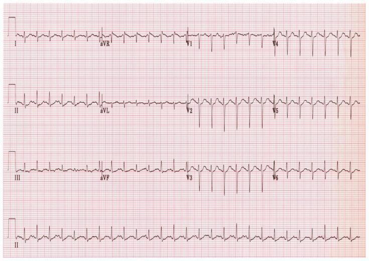
This diagnostic image is a standard 12-lead electrocardiogram (ECG) recorded on a pink grid background. The tracing displays the limb leads (I, II, III), augmented limb leads (aVR, aVL, aVF), and precordial leads (V1-V6) arranged in a traditional four-column format, with a continuous rhythm strip of lead II at the bottom. The horizontal axis represents time at a standard speed (25 mm/sec) and the vertical axis represents voltage (10 mm/mV). The ECG shows a regular sinus rhythm with narrow QRS complexes and distinct P and T waves. Notable morphology includes a normal R-wave progression in the precordial leads, characterized by increasing R-wave amplitude from V1 through V6. In lead V1, the QRS complex is primarily negative (rS pattern), transitioning to a primarily positive deflection by V5-V6. This visual material is used in clinical cardiology education to teach baseline cardiac rhythm analysis, waveform recognition, and lead-specific morphological variations.
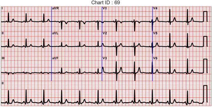
This diagnostic image shows a standard 12-lead electrocardiogram (ECG) displayed on a red-ruled grid. The chart is organized into four rows: the top three rows represent the standard limb leads (I, II, III), augmented limb leads (aVR, aVL, aVF), and precordial leads (V1–V6) in a three-channel format, while the bottom row provides a continuous rhythm strip of Lead II. Each lead demonstrates clear P waves, narrow QRS complexes, and T waves, indicating a normal sinus rhythm. The grid follows standard calibration where the vertical axis represents voltage (10 mm = 1 mV) and the horizontal axis represents time (25 mm = 1 s). Notable morphology includes normal R-wave progression from V1 through V6 and typical negative deflections in aVR. The image serves as a clinical reference for assessing cardiac electrical activity and is used in medical education to teach lead placement, waveform identification, and cardiac rhythm interpretation.
T wave inversion normal leads aVR V1 ECG
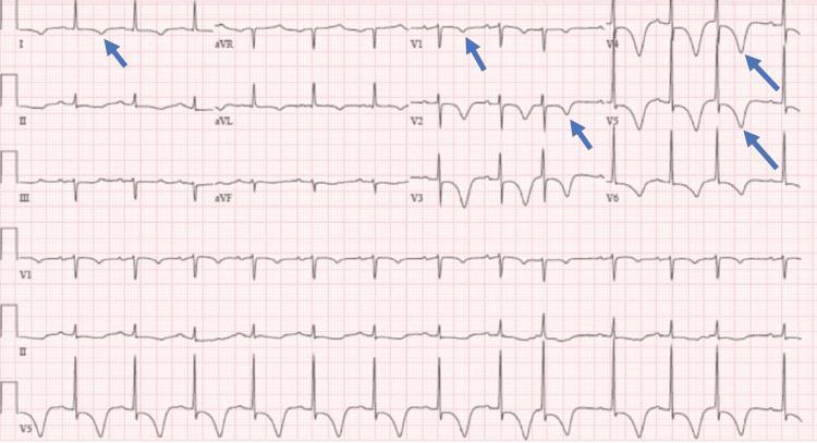
A standard 12-lead diagnostic electrocardiogram (ECG) printed on pink grid paper, demonstrating significant repolarization abnormalities. The primary visual finding is deep, symmetric T-wave inversions (TWIs) localized to the lateral (lead I) and anteroseptal to lateral precordial leads (V1 through V6). Blue arrows highlight these specific inversions in leads I, V1, V2, V4, and V5. The QRS complexes appear to be of normal duration and morphology with regular R-R intervals, suggesting a sinus rhythm. There is evidence of mild ST-segment depression accompanying the T-wave changes in the precordial leads. In contrast, the inferior leads (II, III, aVF) and augmented limb leads (aVR, aVL) show upright or flat T-wave morphology. Clinically, this pattern of widespread anterior and lateral T-wave inversion is highly significant for myocardial ischemia, such as Wellens' syndrome or non-ST elevation myocardial infarction (NSTEMI), or potentially secondary to hypertrophic cardiomyopathy or pulmonary embolism. The educational focus is on identifying patterns of ischemia and differentiating regional repolarization abnormalities.
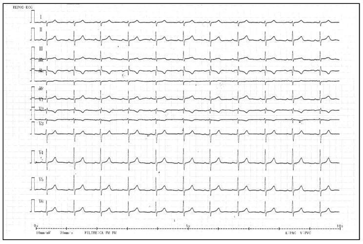
A standard 12-lead resting electrocardiogram (ECG) printed on grid paper, displaying limb leads (I, II, III, aVR, aVL, aVF) and precordial leads (V1-V6). The tracing shows a regular rhythm with visible P-waves, QRS complexes, and T-waves. Key clinical features include T-wave inversions specifically in the right precordial leads V1 and V2. In contrast, T-waves in the lateral precordial leads (V3-V6) and the limb leads (I, II, aVF) are upright and demonstrate normal morphology. The corrected QT (QTc) interval is reported at 432 ms, which is within the normal range. The tracing is set at a standard paper speed of 25 mm/s and voltage of 10 mm/mV. This visual is used to demonstrate cardiac repolarization patterns, where T-wave inversion in V1-V2 can be a normal variant or associated with specific cardiomyopathies such as Arrhythmogenic Right Ventricular Cardiomyopathy (ARVC) or Mitral Valve Prolapse (MVP) syndromes.
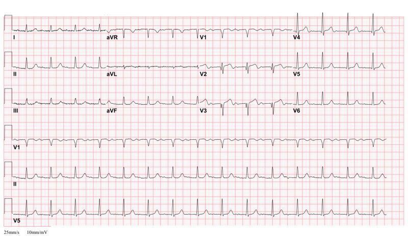
A 12-lead electrocardiogram (ECG) demonstrating the classic 'Type A' Wellens' Syndrome pattern. The recording shows a regular rhythm with normal QRS complexes; however, there is a pathognomonic T-wave morphology in the precordial leads, specifically V2 and V3. These leads exhibit biphasic T-waves characterized by an initial shallow positive deflection followed by a deep, terminal negative deflection. The terminal inversion is more prominent in amplitude than the initial upward phase. Such findings in the context of clinical stability are highly specific for critical stenosis of the proximal left anterior descending (LAD) coronary artery. The ECG also includes standard limb leads (I, II, III), augmented limb leads (aVR, aVL, aVF), and the full precordial series (V1-V6), with rhythm strips for V1, II, and V5 at the bottom. The paper speed is noted at 25mm/s with a sensitivity of 10mm/mV, providing a standard diagnostic reference for ischemic heart disease and acute coronary syndrome (ACS) evaluation.
The Normal 12-Lead ECG
The 12 Leads - What They "See"
- I, II, III - bipolar; look at the heart in the frontal plane
- aVR, aVL, aVF - augmented unipolar limb leads
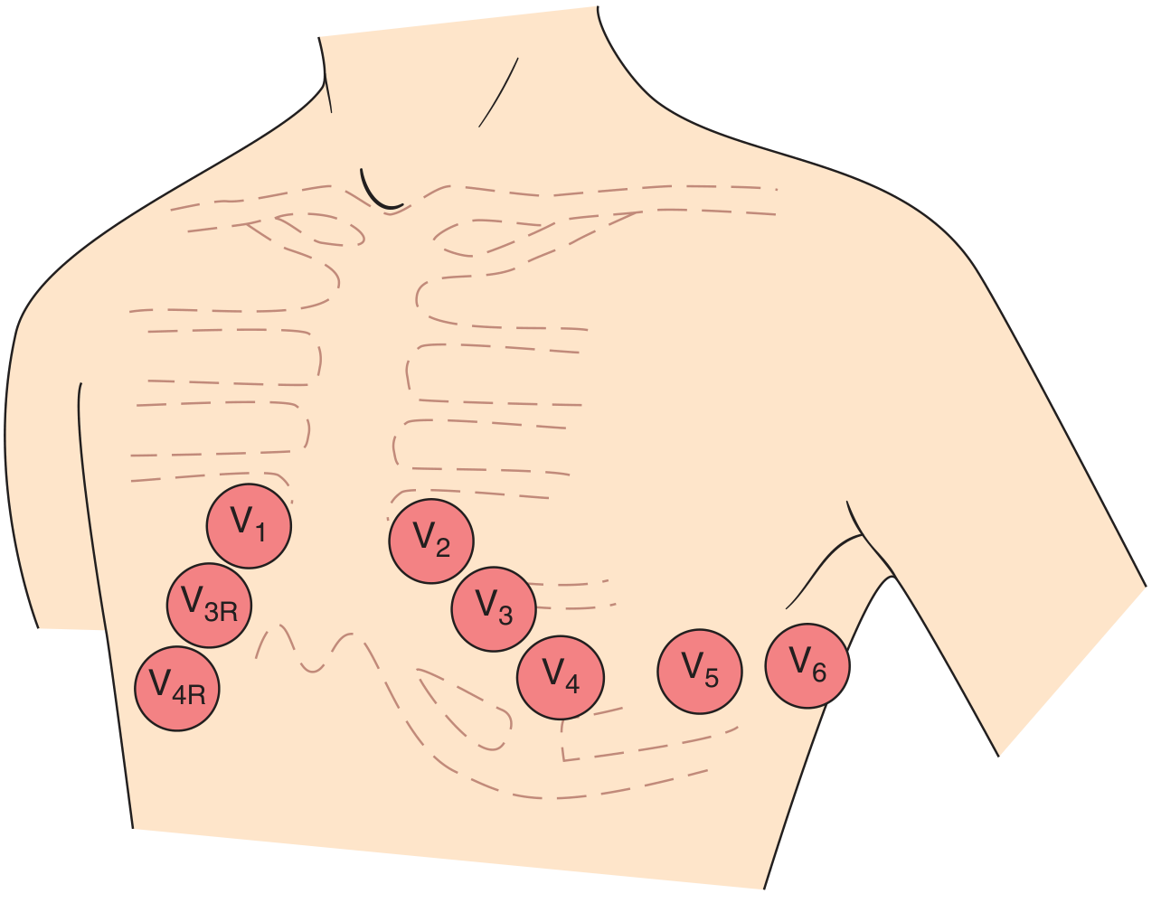
- V1 - right side of the heart (right sternal border, 4th ICS)
- V2 - left sternal border, 4th ICS
- V3 - between V2 and V4
- V4 - mid-clavicular line, 5th ICS
- V5 - anterior axillary line
- V6 - mid-axillary line
Normal ECG Waveforms and Intervals
| Waveform | What it represents | Normal values |
|---|---|---|
| P wave | Atrial depolarization | Duration <120 ms; amplitude <2.5 mm |
| PR interval | AV conduction time | 120-200 ms (3-5 small squares) |
| QRS complex | Ventricular depolarization | <120 ms (3 small squares) |
| ST segment | Start of ventricular repolarization | Isoelectric (at baseline) |
| T wave | Ventricular repolarization | Upright in most leads (see below) |
| QT interval | Total ventricular electrical activity | QTc <440 ms (male), <460 ms (female) |
| U wave | Afterdepolarization (Purkinje?) | Small, same polarity as T wave; most visible in V2-V3 |
The P Wave
- Generated by atrial depolarization spreading from the sinus node (right atrium) leftward and inferiorly
- This vector points toward lead II (positive) and away from aVR (negative)
- Therefore:
- Upright in leads I, II, aVF (sinus P wave)
- Inverted/negative in aVR (always - a key normal finding)
- In V1: may be biphasic - small initial positive component (right atrial depolarization) then small negative component (left atrial depolarization)
The QRS Complex - Ventricular Depolarization
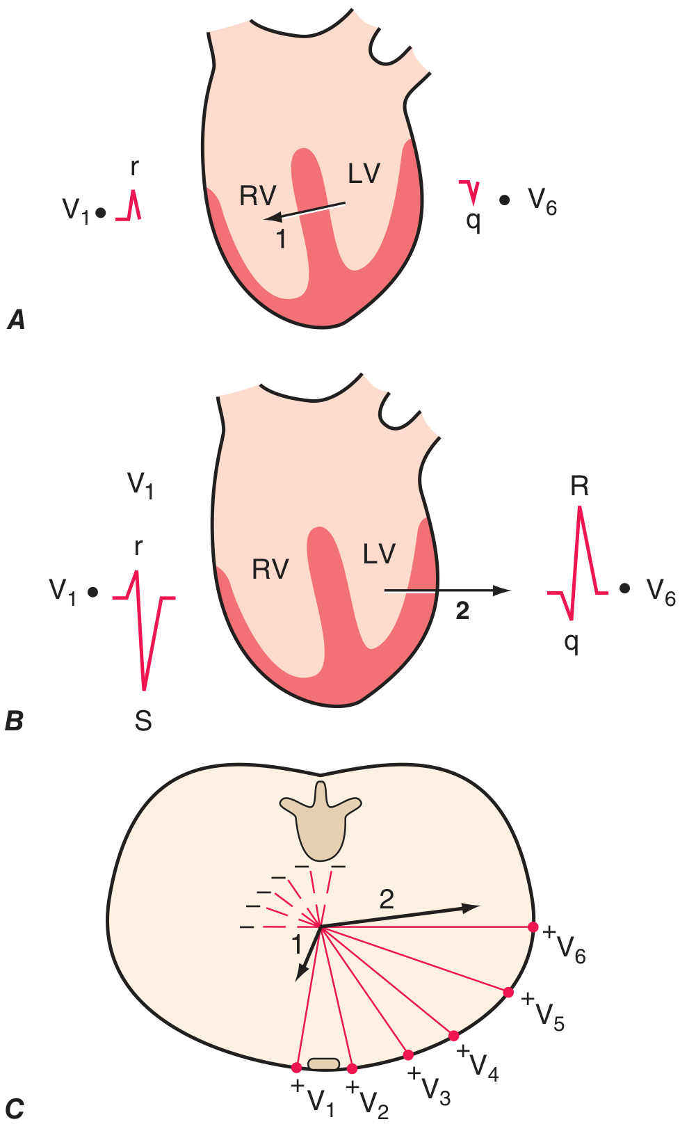
- This small rightward vector creates: a tiny r wave in V1 and a tiny q wave in V6
- This creates: a large S wave in V1 (negative - vector away from V1) and a large R wave in V6 (positive - vector toward V6)
- V1: small r, deep S (rS pattern) - predominantly negative
- V2-V3: growing r wave
- Transition zone (usually V3 or V4): R and S are roughly equal
- V5-V6: tall R, small or absent S (qR pattern) - predominantly positive
Lead-by-Lead Normal QRS Morphology Summary
| Lead | Normal QRS |
|---|---|
| I | Positive (upright R) |
| II | Positive (tallest P and T wave) |
| III | Variable |
| aVR | Predominantly negative (QS or rS) |
| aVL | Variable |
| aVF | Positive |
| V1 | rS (small r, deep S) |
| V2 | rS or RS |
| V3 | Transition |
| V4 | RS or Rs (transition) |
| V5-V6 | qR or qRs (positive) |
Normal Cardiac Axis
"The mean electrical axis of the normal ventricles is 59 degrees." - Guyton and Hall Textbook of Medical Physiology
- Look at leads I and aVF
- If QRS is positive in both I and aVF → normal axis (0° to +90°)
- If positive in I, negative in aVF → left axis deviation (>-30°)
- If negative in I, positive in aVF → right axis deviation (>+90°)
- If negative in both → extreme axis deviation ("northwest axis")
| Axis | Range | Causes |
|---|---|---|
| Normal | -30° to +90° | - |
| Left axis deviation | More negative than -30° | Left anterior fascicular block, LBBB, inferior MI, LVH |
| Right axis deviation | More positive than +90° | Right ventricular overload, RBBB, lateral MI, dextrocardia, normal variant in young |
| Extreme | -90° to +180° | Hyperkalaemia, ventricular rhythms |
The T Wave - Normal Physiology
"The mean T-wave vector is oriented roughly concordant with the mean QRS vector (within about 45° in the frontal plane). Since depolarization and repolarization are electrically opposite processes, this normal QRS-T-wave vector concordance indicates that repolarization normally must proceed in the reverse direction from depolarization (i.e., from ventricular epicardium to endocardium)." - Harrison's Principles of Internal Medicine 22E
- Upright in leads I, II, V3-V6
- Amplitude: <5 mm in limb leads; <10-15 mm in precordial leads
- Asymmetric: gradual upstroke, rapid downstroke
- Always upright in lead II
- Normal T wave = concordant with QRS polarity
Normal Leads Where T-Wave Inversion is Expected
| Lead | T-wave inversion... | Reason |
|---|---|---|
| aVR | Always inverted (normal) | aVR looks at the heart from the right shoulder - QRS is also negative here, so T follows (concordant) |
| V1 | Commonly/normally inverted | V1 faces the right ventricle; the repolarization vector points away from V1 |
| V2 | May be inverted, especially in women | Can be a normal variant; more common in women and athletes |
| III | Can be inverted (normal variant) | Depends on cardiac axis; borderline lead |
| aVL | Can be inverted (normal variant) | Particularly when axis is rightward |
- T-wave inversion in aVR = always normal
- T-wave inversion in V1 = normal (and in V2 in women/children)
- T-wave inversion in V3-V6 = abnormal, suspect ischaemia or other pathology
- T-wave inversion in II and aVF = always abnormal, consider inferior ischaemia
Pathological Causes of T-Wave Inversion
| Pattern | Leads affected | Likely cause |
|---|---|---|
| Anterior T-wave inversions | V1-V4 | Anterior ischaemia, Wellens' syndrome (LAD stenosis), RVOT pathology |
| Widespread deep T inversions | V1-V6 + I, aVL | Massive PE, apical HCM, Takotsubo cardiomyopathy, NSTEMI |
| Inferior T inversions | II, III, aVF | Inferior ischaemia |
| Lateral T inversions | I, aVL, V5-V6 | Lateral ischaemia, LVH ("strain" pattern) |
| Right-sided T inversions | V1-V3 | ARVC (arrhythmogenic right ventricular cardiomyopathy), RVH |
| Diffuse T inversions + Osborn waves | Multiple | Hypothermia |
Summary Card
Normal 12-lead ECG at a glance:
- Sinus rhythm: P before every QRS, P upright in I & II, inverted in aVR
- PR interval: 120-200 ms
- QRS: <120 ms, normal axis -30° to +90°
- R-wave progression: small in V1 → tall in V5-V6, transition at V3-V4
- T waves: upright in I, II, V3-V6
- T waves NORMALLY inverted: aVR (always), V1 (common), V2 (in women/children)
- QTc: <440 ms (men), <460 ms (women)
Neurological examination??
neurological examination dermatomes motor sensory diagram
cranial nerves diagram all 12
dermatome map spinal nerve distribution body
The Neurological Examination
- Mental Status
- Cranial Nerves (CN I-XII)
- Motor System
- Reflexes
- Sensory System
- Coordination
- Gait
"Neurological examination starts during the interview. A patient's lack of facial expression (hypomimia) may suggest parkinsonism or depression...the pattern of speech may suggest dysarthria, aphasia, or spasmodic dysphonia." - Bradley and Daroff's Neurology in Clinical Practice
1. Mental Status
| Domain | What to assess |
|---|---|
| Level of consciousness | Alert, drowsy, confused, comatose |
| Orientation | Person, place, time |
| Attention | Serial 7s, digit span |
| Memory | Immediate recall (repeat 3 words), recent (recall after 5 min), remote |
| Language | Fluency, comprehension, repetition, naming, reading, writing |
| Higher functions | Abstract thinking, judgment, visuospatial ability |
| Mood and affect | Depression, anxiety, irritability |
2. Cranial Nerve Examination
CN I - Olfactory
- Test only if: history of anosmia, suspected inferior frontal lobe lesion (meningioma), or Parkinson's disease
- Ask patient to close eyes, sniff a mild odour (coffee, toothpaste) and identify it - each nostril separately
CN II - Optic
- Visual acuity: Snellen chart (with glasses if worn)
- Visual fields by confrontation: Face the patient at ~1 m, wiggle a finger in each of the 4 quadrants; compare with your own field
- Pupils: Size, symmetry, direct and consensual light reflex; swinging flashlight test for relative afferent pupillary defect (RAPD/Marcus Gunn pupil)
- Fundoscopy: Optic disc colour, margins, cup-to-disc ratio; retinal vessels, haemorrhages, exudates
CN III, IV, VI - Oculomotor, Trochlear, Abducens
- Pupils: Shape, size, reaction to light and accommodation (near reflex - ask patient to follow finger moving toward nose)
- Extraocular movements: Follow finger in an "H" pattern; look for paresis, nystagmus, abnormal pursuit
- Ptosis: CN III palsy causes complete ptosis; Horner's syndrome causes partial ptosis
- CN III - all movements except down-and-out; also pupil constriction
- CN IV - depression of adducted eye (superior oblique)
- CN VI - abduction (lateral rectus)
Diplopia should resolve when one eye is closed - true diplopia is always binocular. - Harrison's 22E
CN V - Trigeminal
- Sensation: Test light touch and temperature (or pinprick) over all 3 divisions:
- V1 (ophthalmic): forehead
- V2 (maxillary): cheek
- V3 (mandibular): jaw/chin
- Corneal reflex: Touch cornea gently with cotton wisp; normal = blink (afferent V, efferent VII)
- Motor: Jaw clench - feel masseter and temporalis; jaw opening against resistance (pterygoids)
- Jaw deviates toward the side of weakness on opening
CN VII - Facial
- Inspect for facial asymmetry at rest and with movement
- Test: eyebrow elevation, forehead wrinkling, eye closure (forced), smiling, cheek puff, whistling
| Finding | Lesion type |
|---|---|
| Lower 2/3 of face weak; forehead spared | Upper motor neuron (contralateral cortex) - forehead has bilateral cortical representation |
| Entire face weak on one side | Lower motor neuron (e.g., Bell's palsy, parotid tumour) |
CN VIII - Vestibulocochlear
- Hearing: Whisper test (whisper numbers 2 feet away, mask other ear by rubbing)
- Weber test: Tuning fork on vertex; normally heard equally; lateralises to good ear in sensorineural loss, bad ear in conductive loss
- Rinne test: Air conduction (fork near ear) vs. bone conduction (fork on mastoid); AC > BC = normal or sensorineural; BC > AC = conductive hearing loss
- Vestibular: Nystagmus, Dix-Hallpike test for BPPV
CN IX, X - Glossopharyngeal and Vagus
- Observe palate at rest and with phonation ("say Ahh") - uvula deviates away from the affected side
- Voice quality - hoarseness (recurrent laryngeal nerve - CN X)
- Gag reflex: Stimulate posterior pharynx with tongue blade; CN IX = afferent, CN X = efferent. Note: may be absent in normal individuals
- Swallowing - ask patient to swallow water
CN XI - Spinal Accessory
- Trapezius: Shoulder shrug against resistance
- Sternocleidomastoid: Head rotation to each side against resistance
CN XII - Hypoglossal
- Inspect tongue at rest: atrophy, fasciculations?
- Protrude tongue - deviates toward the weak side
- Strength: push tongue into each cheek against examiner's finger
3. Motor Examination
Step 1: Inspection
- Muscle bulk (wasting/atrophy vs. hypertrophy)
- Fasciculations (fine twitching at rest - LMN disease)
- Involuntary movements:
- Tremor at rest → Parkinson's (pill-rolling)
- Tremor with posture → essential tremor
- Intention tremor (worsens finger-to-nose) → cerebellar
- Chorea, dystonia, myoclonus, tics
Step 2: Tone
- Upper limb: pronate/supinate wrist, flex/extend elbow and wrist
- Lower limb: patient supine - lift knee rapidly; normal = ankle drags along bed first; increased tone = heel lifts immediately
| Tone abnormality | Type | Cause |
|---|---|---|
| Spasticity | Velocity-dependent resistance; "clasp-knife" | Corticospinal tract (UMN) lesion |
| Rigidity | Resistance in all directions, all velocities | Extrapyramidal (basal ganglia) |
| Cogwheel rigidity | Jerky interruptions during passive movement | Parkinsonism |
| Paratonia (Gegenhalten) | Involuntary variable resistance | Frontal lobe disease |
| Hypotonia (flaccidity) | Reduced resistance | LMN, cerebellar, acute UMN (spinal shock) |
Step 3: Power (MRC Scale)
| Grade | Meaning |
|---|---|
| 0 | No movement at all |
| 1 | Flicker/trace contraction, no joint movement |
| 2 | Movement with gravity eliminated |
| 3 | Movement against gravity, not against resistance |
| 4- | Against mild resistance |
| 4 | Against moderate resistance |
| 4+ | Against strong resistance |
| 5 | Full power |
- Patient holds both arms extended, palms up, eyes closed for 10 seconds
- Pronation or downward drift = contralateral corticospinal tract lesion
| Pattern | Lesion location |
|---|---|
| Hemiplegia (arm + leg, one side) | Contralateral cortex or internal capsule |
| Pyramidal pattern (arm extensors + leg flexors weak) | Corticospinal tract |
| Paraplegia (both legs) | Spinal cord |
| Proximal > distal weakness | Myopathy |
| Distal > proximal weakness | Peripheral neuropathy |
| Fatigable weakness (worse with use) | Neuromuscular junction (myasthenia gravis) |
4. Reflex Examination
Deep Tendon Reflexes (DTRs) - Grading Scale
| Grade | Meaning |
|---|---|
| 0 | Absent |
| 1+ | Present but diminished |
| 2+ | Normal |
| 3+ | Increased (may be normal) |
| 4+ | Clonus (always abnormal) |
Standard DTRs and Their Root Values
| Reflex | Nerve root | Muscle tested |
|---|---|---|
| Biceps jerk | C5, C6 | Biceps brachii |
| Supinator/brachioradialis | C5, C6 | Brachioradialis |
| Triceps jerk | C6, C7 | Triceps |
| Finger flexor (Hoffmann) | C8, T1 | Finger flexors |
| Knee jerk (patellar) | L3, L4 | Quadriceps |
| Ankle jerk (Achilles) | S1, S2 | Gastrocnemius/soleus |
Cutaneous/Superficial Reflexes
- Stroke lateral sole from heel toward little toe, curving medially across the ball
- Normal: plantarflexion of toes
- Abnormal (Babinski sign): extension (dorsiflexion) of big toe + fanning of other toes = UMN lesion above S1
"Despite its popularity, the reliability and validity of the Babinski sign for identifying upper motor neuron weakness are limited - it is far more useful to rely on tests of tone, strength, stretch reflexes, and coordination." - Harrison's 22E
- Stroke each quadrant toward umbilicus; umbilicus should pull toward the stimulus
- Absent with UMN lesions
- Upper abdominal: T8-T10; Lower abdominal: T10-T12
UMN vs. LMN Signs Summary
| Feature | UMN (above spinal cord anterior horn) | LMN (anterior horn, root, nerve) |
|---|---|---|
| Tone | Increased (spastic/rigid) | Decreased (flaccid) |
| Reflexes | Increased (hyperreflexia) | Decreased or absent |
| Plantar | Extensor (Babinski +) | Flexor (normal) |
| Atrophy | Mild/disuse only | Significant |
| Fasciculations | Absent | Present |
| Distribution | Pyramidal pattern or hemiplegia | Focal, nerve/root distribution |
5. Sensory Examination
Primary Sensory Modalities
| Modality | Test method | Pathway |
|---|---|---|
| Pain | Pin-prick (disposable neurological pin) | Spinothalamic tract (crosses at spinal cord level) |
| Temperature | Cold (tuning fork) or hot/cold tubes | Spinothalamic tract |
| Light touch | Cotton wool or fingertip | Dorsal columns (and spinothalamic) |
| Vibration | 128 Hz tuning fork on bony prominences (big toe, medial malleolus, patella, ASIS) | Dorsal columns (ipsilateral) |
| Joint position sense (proprioception) | Hold lateral sides of distal phalanx; move up/down; ask direction | Dorsal columns (ipsilateral) |
| Pattern | Cause |
|---|---|
| Glove-and-stocking | Peripheral neuropathy (length-dependent) |
| Dermatomal | Nerve root (radiculopathy) |
| Sensory level on trunk | Spinal cord |
| Dissociated loss (pain/temp lost, vibration/JPS intact) | Syringomyelia; Brown-Séquard (ipsilateral dorsal column, contralateral spinothalamic) |
| Hemisensory loss (face + arm + leg one side) | Thalamus or cortex |
Higher (Cortical) Sensory Tests
- Graphesthesia: Write a number in the palm; patient identifies with eyes closed
- Stereognosis: Place a familiar object (coin, key) in hand; patient identifies with eyes closed
- Two-point discrimination: Normal fingertip: 3-5 mm
- Sensory extinction: Touch both sides simultaneously; patient with parietal lesion "extinguishes" the contralateral stimulus
6. Coordination
| Test | What to do | Abnormal finding |
|---|---|---|
| Finger-nose-finger | Patient alternates touching own nose and examiner's finger | Intention tremor, past-pointing (dysmetria) |
| Heel-shin test | Patient places heel on opposite knee, slides it down shin | Ataxia, dysmetria |
| Rapid alternating movements (dysdiadochokinesia) | Rapidly pronate/supinate hand on other hand, or tap index finger on thumb | Slowness, inaccuracy = cerebellar |
| Rebound test | Examiner resists arm flexion, then suddenly releases; normal = small rebound only | Excessive rebound = cerebellar |
- Dysdiadochokinesia
- Ataxia (gait)
- Nystagmus
- Intention tremor
- Scanning dysarthria (staccato speech)
- Hypotonia
7. Gait
"Watching the patient walk is the most important part of the neurologic examination. Normal gait requires that multiple systems - including strength, sensation, and coordination - function in a highly integrated fashion." - Harrison's 22E
- Normal walking
- Walking on heels (tests ankle dorsiflexors, L4-L5)
- Walking on toes (tests gastrocnemius, S1)
- Tandem gait (heel-to-toe along straight line) - sensitive for cerebellar and posterior column disease
- Romberg test: Stand feet together; eyes open then closed. Falling with eyes closed (but not open) = dorsal column/proprioceptive loss. If unsteady with eyes open too = cerebellar.
| Gait | Appearance | Cause |
|---|---|---|
| Hemiplegic | Arm flexed, leg circumducts | Corticospinal tract (stroke) |
| Parkinsonian | Shuffling, stooped, reduced arm swing, festination, en bloc turns | Basal ganglia |
| Ataxic (cerebellar) | Wide-based, staggering, lurching | Cerebellum |
| Sensory ataxic | Wide-based, stomping, worse in dark; positive Romberg | Dorsal columns/peripheral neuropathy |
| Foot drop (steppage) | High knee lift, slapping foot | Common peroneal nerve (L4-L5) |
| Waddling | Pelvis drops on swing side | Proximal myopathy, hip girdle weakness |
| Scissors | Legs cross, spastic | Spastic paraplegia |
| Apraxic (magnetic) | Feet appear "glued to floor," wide-based, difficulty initiating | Frontal lobe (NPH) |
Localisation Summary
| Level | Key signs |
|---|---|
| Cerebrum | Altered cognition, seizures, contralateral hemiplegia, visual field defect |
| Brainstem | Crossed signs: ipsilateral cranial nerve + contralateral limb weakness/sensory loss |
| Spinal cord | Sensory level on trunk, UMN signs below, sphincter dysfunction |
| Nerve root | Dermatomal sensory loss, LMN weakness in myotomal distribution, loss of single reflex |
| Peripheral nerve | Stocking-glove sensory loss, LMN weakness, absent reflexes |
| Neuromuscular junction | Fatigable weakness, no sensory loss |
| Muscle | Proximal or distal weakness, no sensory loss |
What suture material should I use to stitch forehead and how much lignocaine can I inject in subcutaneous tissue