
Management of pelvic organ prolapse in reproductive age group
"pelvic organ prolapse" AND "reproductive age" AND management
"pelvic organ prolapse" AND treatment AND "young women"
"pelvic organ prolapse" AND "pelvic floor muscle training"
pelvic organ prolapse anatomy stages diagram
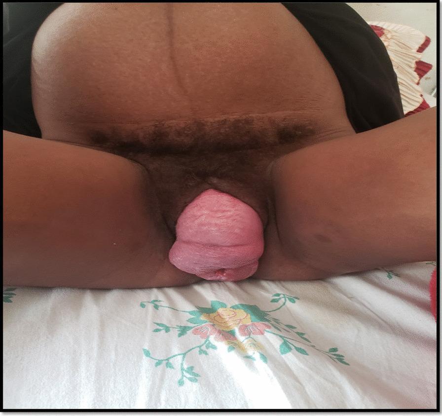
This clinical photograph demonstrates a severe case of stage IV pelvic organ prolapse (procidentia) in a patient. The image shows a large, cylindrical, pinkish-red mass of tissue protruding entirely beyond the vaginal introitus and lying on the vulva. The visible structures include the everted anterior and posterior vaginal walls and the uterine cervix, which occupies the most distal portion of the prolapsed mass. The surface texture of the prolapsed tissue appears moist and slightly lobulated with visible rugae. This visual representation illustrates maximal descent of pelvic organs as defined by the Pelvic Organ Prolapse Quantification (POP-Q) system, where the entire vaginal length is essentially everted. The clinical context suggests a profound failure of the pelvic floor support structures, including the cardinal and uterosacral ligaments. This material is highly relevant for medical education in gynecology, specifically for identifying advanced stages of pelvic floor dysfunction and understanding the anatomical landmarks involved in total vault prolapse.
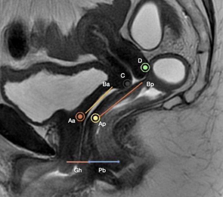
A mid-sagittal T2-weighted Magnetic Resonance Image (MRI) of the female pelvis, illustrating the anatomical landmarks used in the Pelvic Organ Prolapse Quantification (POP-Q) system. The image clearly depicts the uterus, a partially filled bladder, the vaginal canal, and the rectum. Superimposed on the anatomy are standardized POP-Q reference points: Aa and Ba (anterior vaginal wall), Ap and Bp (posterior vaginal wall), C (cervix/apex), and D (posterior fornix). Linear measurements at the pelvic outlet indicate the Genital Hiatus (Gh) and Perineal Body (Pb). Lines connect these points to demonstrate vaginal topography and relative descent. This diagnostic image serves as a clinical education tool for correlating radiological findings with physical examination stages of pelvic floor dysfunction, specifically identifying the relationship of pelvic organs to the plane of the hymen for prolapse assessment.
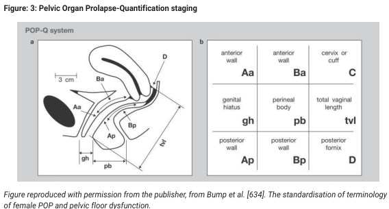
Summary : This figure illustrates the Pelvic Organ Prolapse Quantification (POP-Q) system, which is used for staging and standardizing the assessment of pelvic organ prolapse in women. It includes both an anatomical diagram and a terminology grid for reference points used in the POP-Q system. illustration: # POP-Q System Diagram : • Sagittal anatomical illustration of the female pelvis showing the vagina, bladder, and rectum. • Key reference points are labeled: Aa, Ba, Ap, Bp, C, D, gh, pb, tvl. • Measurements such as "3 cm" and distances for genital hiatus (gh) and perineal body (pb) are indicated with double-headed arrows. • The diagram visually demonstrates where each POP-Q point is located along the vaginal walls and perineal structures. # POP-Q Terminology Grid : • 3x3 grid listing anatomical reference points and their abbreviations: – Anterior wall: Aa, Ba – Cervix or cuff: C – Genital hiatus: gh – Perineal body: pb – Total vaginal length: tvl – Posterior wall: Ap, Bp – Posterior fornix: D • Each cell contains the abbreviation and the corresponding anatomical term. # Technical Details : • Figure is adapted from Bump et al., referencing the standardization of terminology for female pelvic organ prolapse and pelvic floor dysfunction. • No scale bar, but a 3 cm reference is shown for anatomical context. # Spatial Relationships : • The anatomical diagram is oriented in the sagittal plane, with the anterior wall (bladder side) on the left and the posterior wall (rectum side) on the right. • The grid provides a cross-reference for the anatomical points shown in the diagram. # Analysis : • The figure provides a comprehensive visual guide to the POP-Q system, clarifying the location and terminology of key reference points used in clinical staging of pelvic organ prolapse. • The combination of diagram and grid supports both anatomical understanding and standardized clinical communication.
pessary types ring shelf Gellhorn pelvic organ prolapse
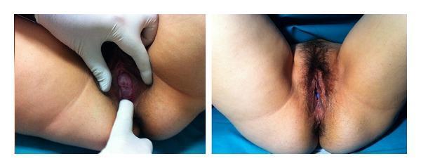
A side-by-side clinical photograph documenting uterine prolapse and its subsequent management with a pessary in a pregnant patient. The left panel shows a clinical examination of the vulva with a gloved hand retracting the labia. The cervix is visibly descended and protruding through the vaginal introitus, characteristic of grade 2 pelvic organ prolapse. The cervical tissue appears hyperemic and edematous. The right panel displays the external female genitalia following the placement of an Arabin cerclage pessary. In this view, the prolapse is reduced, and a portion of the blue silicone pessary is visible within the vaginal canal, positioned to support the cervix and maintain its placement. The image illustrates a non-surgical management strategy for symptomatic uterine prolapse and acute urinary retention during pregnancy. Educational concepts include pelvic floor anatomy, grading of organ prolapse, and mechanical support devices in obstetrics and gynecology.
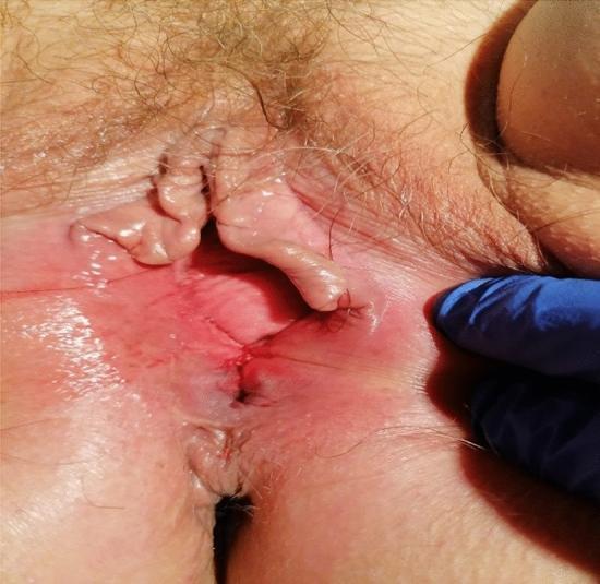
This clinical photograph shows the post-procedure appearance of the female perineum following the insertion of a pessary for pelvic organ prolapse management. The image displays the external genitalia with three independent resorbable sutures placed at the perineum to narrow the vaginal introitus and prevent pessary expulsion. The surgical site exhibits expected post-operative changes, including moderate erythema, localized edema of the labia, and slightly raised tissue along the suture line. A blue-gloved hand is visible on the right lateral aspect, providing retraction to optimize visualization of the approximated tissues and suture integrity. The surrounding skin shows normal follicular distribution with mild signs of procedure-related irritation. This visual demonstrates a conservative surgical technique for stabilizing mechanical treatments in advanced pelvic floor dysfunction.
pelvic organ prolapse management reproductive age fertility preservation 2024 guidelines
Management of Pelvic Organ Prolapse (POP) in the Reproductive Age Group
1. Initial Assessment
- History: vaginal bulge (the most predictive symptom), pelvic pressure/heaviness, changes in bladder/bowel function, sexual dysfunction, desire for future pregnancy
- Physical exam: POP-Q staging (the standardised quantification system), identification of affected compartment(s) - anterior (cystocele), posterior (rectocele/enterocele), or apical (uterovaginal prolapse)
- Bladder function evaluation: rule out occult stress urinary incontinence (masked by urethral kinking from prolapse)
- Bowel function: assess for defecatory dysfunction, obstructed defecation, splinting
- Imaging: MRI pelvic floor if complex or multi-compartment disease
2. Conservative (Non-Surgical) Management
- Younger age is an independent risk factor for surgical recurrence (~16% at 12 months after native tissue repair)
- Pregnancy after repair risks re-prolapse
- Obliterative procedures are contraindicated in this group
A. Lifestyle Modifications
- Weight reduction in obese patients
- Treat chronic constipation (high-fibre diet, hydration, avoid straining)
- Smoking cessation (reduces chronic cough contributing to raised intra-abdominal pressure)
- Avoid heavy lifting and high-impact exercise during symptomatic periods
B. Pelvic Floor Muscle Training (PFMT)
- Strengthens the levator ani, pubococcygeus, and iliococcygeus
- Reduces prolapse symptoms and can improve POP-Q stage
- A 2023 systematic review (PMID 37849316) confirmed effectiveness in reducing POP and urinary incontinence in women of childbearing age
- A 2025 systematic review on postpartum exercise (PMID 39694630) found pelvic floor exercise programs significantly reduce pelvic floor disorders after delivery
- Supervised PFMT by a pelvic floor physiotherapist achieves better results than self-guided exercise
- Minimum 3 months of structured training before reassessment
- Hypopressive exercises (low-pressure fitness techniques) are also being studied as adjuncts - a 2024 systematic review (PMID 38432833) found they improve pelvic floor and abdominal muscle function
C. Vaginal Pessaries
- Types: Ring pessary (mild-moderate prolapse), Gellhorn pessary or shelf pessary (moderate-severe prolapse requiring more support)
- Successfully fitted in the majority of women; may require 1-2 trial fittings
- Mechanism: mechanical reduction of prolapse, restoring normal anatomy
- Particularly useful while awaiting surgery, during pregnancy, or when surgery is declined
- Note: Younger age, prior hysterectomy, and increasing parity are risk factors for pessary failure/expulsion (IUGA 2024)
- Maintenance: Remove and clean every 3-6 months; inspect for erosions. Self-management (patient removes/cleans herself) is preferred
- Concomitant vaginal oestrogen (topical) helps prevent atrophy-related erosions
- Complications: vaginal discharge, erosion, expulsion, discomfort
3. Surgical Management
Apical Compartment (Uterovaginal Prolapse)
Uterine-Preserving Procedures (Preferred in Reproductive Age)
| Procedure | Route | Key Features |
|---|---|---|
| Sacrospinous hysteropexy (SSH) | Vaginal | Suspension of cervix to sacrospinous ligament; non-inferior to vaginal hysterectomy + USL suspension for apical prolapse; risk of pudendal neurovascular injury |
| Manchester (Fothergill) Procedure | Vaginal | Amputation of elongated cervix + cardinal ligament plication + anterior colporrhaphy; suitable when cervical elongation is prominent |
| Uterosacral ligament hysteropexy | Vaginal/laparoscopic | Shortening and fixation of uterosacral ligaments; monitors ureter during procedure |
| Laparoscopic/robotic sacrohysteropexy | Abdominal | Mesh attached from uterus/cervix to sacrum; lowest recurrence; preferred if future pregnancy not desired but uterus to be conserved |
- The AUGS April 2025 Clinical Practice Statement specifically addresses the role of uterine preservation at the time of POP - reflecting growing consensus for conserving the uterus when technically feasible.
If Hysterectomy Cannot Be Avoided
- Vaginal hysterectomy + uterosacral ligament suspension (USLF) or sacrospinous ligament fixation (SSLF)
- Abdominal sacrocolpopexy (open, laparoscopic, or robotic): gold standard for apical support with lowest recurrence; mesh placed from vaginal apex to sacrum; longer operative time but superior anatomic outcomes - Sabiston Textbook of Surgery, p. 2789
Anterior Compartment (Cystocele)
- Anterior colporrhaphy (anterior repair): midline plication of pubocervical fascia; anatomic cure 76-100% but failure rates higher using vaginal approach alone
- Combined central + lateral defects are common; anterior colporrhaphy may be combined with paravaginal repair for lateral defects
- Vaginal mesh: FDA has significantly restricted transvaginal mesh use for prolapse repair due to mesh erosion, pelvic pain, and dyspareunia complications - not routinely recommended, especially in younger women
Posterior Compartment (Rectocele/Enterocele)
- Posterior colporrhaphy: plication of rectovaginal septum; anatomic cure 76-96%; however, traditionally associated with de novo dyspareunia in 7-26% (up to 50% with levator plication)
- Avoid levator plication in reproductive-age women - markedly increases dyspareunia risk
- Use fascial plication without levator plication + perineal body reconstruction rather than routine perineorrhaphy to reduce sexual dysfunction
- Rectocele repair is indicated for bulge symptoms only - not for constipation (evidence does not support repair for defecatory dysfunction)
- For enterocele: culdoplasty (McCall, Moschcowitz) at time of apical repair
- Berek & Novak's Gynecology, pp. 1723-1724
Obliterative Procedures (Contraindicated in Reproductive Age)
- Colpocleisis (LeFort): vaginal canal obliteration; reserved for elderly, medically unfit women who do not desire coital function
- Contraindicated in reproductive-age women
4. Special Considerations in Reproductive Age
Fertility Preservation
- Postpone definitive surgery until childbearing is complete whenever possible
- If surgery is needed before completed family: use uterine-preserving procedure; counsel about recurrence risk with subsequent pregnancy and delivery
- Mode of delivery after prolapse repair: elective caesarean section is generally recommended to protect the repair
- Pessary can serve as a long-term bridge until childbearing is complete
During Pregnancy
- Uterine prolapse in pregnancy (rare): ring or Arabin cerclage pessary is safe and effective for reduction and support
- Avoid definitive surgical repair during pregnancy
Recurrence Risk
- Younger age is a risk factor for recurrence after native tissue repair (~16% recurrence at 12 months, higher long-term)
- Recurrence rates: approximately 62-70% objective failure at 5 years for USLF and SSLF respectively, though symptom scores remain improved
- Long-term follow-up is essential; patients must understand that recurrence requiring re-operation is possible
Concomitant Stress Urinary Incontinence (SUI)
- Prolapse repair can unmask occult SUI (previously masked by urethral kinking)
- Pre-operative urodynamic assessment or pessary test to identify occult SUI
- If demonstrated, add a mid-urethral sling at the time of prolapse repair
- Berek & Novak's Gynecology, p. 1882
5. Management Algorithm Summary
Symptomatic POP (Reproductive Age)
         |
         ├── Asymptomatic? --> Expectant management + lifestyle advice
         |
         ├── Stage I-II (mild-moderate):
         |     --> PFMT (minimum 3 months, supervised)
         |     --> Lifestyle modifications
         |     --> Pessary (ring type) if PFMT inadequate
         |
         ├── Stage II-III (moderate):
         |     --> Pessary trial first
         |     --> PFMT + pessary
         |     --> Surgery if conservative fails
         |
         └── Stage III-IV (severe) / complications / conservative failure:
               --> Surgery
               --> Uterine-preserving (hysteropexy) preferred
               --> Reconstruct all affected compartments
               --> Address apical support as primary priority
               --> Abdominal sacrocolpopexy if low recurrence paramount
               --> Avoid obliterative procedures
               --> Discuss recurrence risk + future pregnancy
Key Takeaways
- Conservative first: PFMT and pessary are effective and should precede surgery, especially with desire for future fertility.
- Apical support is the keystone: Correcting apical prolapse often resolves anterior and posterior defects simultaneously.
- Uterine conservation is preferred: Sacrospinous hysteropexy, Manchester procedure, or laparoscopic sacrohysteropexy in preference to hysterectomy.
- No obliterative procedures: Colpocleisis is contraindicated in sexually active or potentially fertile women.
- Avoid vaginal mesh for cystocele repair: FDA restrictions apply; native tissue repair is preferred.
- Recurrence risk is higher in younger women: Counsel thoroughly; delay surgery until family is complete when possible.
- Dyspareunia prevention: Avoid levator plication; perform perineal body reconstruction rather than routine perineorrhaphy.