
Explain Chronic Renal Failure. What is it’s cause, Pathology, clinical features, Differential diagnosis, Management, treatment. Write answer for university exam
Chronic renal failure
"chronic kidney disease" management
chronic kidney disease pathology histology stages
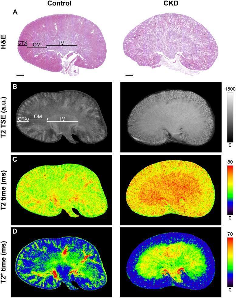
A comparison of renal pathology and imaging modalities between a control kidney and a Chronic Kidney Disease (CKD) model. The figure is organized into four rows representing different assessment techniques: (A) Hematoxylin & Eosin (H&E) histology, (B) T2-weighted Turbo Spin Echo (TSE) MRI, (C) T2 relaxation time maps (ms), and (D) T2* relaxation time maps (ms). Three distinct anatomical regions are demarcated: cortex (CTX), outer medulla (OM), and inner medulla (IM). In H&E staining, the control displays clear corticomedullary differentiation, whereas the CKD kidney shows tissue architectural disruption and diffuse staining. In MRI modalities, the control kidney exhibits high contrast between the CTX, OM, and IM. Conversely, the CKD kidney shows a loss of this regional contrast, characterized by more uniform high signal intensity in T2 TSE and significantly increased T2 relaxation times (yellow-orange shift) on parametric maps. T2* maps demonstrate altered oxygenation or tissue composition in the CKD model compared to the complex patterns in the control. This infographic serves as an educational tool for identifying renal disease progression through correlative histology and functional MRI techniques.
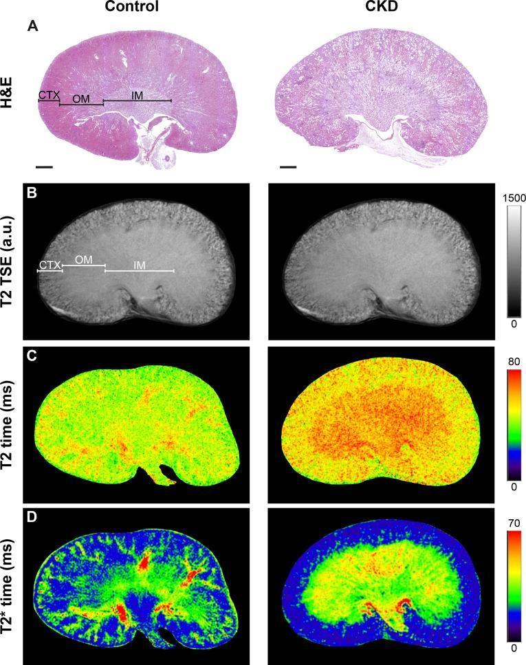
This multi-panel figure compares renal histology and magnetic resonance imaging (MRI) between a control kidney and a Chronic Kidney Disease (CKD) model. Row A presents Hematoxylin & Eosin (H&E) stained sections, highlighting the renal cortex (CTX), outer medulla (OM), and inner medulla (IM). In the CKD kidney, there is visible cortical thinning and loss of clear structural demarcation between medullary layers. Row B displays T2-weighted Turbo Spin Echo (TSE) images, showing morphological changes and signal intensity variations corresponding to the tissue layers. Rows C and D provide functional mapping through T2 and T2* relaxation time maps using pseudo-color scales (0-80ms and 0-70ms respectively). These maps demonstrate significantly elevated relaxation times in the CKD kidney, particularly within the outer medulla, visualized by a shift from cooler green/blue tones to warmer yellow/red tones. The composite image illustrates the correlation between histological tissue remodeling and functional MRI biomarkers in renal pathology.
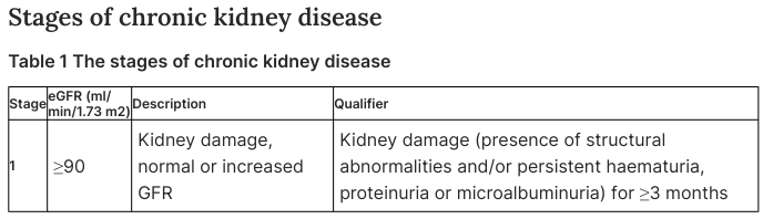
Stages of chronic kidney disease Table 1 The stages of chronic kidney disease <table><thead><tr><th>Stage</th><th>eGFR (ml/min/1.73 m2)</th><th>Description</th><th>Qualifier</th></tr></thead><tbody><tr><td>1</td><td>≥90</td><td>Kidney damage, normal or increased GFR</td><td>Kidney damage (presence of structural abnormalities and/or persistent haematuria, proteinuria or microalbuminuria) for ≥3 months</td></tr></tbody></table>
CHRONIC RENAL FAILURE (CHRONIC KIDNEY DISEASE)
DEFINITION
- Albuminuria > 30 mg/day
- GFR < 60 mL/min/1.73 m²
- Abnormal urine sediment, imaging abnormalities, renal tubular syndromes, or histological evidence of kidney damage
STAGING (KDIGO Classification)
| Stage | GFR (mL/min/1.73 m²) | Description |
|---|---|---|
| G1 | ≥ 90 | Normal or high GFR, with kidney damage markers |
| G2 | 60-89 | Mildly decreased |
| G3a | 45-59 | Mildly to moderately decreased |
| G3b | 30-44 | Moderately to severely decreased |
| G4 | 15-29 | Severely decreased |
| G5 | < 15 | Kidney failure (ESRD) |
AETIOLOGY (CAUSES)
Primary (Intrinsic Renal) Causes
- Diabetic nephropathy - the leading cause worldwide; due to diabetic glomerulosclerosis
- Hypertensive nephrosclerosis - second most common; due to long-standing hypertension damaging the renal vasculature
- Chronic glomerulonephritis - e.g., IgA nephropathy, focal segmental glomerulosclerosis (FSGS), membranous nephropathy
- Polycystic kidney disease (PKD) - autosomal dominant (ADPKD) most common hereditary cause; GFR begins declining in the 4th-6th decade
- Chronic tubulointerstitial nephritis - due to analgesic abuse, heavy metals, toxins, recurrent infections
- Reflux nephropathy - chronic vesicoureteral reflux causing recurrent pyelonephritis and scarring
Secondary Causes
- Systemic lupus erythematosus (lupus nephritis)
- Amyloidosis
- Myeloma kidney
- Sickle cell nephropathy
- Renovascular disease / renal artery stenosis
PATHOLOGY
A. Macroscopic Appearance
- Kidneys are small, shrunken, and fibrotic (bilateral symmetric reduction in size)
- Cortical thinning with loss of corticomedullary differentiation
- Granular surface (in hypertensive nephrosclerosis) or smooth surface (in polycystic disease)
B. Microscopic / Histological Changes
- Glomerular changes: global glomerulosclerosis, mesangial expansion, basement membrane thickening
- Tubular changes: tubular atrophy, loss of tubular epithelium, simplified lining
- Interstitial changes: interstitial fibrosis and inflammation (hallmark of progression)
- Vascular changes: arteriosclerosis and arteriolar hyalinosis
- Widespread loss of nephron mass with compensatory hypertrophy of remaining nephrons
C. Pathophysiology - The "Intact Nephron" Hypothesis
- Increased intraglomerular pressure → proteinuria
- Angiotensin II activation → efferent arteriolar constriction → further glomerular damage
- Each lost nephron places greater hemodynamic stress on remaining nephrons, accelerating progression
- Normal GFR (~125 mL/min/1.73 m²) is maintained until nephron loss precludes complete compensation, at which point GFR falls sharply
D. Uremic Syndrome (GFR < 15 mL/min)
| Mechanism | Consequence |
|---|---|
| ↓ Erythropoietin production | Normochromic, normocytic anaemia |
| ↓ 1,25-(OH)₂ Vitamin D₃ | Hypocalcaemia, secondary hyperparathyroidism, renal osteodystrophy |
| ↑ Phosphate retention | Stimulates PTH → bone resorption |
| ↓ Ammonia production | Metabolic acidosis (uremic acidosis) |
| ↓ Nitric oxide, ↑ renin-angiotensin | Hypertension |
| Hepcidin accumulation | Worsens anaemia (iron restriction) |
CLINICAL FEATURES
1. General
- Fatigue, weakness, malaise
- Weight loss, muscle and fat wasting (protein-energy malnutrition)
- Anorexia, nausea, vomiting
2. Cardiovascular (leading cause of death in CKD)
- Hypertension (due to Na/fluid retention + ↑ renin-angiotensin-sympathetic activity)
- Congestive heart failure, fluid overload, oedema
- Pericarditis (uraemic pericarditis - pathognomonic of advanced uraemia)
- Accelerated atherosclerosis
- Arrhythmias, conduction blocks
- Vascular calcification
3. Haematological
- Normochromic, normocytic anaemia (due to ↓ EPO, ↑ hepcidin, ↓ RBC survival)
- Platelet dysfunction → bleeding tendency (prolonged bleeding time)
- Leukocyte dysfunction → increased susceptibility to infection
4. Neurological
- Peripheral neuropathy (stocking-glove distribution; earliest = restless legs syndrome)
- Autonomic neuropathy
- Encephalopathy, confusion, lethargy
- Asterixis (metabolic flap)
- Myoclonus, muscle twitching
- Seizures, coma (terminal)
5. Gastrointestinal
- Anorexia, nausea, vomiting (early symptoms)
- Uraemic fetor (ammonia breath)
- Delayed gastric emptying, hyperacidity
- Mucosal ulcerations, GI haemorrhage
- Adynamic ileus
6. Skeletal / Endocrine
- Renal osteodystrophy: combination of osteitis fibrosa cystica (from ↑ PTH) and osteomalacia (from ↓ active Vitamin D)
- Periarticular calcification, metastatic calcification
- Secondary → tertiary hyperparathyroidism
- Glucose intolerance
- Hypertriglyceridaemia
- Sexual dysfunction: amenorrhoea, menorrhagia, impotence, oligospermia, galactorrhoea
7. Pulmonary
- Hyperventilation (compensatory for metabolic acidosis)
- Pulmonary oedema ("uraemic lung" - butterfly pattern on CXR)
- Pleural effusion
8. Metabolic Abnormalities
- Metabolic acidosis (anion gap elevated at G4-G5)
- Hyperkalaemia (K⁺ >6.5 mEq/L in advanced CKD - life-threatening)
- Hyperphosphataemia, hypocalcaemia
- Hypermagnesaemia, hyperuricaemia
- Hyponatraemia, hypoalbuminaemia
9. Skin
- Uraemic pruritus (intractable itching)
- Hyperpigmentation (yellowish-brown discolouration)
- Ecchymoses
- "Uraemic frost" (urea crystals on skin - rare, terminal)
INVESTIGATIONS / DIAGNOSIS
Blood Tests
- Serum creatinine and BUN: elevated; BUN:creatinine ratio ~10:1 (vs >20:1 in pre-renal)
- eGFR: calculated using CKD-EPI equation (using creatinine ± cystatin C)
- FBC: normochromic normocytic anaemia
- Electrolytes: hyperkalaemia, hyponatraemia, hyperphosphataemia, hypocalcaemia
- ABG: metabolic acidosis (↓ HCO₃⁻, ↓ pH)
- PTH: elevated (secondary hyperparathyroidism)
- Serum albumin: low (malnutrition)
- Lipid profile: hypertriglyceridaemia
Urine Tests
- Urinalysis: proteinuria (albuminuria), haematuria
- Urine albumin:creatinine ratio (ACR) > 30 mg/g
- Urine sediment: granular casts, RBC casts (glomerulonephritis), WBC casts (interstitial nephritis), oval fat bodies
Imaging
- Renal ultrasound: small, echogenic kidneys bilaterally (most characteristic finding in CKD); loss of corticomedullary differentiation
- CT / MRI if structural cause suspected
- Chest X-ray: cardiomegaly, pulmonary oedema
Renal Biopsy
- Indicated when cause is uncertain and histology will change management
- Findings: glomerulosclerosis, tubular atrophy, interstitial fibrosis
DIFFERENTIAL DIAGNOSIS
| Condition | Key Distinguishing Features |
|---|---|
| Acute Kidney Injury (AKI) | Abrupt onset (< 48 hrs), reversible; urine Na < 20, FENa < 1% (pre-renal); kidney size normal |
| Acute-on-Chronic CKD | Pre-existing CKD with acute deterioration; identify reversible precipitant |
| Nephrotic syndrome | Massive proteinuria (>3.5 g/day), hypoalbuminaemia, oedema, hyperlipidaemia; GFR may be normal initially |
| Renovascular hypertension | Renal artery stenosis; asymmetric kidneys, deterioration with ACE inhibitors |
| Obstructive uropathy | Hydronephrosis on ultrasound; reversible if relieved early |
| Myeloma kidney | Age >50, bone pain, hypercalcaemia, Bence-Jones proteinuria, SPEP/immunofixation abnormal |
| Lupus nephritis | Young female, ANA/dsDNA positive, multi-system features |
| Amyloidosis | Congo red staining, nephrotic range proteinuria, systemic involvement |
- CKD: bilateral small kidneys on ultrasound, anaemia established, hyperphosphataemia, secondary hyperparathyroidism, prolonged elevated creatinine history
- AKI: normal kidney size, recent onset, absence of renal osteodystrophy
MANAGEMENT
- Slow progression of CKD
- Treat complications
- Prepare for renal replacement therapy (RRT) when needed
A. Conservative (Non-Pharmacological) Management
- Dietary protein restriction: 0.6-0.8 g/kg/day (reduces nitrogen load, slows progression)
- Sodium restriction: < 2 g/day (controls hypertension and fluid retention)
- Potassium restriction: in hyperkalaemia (avoid high K⁺ foods: bananas, oranges)
- Phosphate restriction: dairy products, processed foods
- Fluid management: restrict in oliguric patients
- Smoking cessation (accelerates CKD progression)
- Weight loss and regular exercise
B. Slowing Progression (Disease-Modifying Treatment)
- ACE Inhibitors (e.g., ramipril) or ARBs (e.g., losartan): First-line in all CKD patients with proteinuria. Reduce intraglomerular pressure, decrease proteinuria, slow GFR decline. Particularly beneficial in diabetic nephropathy.
- SGLT2 inhibitors (e.g., empagliflozin, dapagliflozin): Major advance - reduce CKD progression and cardiovascular events in both diabetic and non-diabetic CKD
- GLP-1 receptor agonists: Reduce cardiovascular risk in diabetic CKD
- Mineralocorticoid receptor antagonists (finerenone): Reduce progression and cardiovascular risk in diabetic CKD
- Tight blood pressure control: Target <130/80 mmHg
- Glycaemic control in diabetic CKD: target HbA1c ~7% (53 mmol/mol)
- Statins: for cardiovascular risk reduction
C. Management of Complications
| Complication | Treatment |
|---|---|
| Anaemia | Erythropoiesis-stimulating agents (ESA: epoetin alfa, darbepoetin); IV/oral iron supplementation; target Hb 10-12 g/dL |
| Hypertension | ACE inhibitor/ARB first-line; add amlodipine, diuretics as needed |
| Hyperkalaemia | Dietary restriction; diuretics; sodium bicarbonate; potassium binders (patiromer, sodium zirconium cyclosilicate); emergent dialysis if severe |
| Metabolic acidosis | Oral sodium bicarbonate supplementation (target serum HCO₃⁻ ≥22 mEq/L) |
| Renal osteodystrophy | Phosphate binders (calcium carbonate, sevelamer); active vitamin D (calcitriol); cinacalcet for tertiary hyperparathyroidism |
| Fluid overload | Loop diuretics (furosemide); salt restriction; dialysis if refractory |
| Dyslipidaemia | Statins (statin + ezetimibe in advanced CKD) |
| Uraemic pruritus | Antihistamines; gabapentin; adequate dialysis |
D. Renal Replacement Therapy (RRT)
- Most common form of RRT
- Blood is filtered extracorporeally through a semipermeable membrane 3 times/week (~4 hours per session)
- Complications: hypotension, disequilibrium syndrome, arrhythmias, access infections
- Uses peritoneal membrane as the dialysis membrane
- Continuous ambulatory PD (CAPD) or automated PD (APD)
- Advantage: home-based, preserves residual renal function
- Complications: peritonitis, catheter infections, poor ultrafiltration
- Best option for eligible patients - superior survival and quality of life compared to dialysis
- Live-donor preferred over cadaveric donor
- Requires lifelong immunosuppression (calcineurin inhibitors + mycophenolate + steroids)
- Complications: rejection, opportunistic infections, drug nephrotoxicity
INDICATIONS FOR URGENT DIALYSIS (AEIOU Mnemonic)
| Letter | Indication |
|---|---|
| A | Acidosis (metabolic, refractory) |
| E | Electrolyte abnormality (severe hyperkalaemia, K⁺ >6.5) |
| I | Intoxication/Ingestion (dialysable toxin) |
| O | Overload (fluid overload refractory to diuretics) |
| U | Uraemia (pericarditis, encephalopathy, bleeding) |
PROGNOSIS
- CKD is progressive; rate of decline varies by cause, degree of proteinuria, and BP control
- Average GFR decline: ~4-5 mL/min/year in advanced disease
- Leading cause of death: cardiovascular disease (not renal failure itself)
- 5-year survival on haemodialysis: ~35-40%; kidney transplant significantly improves this
- Risk stratification combines GFR category + albuminuria category (CGA staging)
SUMMARY TABLE
| Feature | Details |
|---|---|
| Definition | GFR < 60 mL/min/1.73 m² for >3 months OR kidney damage markers |
| Top causes | Diabetic nephropathy, hypertension, chronic GN, ADPKD |
| Key pathology | Nephron loss, glomerulosclerosis, interstitial fibrosis |
| Cardinal symptoms | Fatigue, anaemia, oedema, hypertension, uraemia |
| Most specific sign | Uraemic pericarditis, small echogenic kidneys on ultrasound |
| Key investigations | eGFR (CKD-EPI), proteinuria (ACR), renal ultrasound |
| Progression slowed by | ACE-I/ARB, SGLT2 inhibitors, BP control, protein restriction |
| RRT options | Haemodialysis, peritoneal dialysis, renal transplantation |
| Main cause of death | Cardiovascular disease |