
Cultivation of viruses microbiology for my exam
Reading File
Viruses are obligate intracellular parasites and cannot grow on artificial media - they require living cells. There are three main methods: embryonated eggs, cell/tissue cultures, and animal inoculation.
Searching sources...
Finding Sources
Finding Sources
Reading File
Finding Sources
Reading File
Finding Sources
Finding Sources
Reading File
Finding Sources
Finding Sources
Finding Sources
Loading Image
Now I have comprehensive material from Jawetz's Medical Microbiology. Let me compile a thorough exam-oriented answer.
Cultivation of Viruses - Microbiology Exam Notes
Source: Jawetz, Melnick & Adelberg's Medical Microbiology, 28th Ed.
Why Special Methods Are Needed
Viruses are obligate intracellular parasites - they cannot replicate on artificial (cell-free) media. They require living host cells with intact metabolic machinery to replicate. Three main systems are used:
- Cell (tissue) culture
- Embryonated (fertile) hen's eggs
- Animal inoculation
1. Cell Culture (Most Important - Central to Modern Virology)
Types of Cell Cultures
| Type | Features | Passages | Example |
|---|---|---|---|
| Primary cultures | Dispersed from fresh host tissue (usually with trypsin); closest to normal tissue | Only a few passages | Primary monkey kidney cells |
| Diploid cell lines | Underwent a change allowing limited culture; retain normal chromosomes | Up to 50 passages | Human embryonic lung fibroblasts |
| Continuous cell lines | Derived from diploid lines or malignant tissue; indefinite growth; abnormal, irregular chromosome numbers | Indefinite | HeLa, Vero, HEp-2 |
The choice of cell line depends on the sensitivity of that cell type to the particular virus.
Detection of Virus Growth in Cell Culture
Growth is monitored by several means:
1. Cytopathic Effects (CPE) - morphologic changes in infected cells (most important indicator):
- Cell lysis/necrosis - e.g., enteroviruses (rapid rounding, complete destruction)
- Inclusion body formation - virus-specific structures far larger than individual virions, often eosinophilic
- Giant cell / syncytia formation - e.g., paramyxoviruses (fused cells)
- Cytoplasmic vacuolization
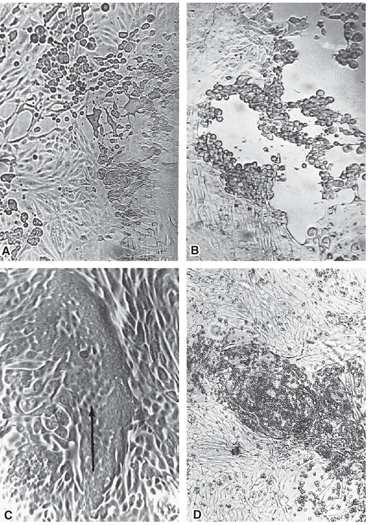
2. Hemadsorption - virus-encoded hemagglutinin (HA) in the cell membrane causes erythrocytes to adsorb to infected cells. Seen with parainfluenza and influenza. Becomes positive before CPE is visible.
3. Viral protein detection - specific antisera detect viral proteins (e.g., HA of influenza) in infected cells via immunofluorescence.
4. Nucleic acid detection - PCR provides rapid, sensitive, specific detection of viral genome.
Inclusion Bodies (Exam Favourite)
Virus-specific structures formed during viral replication inside cells. Larger than virions, often eosinophilic (acidophilic).
| Location | Examples |
|---|---|
| Nuclear | Herpesvirus |
| Cytoplasmic | Poxvirus, Rabies (Negri bodies) |
| Both nucleus and cytoplasm | Measles virus |
2. Embryonated Hen's Egg
A self-contained living system with multiple sites for inoculation. Signs of viral growth:
| Finding | Example Viruses |
|---|---|
| Death of embryo | Encephalitis viruses |
| Pocks/plaques on chorioallantoic membrane (CAM) | Herpes, Smallpox, Vaccinia |
| Hemagglutinins in embryonic fluids/tissues | Influenza |
- Pocks on CAM can also be used for quantitation (relating pock count to viral dilution)
3. Animal Inoculation
- Still used for primary isolation of certain viruses (e.g., some arboviruses, coxsackieviruses in suckling mice)
- Used for studying pathogenesis of viral diseases
- Used for studies of viral oncogenesis
- Expensive, raises ethical concerns; largely replaced by cell culture
Quantitation of Viruses
Physical Methods
| Method | Principle | Notes |
|---|---|---|
| Quantitative PCR | Detects genome copies | Detects both infectious and non-infectious particles |
| Serologic assays (RIA, ELISA) | Detect viral proteins | Cannot distinguish infectious from non-infectious |
| Hemagglutination (HA) assay | Measures HA protein | Quantitates HA-containing viruses (e.g., influenza) |
| Electron microscopy | Direct particle count vs. latex bead standard | Needs concentrated prep; cannot distinguish infectivity |
Biological Methods
50% Infectious Dose (ID₅₀):
- End-point assay using serial dilutions
- Titer = reciprocal of the dilution causing infection in 50% of inoculated cells/animals
- Ratio of infectious to total particles varies widely (from ~1:1 to < 1:1000)
Plaque Assay (Gold Standard for Infectious Virus Titration):
- Inoculate monolayer of host cells with serial dilutions of virus
- After adsorption, overlay with agar or carboxymethylcellulose (prevents widespread diffusion)
- Virus spreads only to adjacent cells → produces a visible clearing = plaque
- 1 plaque = 1 plaque-forming unit (PFU) (from a single clonal virus particle)
- Time to visible plaques: days (e.g., poliovirus) to >2 weeks (e.g., SV40)
- Can only be used for viruses that grow well in tissue culture
Purification of Viruses
After cultivation, viruses are purified by:
- Concentration: precipitation (ammonium sulfate, ethanol, PEG) or ultrafiltration
- Differential centrifugation: separates by size/sedimentation rate
- Density gradient centrifugation (most important final step):
- Rate-zonal: virus bands by sedimentation rate in sucrose/glycerol gradient
- Equilibrium/isopycnic: in CsCl gradients, virus migrates to position where solution density = buoyant density of virus
- Column chromatography: virus bound and eluted by pH/salt changes
- Zone electrophoresis: separation by surface charge
Note: Icosahedral viruses are easier to purify than enveloped viruses (enveloped viruses have variable envelope amounts, making the population heterogeneous in size and density).
Identification Criteria for a Virus Particle
A purified particle is only confirmed as a virus if:
- It can be obtained only from infected cells/tissues
- It transmits infection to susceptible hosts
- It reproduces itself (i.e., replicates)
Quick Summary Table for Exam
| Method | Best For | Key Signs of Growth |
|---|---|---|
| Cell culture (primary) | Most viruses | CPE, hemadsorption, immunofluorescence, PCR |
| Cell culture (continuous) | Many viruses, vaccine production | CPE, plaque formation |
| Embryonated egg | Influenza, herpesviruses, poxviruses | Embryo death, pocks on CAM, HA in fluids |
| Animal inoculation | Arboviruses, rabies, coxsackieviruses | Disease signs, death |
Key exam points to remember:
- Viruses = obligate intracellular parasites; cannot grow on artificial media
- Three types of cell cultures: primary, diploid, continuous
- CPE types: lysis, inclusion bodies, syncytia, vacuolization
- Hemadsorption detects influenza/parainfluenza before CPE appears
- Inclusion bodies: nuclear (herpes), cytoplasmic (rabies/Negri, poxvirus), both (measles)
- Plaque assay = gold standard for infectious virus titer; 1 plaque = 1 PFU
- ID₅₀ = dilution infecting 50% of test subjects
id50 nhi smjha
ID₅₀ - Aasaan Bhasha Mein Samjhata Hoon 😊
Pehle Ek Real-Life Example Lo
Socho tumhare paas ek virus ka sample hai aur tumhe pata karna hai - kitna concentrated hai yeh virus? Kitna powerful hai?
Tum directly dekh nahi sakte, toh ek experiment karte ho.
ID₅₀ Kya Hai?
ID₅₀ = Infectious Dose 50%
Woh minimum amount of virus jo 50% cells ya animals ko infect kar sake.
Step-by-Step Experiment Samjho
Virus ka Serial Dilution banao:
Original virus → 1/10 → 1/100 → 1/1000 → 1/10,000
Ab har dilution ko 10 animals/cell cultures mein dalo aur dekho kaun infect hota hai:
| Dilution | Animals Infected | % Infected |
|---|---|---|
| 1/10 | 10/10 | 100% ✅ |
| 1/100 | 9/10 | 90% ✅ |
| 1/1000 | 5/10 | 50% ← YEH HAI ID₅₀ |
| 1/10,000 | 1/10 | 10% |
| 1/100,000 | 0/10 | 0% |
Toh is virus ka ID₅₀ = 1/1000 dilution
Titer Kya Hota Hai?
Titer = Dilution ka reciprocal (ulta)
$$\text{Titer} = \frac{1}{1/1000} = \mathbf{1000}$$
Matlab: 1 mL original sample mein 1000 infectious units hain.
Simple Logic - "50% Kyun?"
Kyunki biological experiments mein:
- 100% infection ke liye bahut zyada virus chahiye
- 0% infection bahut kam se hoti hai
- 50% point sabse accurate aur reproducible hota hai - na zyada na kam, beech ka point statistically best hota hai
ID₅₀ vs Titer - Ek Line Mein
| Term | Matlab |
|---|---|
| ID₅₀ | Woh dilution jisme 50% infect ho |
| Titer | Uska reciprocal = original sample ki strength |
| High titer | Virus zyada concentrated/powerful |
| Low titer | Virus kam concentrated |
Reed-Muench Method (Exam Mein Aata Hai)
Jab exactly 50% wali dilution na mile, toh Reed-Muench formula use karte hain interpolation ke liye. Yeh mathematically 50% point calculate karta hai do observations ke beech se.
Yaad Rakhne Wali Lines
- ID₅₀ = 50% animals/cells ko infect karne wali dose
- Titer = ID₅₀ ka reciprocal
- Jitna zyada titer → utna zyada potent virus
- Statistical accuracy ke liye 50% choose karte hain
- Plaque assay mein isko PFU/mL bolte hain
Koi aur confusing topic ho toh batao! 🎯
Plaque assay
Plaque assay virus titration ka gold standard method hai - ek virus particle se ek plaque banta hai.
Searching sources...
Finding Sources
Finding Sources
Reading File
Reading File
Textbook se poora content mil gaya. Ab ekdum clearly samjhata hoon!
Plaque Assay - Seedha Samjho 🎯
Source: Jawetz, Melnick & Adelberg's Medical Microbiology, 28th Ed. + Murray's Medical Microbiology 9e
Plaque Assay Kya Hai?
Yeh infectious virus count karne ka gold standard method hai.
1 Plaque = 1 Infectious Virus Particle = 1 PFU (Plaque-Forming Unit)
Step-by-Step Process - Bilkul Simple
Step 1: Serial Dilution Banao
Virus sample → 1/10 → 1/100 → 1/1000 → 1/10,000
Har dilution ek alag plate mein jaayegi.
Step 2: Cell Monolayer Mein Dalo
- Ek flat dish mein host cells ki ek layer (monolayer) grow karo
- Is monolayer pe diluted virus dalo
- Thodi der baad (adsorption ke liye) virus cells se chipak jaata hai
🔲🔲🔲🔲🔲🔲🔲🔲   ← Healthy cell monolayer
         ↓
       Virus daalo
         ↓
🔲🔲🔲🦠🔲🔲🔲🔲   ← Ek cell infect ho gayi
Step 3: Agar/CMC Overlay Daalo ⬅️ MOST IMPORTANT STEP
Adsorption ke baad upar se agar ya carboxymethylcellulose (CMC) ki layer daalo.
Kyun? Taaki virus sirf aas-paas ki cells mein spread ho sake - poori plate mein nahi faile. Yeh virus ko ek jagah tak limit kar deta hai.
━━━━━━━━━━━━━━━━  ← Agar/CMC layer (virus ko rokta hai)
🔲🔲🔲🦠🔲🔲🔲🔲   ← Cell monolayer
Step 4: Incubate Karo
Kuch din baad:
- Infected cell → virus replicate karta hai → cell marti hai
- Virus sirf adjacent cells mein spread hota hai
- Multiple cycles of infection + cell death = ek circular clearing zone = PLAQUE
🔲🔲🔲⬜⬜🔲🔲🔲
🔲🔲⬜⬜⬜⬜🔲🔲   ← PLAQUE (cells mar gayi = white/clear area)
🔲🔲🔲⬜⬜🔲🔲🔲
🔲🔲🔲🔲🔲🔲🔲🔲   ← Healthy cells (pink/dark)
Step 5: Plaques Gino aur Titer Calculate Karo
Formula:
$$\text{Viral Titer (PFU/mL)} = \frac{\text{Number of plaques}}{\text{Dilution factor} \times \text{Volume inoculated (mL)}}$$
Example:
- Dilution: 1/10,000
- Volume: 0.1 mL
- Plaques counted: 50
$$\text{Titer} = \frac{50}{(1/10,000) \times 0.1} = \frac{50}{0.00001} = 5 \times 10^6 \text{ PFU/mL}$$
Plaque Dikhne Mein Kitna Time Lagta Hai?
| Virus | Time |
|---|---|
| Poliovirus | Kuch din (fast replicator) |
| SV40 | 2 weeks ya zyada (slow) |
Important Facts (Exam Mein Aate Hain)
| Point | Detail |
|---|---|
| 1 Plaque = 1 PFU | Ek single clonal infectious particle se banta hai |
| Agar/CMC kyun? | Virus ko freely diffuse hone se rokta hai |
| Kab use karte hain? | Sirf un viruses ke liye jo cell culture mein achhe se grow karte hain |
| Particle:PFU ratio | Hamesha > 1 (matlab bahut saare defective/non-infectious particles bhi hote hain) |
| ID₅₀ se better kyun? | Direct count karta hai (50% endpoint nahi chahiye) |
Plaque Assay vs ID₅₀ - Fark Samjho
| Plaque Assay | ID₅₀ | |
|---|---|---|
| Kya count karta hai | Actual infectious units (PFU) | 50% endpoint dilution |
| Method | Direct counting | Statistical calculation |
| Accuracy | Zyada precise | Kam precise |
| Result | PFU/mL | Reciprocal of dilution |
| Use | Viruses jo CPE karte hain | Viruses/animals dono mein |
Ek Line Mein Yaad Karo
"Virus daalo → Agar se dhako → Plaque bane → Gino → Titer nikaalo" 1 Plaque = 1 PFU = 1 Infectious Virus
Koi aur concept confuse kar raha ho toh batao! 💪
This is a shared conversation. Sign in to Orris to start your own chat.