
How heart is preserved during transplantation
heart transplantation preservation myocardial protection
heart transplant preservation cold storage cardioplegia
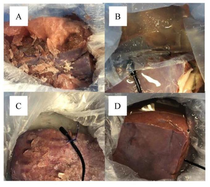
This composite of four clinical photographs (labeled A–D) demonstrates the macroscopic findings of experimental liver grafts following six hours of cold storage under different preservation protocols. The images contrast the impact of freezing temperatures on organ integrity and University of Wisconsin (UW) solution consistency. Panel A shows severe freezing injury with a fragmented liver graft surrounded by extensive ice crystal formation. Panel B displays a graft with surface condensation and a wet appearance, suggesting a partially frozen or thawing state. Panel C illustrates an intact liver graft but with visible superficial frosting and condensation, indicating moderate ice crystal formation. Panel D serves as a successful preservation model, showing an intact liver graft with a smooth, glossy surface and normal anatomical color, entirely devoid of visible ice or freezing injury. This comparison is clinically relevant for evaluating organ packaging techniques and the efficacy of thermal barriers, such as lactated Ringer’s solution layers, in preventing accidental graft freezing during transport and storage in transplant medicine.
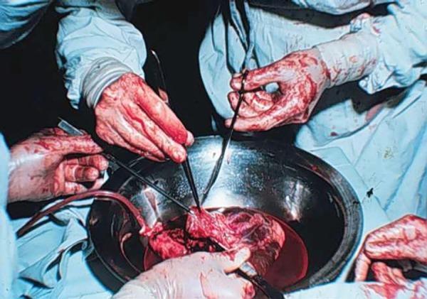
This historical clinical photograph illustrates a key stage in the first Brazilian heart transplant procedure, specifically the preparation of the donor heart. The image shows a donor heart placed within a stainless steel surgical basin, partially submerged in a blood-filled solution. Multiple surgical hands are visible, some gloved and others appearing bare and heavily stained with blood, reflecting historical surgical practices. The surgeons are utilizing instruments, including forceps and a cannula inserted into the vascular structures of the heart, to facilitate continuous normothermic blood perfusion of the organ prior to transplantation. The heart tissue appears intact with visible anatomical structures, including major vessels and atrial/ventricular walls. This visual documents the early technical challenges and methodologies of cardiac transplantation, specifically the preservation and preparation of the graft using warm blood perfusion techniques rather than modern cold storage methods.
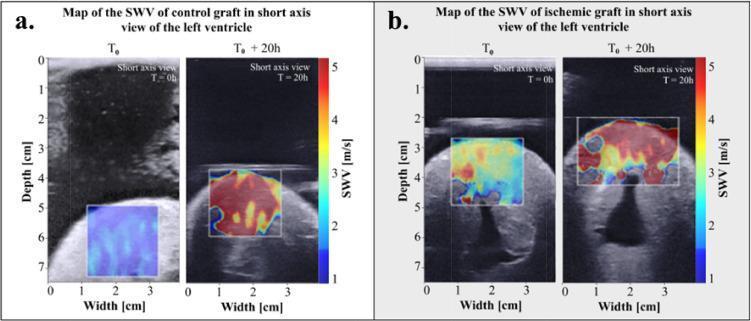
This diagnostic image display consists of two panels (a and b) comparing Shear Wave Velocity (SWV) elastography maps superimposed on B-mode ultrasound images of cardiac grafts. Panel A illustrates a control graft, while Panel B displays an ischemic heart graft, both in short-axis views of the left ventricle at baseline (T0) and after 20 hours (T0 + 20h) of preservation. The SWV color scale on the right of each panel ranges from 1 m/s (blue) to 5 m/s (red), indicating tissue stiffness. At T0, both grafts show predominantly blue and cyan hues, reflecting low initial myocardial stiffness. At T0 + 20h, the control graft shows a shift toward yellow and red, indicating increased stiffness due to cold storage. However, the ischemic graft (Panel B) at T0 + 20h demonstrates a much more pronounced and heterogeneous increase in stiffness, represented by widespread deep red colors across the Region of Interest (ROI). This visual comparison highlights the accelerated stiffening and myocardial damage associated with ischemic injury during organ preservation.
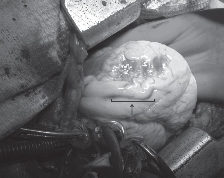
This intraoperative clinical photograph captures a surgical view of a human heart during an emergency coronary artery bypass graft (CABG) procedure. The heart is in a state of cardiac arrest, appearing pale and displaying a wrinkled, non-smooth surface texture consistent with the administration of cold cardioplegia. An arrow and bracket indicate a specific segment of the left anterior descending (LAD) artery, identifying the site of a coronary artery dissection that extends from the left main coronary artery (LMCA). In the foreground and surrounding the heart, several surgical instruments are visible, including metallic clamps and forceps, along with surgical cannulae and tubing used for cardiopulmonary bypass. The image demonstrates the surgical presentation of a Type B coronary dissection requiring urgent revascularization. This visual serves as an educational reference for cardiothoracic surgeons and medical students to understand the gross appearance of coronary pathology and the intraoperative environment during major cardiac surgery.
normothermic machine perfusion heart transplantation organ preservation
PMID: 37636303
PMID: 38069017