
Explain erythropoiesis
I'll start by reading the medical library skill, then search for erythropoiesis content in parallel.
Reading File
Now I'll search the library for erythropoiesis content in parallel:
Finding Sources
Finding Sources
Excellent results. Now I'll read the most relevant sections in parallel - the Histology textbook (which has a dedicated erythropoiesis section) and the Biochemistry textbook, plus search for images:
Reading File
Reading File
Reading File
Good. Now let me read the histology textbook's dedicated erythropoiesis section and fetch the key diagram in parallel:
Reading File
Reading File
Loading Image
Now let me get the histology section on erythrocyte development stages and fetch one more histology image:
Finding Sources
Finding Sources
Now let me read the detailed erythrocyte maturation stages from Henry's and the histology erythropoiesis plate:
Reading File
Reading File
Now let me fetch the histology erythropoiesis plate image and get the maturation stages section:
Reading File
Reading File
I have rich content. Let me fetch the hemopoiesis diagram and the erythropoiesis plate image to include in the answer:
Loading Image
Reading File
I now have comprehensive content from multiple authoritative textbooks. Let me compile the full answer.
Erythropoiesis
Erythropoiesis is the process by which red blood cells (erythrocytes) are continuously produced in the bone marrow to maintain a steady state of oxygen delivery. Under normal conditions, approximately 1% of circulating erythrocytes is replaced daily - equating to about 250 billion erythrocytes, with 2.5 to 3.0 million produced every second.
- Brenner and Rector's The Kidney, p. 2465
- Histology: A Text and Atlas, p. 746
1. Overview of Hemopoiesis
All blood cells derive from a single hemopoietic stem cell (HSC) in the bone marrow. This is the monophyletic theory of hemopoiesis. The erythroid lineage branches off through the megakaryocyte/erythrocyte progenitor (MEP):
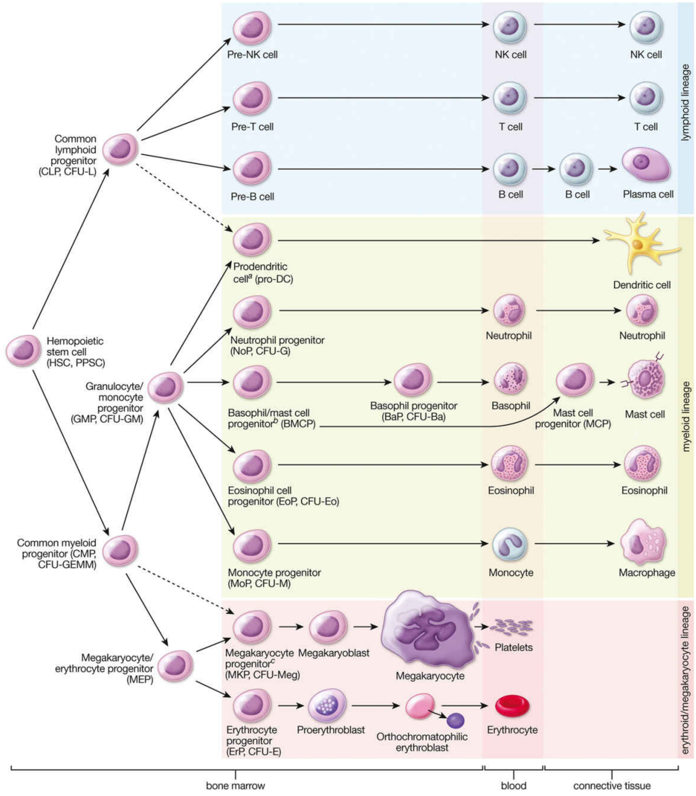
FIGURE 10.19 - Hemopoiesis. From: Histology A Text and Atlas.
2. Regulation by Erythropoietin (EPO)
The primary regulator of erythropoiesis is erythropoietin (EPO), a 30.4-kDa glycoprotein hormone produced mainly by the kidneys. The control mechanism is a classic negative feedback loop:
- Decreased tissue oxygenation (hypoxia) is sensed by renal peritubular cells
- The kidney releases EPO into the bloodstream
- EPO binds to EPO receptors (EPO-Rs) on erythroid progenitors in the bone marrow
- EPO stimulates proliferation and differentiation of these progenitors
- Rising red cell mass restores oxygen delivery, switching off the EPO signal
EPO production begins within 24-48 hours of hypoxia and declines over ~3 weeks as hematocrit rises. For EPO to work, adequate iron, vitamin B12, and folic acid are also required as cofactors.
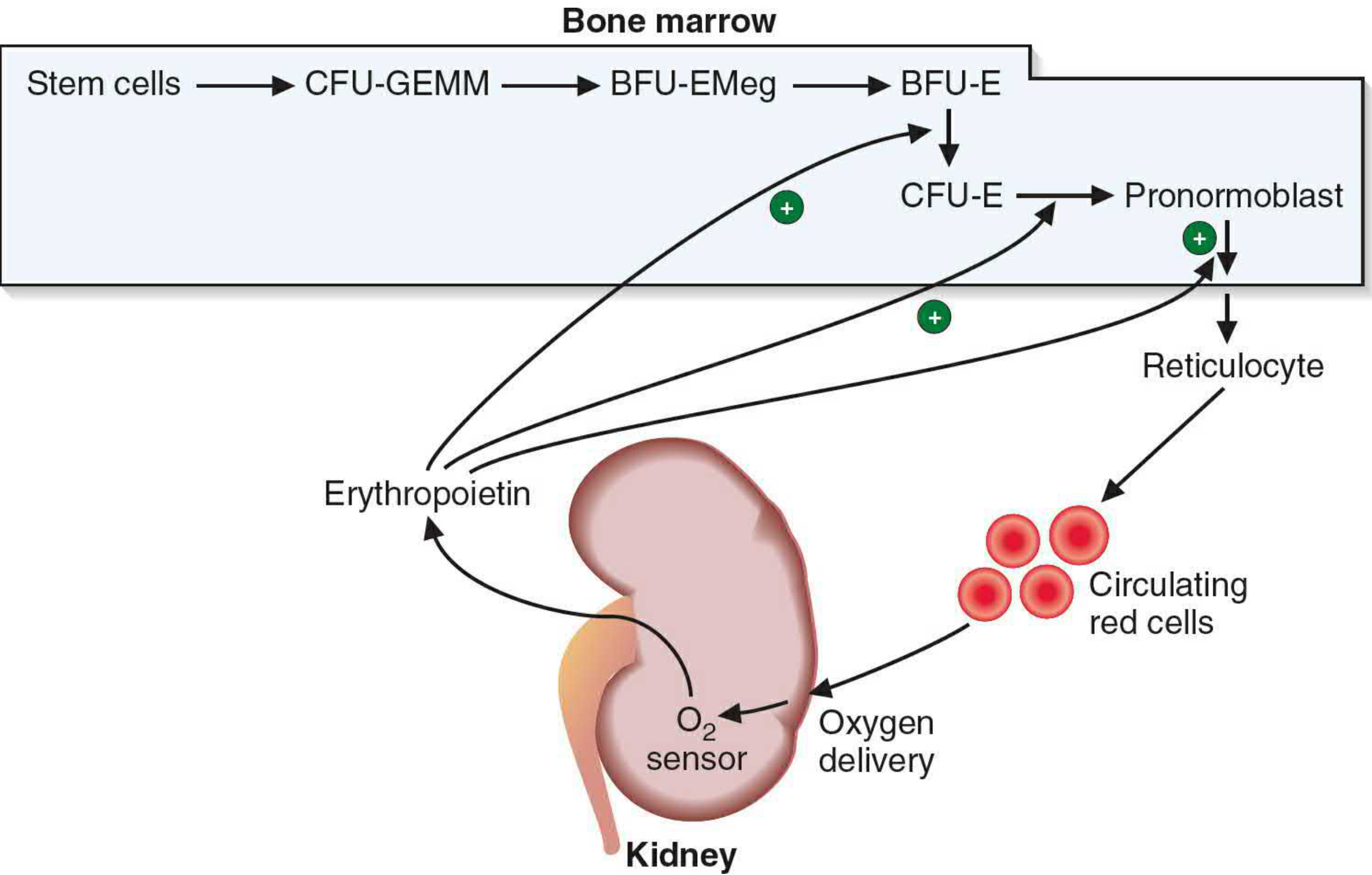
FIGURE 42.13 - Erythropoietin stimulation of erythrocyte maturation. From: Basic Medical Biochemistry, 6e.
3. Progenitor Cell Hierarchy
The committed erythroid pathway proceeds through the following progenitors (which cannot be distinguished morphologically on a standard smear):
| Stage | Name | Notes |
|---|---|---|
| Pluripotent | HSC | Self-renewing; gives rise to all blood lineages |
| Mixed myeloid | CFU-GEMM | Colony-forming unit: Granulocyte, Erythroid, Monocyte, Megakaryocyte |
| Early erythroid | BFU-E (Burst-Forming Unit - Erythroid) | Earliest EPO-responsive cell |
| Late erythroid | CFU-E (Colony-Forming Unit - Erythroid) | Highly EPO-sensitive; gives rise to pronormoblast |
- Basic Medical Biochemistry, 6e, p. 1536
4. Morphologically Recognizable Maturation Stages
From the CFU-E onward, cells can be identified under the microscope. As erythrocytes mature, four consistent changes occur:
- Cell size decreases (from 12-20 µm to 7.8 µm)
- Nucleus shrinks and condenses (nucleus-to-cytoplasm ratio increases from 1:8 to 1:2)
- Nucleoli disappear (reflecting cessation of rRNA synthesis)
- Cytoplasm shifts from basophilic (blue) to eosinophilic (pink) as RNA is replaced by hemoglobin
Stage-by-stage:
1. Pronormoblast / Proerythroblast
- Largest cell in the series (12-20 µm diameter)
- Large nucleus with open chromatin and 1-2 prominent nucleoli
- Deeply basophilic cytoplasm (high RNA content)
- No hemoglobin yet
- Divides to form basophilic erythroblasts
2. Basophilic Erythroblast (Basophilic Normoblast)
- Smaller than proerythroblast
- Nucleus smaller; nucleoli typically absent
- Deeply basophilic cytoplasm - increasing ribosomes for hemoglobin synthesis
- Mitotic divisions occur at this stage
3. Polychromatophilic Erythroblast (Polychromatophilic Normoblast)
- Intermediate size
- Nucleus shows coarser, clumped heterochromatin
- Cytoplasm is blue-gray, with increasing eosinophilia as hemoglobin accumulates
- RNA content is declining; hemoglobin content rising
- Mitoses occur here too
4. Orthochromatic Erythroblast (Orthochromatic Normoblast)
- Smallest nucleated red cell precursor
- Nucleus is very small, deeply condensed (pyknotic) - no visible chromatin pattern
- Cytoplasm is predominantly eosinophilic (full of hemoglobin)
- The nucleus is then extruded, forming the reticulocyte
5. Reticulocyte (Polychromatophilic Erythrocyte)
- Anucleate (nucleus extruded)
- Still contains residual ribosomes, mRNA, and mitochondria - capable of synthesizing hemoglobin
- Appears slightly blue-gray on Wright-Giemsa stain due to residual RNA
- Spends 1-2 days in bone marrow stroma, then is released into blood
- Matures further in the spleen, where organelles are removed
6. Mature Erythrocyte
-
Anucleate, biconcave disc, ~7.8 µm
-
Packed with hemoglobin
-
No ribosomes, mitochondria, or organelles
-
Life span: ~120 days
-
Histology: A Text and Atlas, pp. 745-746
-
Henry's Clinical Diagnosis and Management by Laboratory Methods, p. 659
5. Erythroblastic Islands
In the marrow, developing erythroid cells cluster around central macrophages in structures called erythroblastic islands. These macrophages:
-
Supply iron directly to developing erythroblasts via transferrin
-
Phagocytose the extruded nuclei
-
Provide growth support signals
-
Henry's Clinical Diagnosis and Management, p. 659
6. Division Kinetics
Each pronormoblast undergoes 3-4 mitotic divisions over approximately 3 days, potentially generating up to 16 reticulocytes from a single pronormoblast. Mitoses occur in the proerythroblast, basophilic, and polychromatophilic erythroblast stages - but not in the orthochromatic stage onward. It takes roughly 1 week from a newly formed basophilic erythroblast to a circulating reticulocyte.
7. Iron and Hemoglobin Synthesis
- Iron is transferred from plasma transferrin to erythroid cells during maturation
- RNA content is highest in pronormoblasts and basophilic normoblasts, declining progressively
- Hemoglobin content rises throughout maturation, inversely to RNA
- In reticulocytes, RNA synthesis has ceased but residual RNA supports continued hemoglobin synthesis for another 1-2 days in circulation
8. Red Cell Destruction (Senescence)
After ~120 days, erythrocytes become senescent. Macrophages in the spleen, bone marrow, and liver (the reticuloendothelial system) phagocytose and degrade them:
- Globin → hydrolyzed to amino acids (recycled)
- Iron → stored as ferritin/hemosiderin in the spleen; recycled for new hemoglobin synthesis
- Heme → converted to bilirubin → bound to albumin → transported to liver → conjugated and excreted in bile
Summary Table
| Stage | Nucleus | Cytoplasm | Hemoglobin | Divides? |
|---|---|---|---|---|
| Proerythroblast | Large, nucleoli present | Deeply basophilic | Absent | Yes |
| Basophilic erythroblast | Smaller, no nucleoli | Deeply basophilic | Traces | Yes |
| Polychromatophilic erythroblast | Coarse chromatin | Blue-gray (mixed) | Increasing | Yes |
| Orthochromatic erythroblast | Pyknotic, then extruded | Eosinophilic (pink) | Nearly full | No |
| Reticulocyte | Absent | Slightly blue-gray | Yes (still synthesizing) | No |
| Mature erythrocyte | Absent | Pink/eosinophilic | Full | No |
Wbc
Asking for Preferences
What aspect of WBCs would you like covered?
Searching the medical library for leukopoiesis content now.
Finding Sources
Finding Sources
Finding Sources
Finding Sources
Finding Sources
Reading File
Excellent content. Let me now read more on kinetics/maturation stages and fetch the key WBC diagram:
Reading File
Loading Image
Now let me read the individual WBC type sections and the lymphopoiesis/monocyte sections:
Reading File
Finding Sources
Let me now read the individual WBC type morphology from the histology text and the leukocyte table from Surgery:
Reading File
Reading File
Now let me get the individual WBC morphology sections - neutrophil, eosinophil, basophil, monocyte, lymphocyte:
Reading File
Let me get the neutrophil/eosinophil/basophil/monocyte/lymphocyte morphology sections:
Finding Sources
Reading File
Excellent - this read gave me the neutrophil, eosinophil, basophil, monocyte, and lymphocyte sections all at once. I now have enough comprehensive content to write a full answer.
White Blood Cells (Leukocytes) - Overview and Leukopoiesis
White blood cells (WBCs), or leukocytes, are nucleated blood cells that together form the cellular arm of the immune defense system. Unlike erythrocytes, they leave the bloodstream to perform most of their functions in tissues.
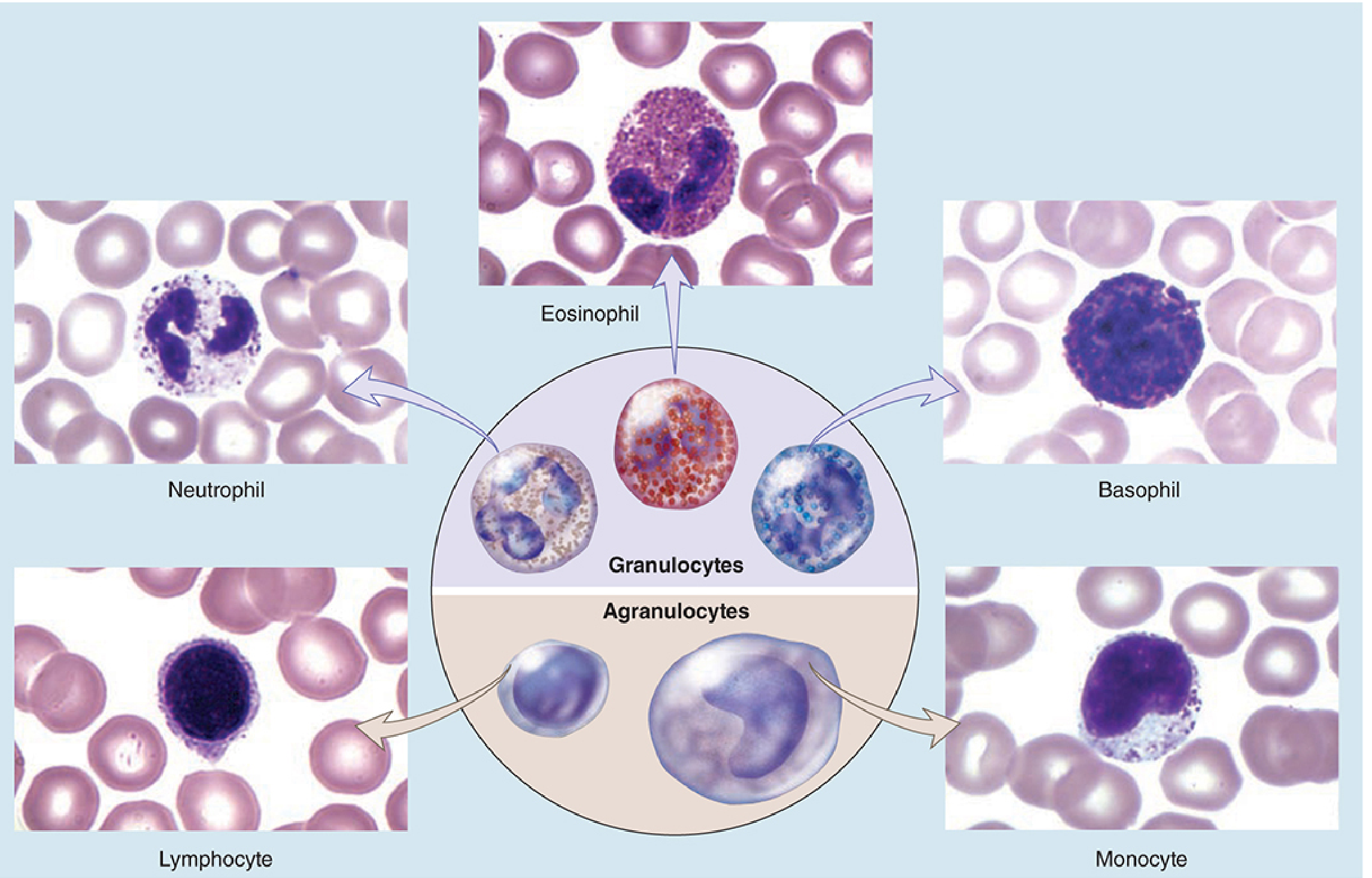
From: Junqueira's Basic Histology, 17e
Classification of WBCs
WBCs are divided into two major groups based on whether they contain visible granules in their cytoplasm:
| Group | Cell Types |
|---|---|
| Granulocytes | Neutrophil, Eosinophil, Basophil |
| Agranulocytes | Monocyte, Lymphocyte |
Normal Reference Values
| Cell | Size (µm) | Differential (%) | Life Span | Key Activators |
|---|---|---|---|---|
| Neutrophil | 13 | 40-75% | 6 h to 7 days | G-CSF, IL-8 |
| Monocyte | 16-20 | 2-6% | 1 day to years | M-CSF, GM-CSF, IFN-γ, TNF-α |
| Lymphocyte | 9-16 | 20-45% | Months to years | IL-2, IL-12 (Th1), IL-4 (Th2) |
| Eosinophil | 12-16 | 1-6% | 8-12 days | G-CSF, IL-5 |
| Basophil | 15 | <1% | ~1 year | G-CSF, IL-3 |
- Mulholland and Greenfield's Surgery, 7e, Table 7.1
Individual WBC Types
1. Neutrophils
The most numerous WBC (40-75% of differential). They are the first responders to bacterial infection.
Morphology:
- 10-12 µm in diameter
- Multi-lobed nucleus (2-4 lobes joined by thin strands) - hence called polymorphonuclear (PMN) or polymorphs
- Cytoplasm stains neutral (neither strongly basophilic nor eosinophilic)
- In women, a drumstick-shaped Barr body (inactive X chromosome) is visible as a nuclear appendage
- Two granule types:
- Azurophilic (primary) granules - contain myeloperoxidase, defensins, lysozyme
- Specific (secondary) granules - contain collagenase, lactoferrin, NADPH oxidase components
- Tertiary granules - contain gelatinase; facilitate extravasation
Function: Phagocytosis and killing of bacteria via oxidative burst (reactive oxygen species), degranulation, and NET (neutrophil extracellular trap) formation.
Recruitment: In infection, neutrophils roll on endothelium via selectin-sialyl-Lewisx interactions, then firmly adhere via integrin-ICAM-1 binding, then migrate through the endothelium by diapedesis guided by chemokines (e.g., IL-8).
2. Eosinophils
Account for 1-6% of circulating WBCs.
Morphology:
- ~12-16 µm
- Bilobed nucleus (two lobes connected by a thin strand)
- Cytoplasm packed with large, refractile eosinophilic (red-orange) granules on H&E stain
- Granules contain a crystalloid body (Charcot-Leyden crystals) within a less electron-dense matrix
- Two granule types: large specific granules and azurophilic granules
Function: Defense against parasites (too large for phagocytosis); release major basic protein (MBP), eosinophil cationic protein (ECP), and peroxidase to damage parasites. Also involved in allergic reactions and asthma (recruited by IL-5, eotaxin/CCL11).
3. Basophils
The least numerous WBC (<1% of differential).
Morphology:
- ~15 µm
- S-shaped or bilobed nucleus often obscured by densely staining granules
- Large, deeply basophilic (purple-blue) granules containing heparin, histamine, leukotrienes, and serotonin
- Morphologically similar to mast cells (tissue counterpart) but circulate in blood
Function: Mediate immediate hypersensitivity (Type I allergic) reactions. IgE binds to high-affinity Fc receptors on their surface; re-exposure to allergen cross-links IgE → degranulation → histamine and leukotrienes released → vasodilation, bronchoconstriction.
4. Monocytes
Account for 2-6% of circulating WBCs. They are the largest circulating WBC.
Morphology:
- 16-20 µm
- Kidney bean- or horseshoe-shaped nucleus (indented, not lobulated)
- Abundant grayish, pale cytoplasm with fine azurophilic granules
- Cytoplasm may show vacuoles
Function: Monocytes are precursors to tissue macrophages and dendritic cells. They migrate into tissues and differentiate - becoming Kupffer cells in liver, microglia in the CNS, alveolar macrophages in lung, and osteoclasts in bone. Functions include phagocytosis, antigen presentation, and cytokine secretion (IL-1, IL-6, TNF-α).
5. Lymphocytes
Account for 20-45% of circulating WBCs.
Morphology:
- 9-16 µm (small to large varieties)
- Large, round, dark nucleus occupying nearly all of the cell
- Thin rim of pale blue cytoplasm
- Agranular (or very sparse granules in NK cells)
Subtypes (not distinguishable by light microscopy - require immunophenotyping):
- T lymphocytes (T cells): Mature in the thymus; mediate cellular immunity (cytotoxic CD8+, helper CD4+, regulatory T cells)
- B lymphocytes (B cells): Mature in bone marrow; mediate humoral immunity; differentiate into plasma cells that produce antibodies
- NK (Natural Killer) cells: Innate cytotoxic cells; kill virus-infected and tumor cells without prior sensitization
Leukopoiesis - Development in Bone Marrow
All WBCs arise from the hemopoietic stem cell (HSC) via two major pathways:
Myeloid Pathway (Granulopoiesis + Monocytopoiesis)
HSC → CMP (Common Myeloid Progenitor) → GMP (Granulocyte/Monocyte Progenitor)
Under the influence of GM-CSF, G-CSF, and IL-3:
For neutrophils, eosinophils, basophils - the NoP (neutrophil progenitor) undergoes 6 morphologically identifiable stages:
| Stage | Key Features | Divides? |
|---|---|---|
| Myeloblast | 14-20 µm; large euchromatic nucleus with 3-5 nucleoli; agranular basophilic cytoplasm | Yes |
| Promyelocyte | Large; azurophilic (primary) granules appear - only stage that produces them | Yes |
| Myelocyte | Specific (secondary) granules appear - lineage now identifiable (neutrophil vs. eosinophil vs. basophil); nucleus begins to indent | Yes |
| Metamyelocyte | Kidney bean-shaped nucleus; specific granules outnumber azurophilic ~2:1; last stage before band | No |
| Band (Stab) cell | Horseshoe-shaped nucleus of uniform width | No |
| Mature granulocyte | Multi-lobed (neutrophil: 2-4 lobes); fully functional | No |
Key rule: Azurophilic granules are ONLY made in promyelocytes. Subsequent divisions dilute them. Specific granules appear at the myelocyte stage and allow identification of granulocyte lineage.
For monocytes: GMP → Monocyte progenitor (MoP) → Monocyte → Tissue macrophage/Dendritic cell (under M-CSF, GM-CSF)
Lymphoid Pathway (Lymphopoiesis)
HSC → CLP (Common Lymphoid Progenitor)
- T cells: CLP → Pre-T cell → migrate to thymus → positive and negative selection → mature T cell re-enters circulation
- B cells: CLP → Pre-B cell → mature in bone marrow (bursa-equivalent organs: bone marrow, gut-associated lymphoid tissue, spleen) under Pax5 transcription factor activation
- NK cells: Differentiate under IL-2 and IL-15 influence; produced mainly in bone marrow
Kinetics of Granulopoiesis
- Total time from myeloblast to mature granulocyte: ~2 weeks
- Mitotic (proliferative) phase: ~1 week (myeloblast → late myelocyte)
- Post-mitotic (maturation) phase: ~1 week (metamyelocyte → mature granulocyte)
- The bone marrow produces >10¹¹ neutrophils per day
- A large storage pool of mature neutrophils is retained in bone marrow for 4-6 days
- In infection, this pool is rapidly mobilized (within <1 hour) via loss of CXCL12/CXCR4 retention signaling
Summary Diagram - Complete Hemopoiesis
The hemopoiesis diagram from earlier (see erythropoiesis response) shows the full lineage tree, with the lymphoid lineage (blue) at top, myeloid lineage (yellow) in the middle, and erythroid/megakaryocyte lineage (pink) at bottom - all converging on a single hemopoietic stem cell.
- Histology: A Text and Atlas with Correlated Cell and Molecular Biology, pp. 748-758
- Mulholland and Greenfield's Surgery, 7e, p. 285
This is a shared conversation. Sign in to Orris to start your own chat.