
Thyroid gland and it's arteries
Reading File
The thyroid gland receives blood from three main arteries: the superior thyroid artery (from the external carotid), the inferior thyroid artery (from the thyrocervical trunk of the subclavian), and occasionally a thyroid ima artery (from the aorta or brachiocephalic trunk). Now let me pull the full anatomical detail from the medical library.
Searching sources...
Finding Sources
Finding Sources
Good results. Now let me read the most detailed anatomical sections in parallel.
Reading File
Reading File
Reading File
Excellent content. Now let me read the remainder of Gray's Anatomy arterial supply section and fetch the key diagrams.
Reading File
Loading Image
Loading Image
I now have comprehensive content and clear diagrams. Let me fetch the remaining two anatomical diagrams showing the arterial supply in detail.
Loading Image
Loading Image
I now have all the content and diagrams needed for a complete answer.
Thyroid Gland and Its Arteries
Overview of the Thyroid Gland
The thyroid gland sits anterior in the neck, below and lateral to the thyroid cartilage. It has two lateral lobes connected by an isthmus, which crosses the anterior surface of the 2nd and 3rd tracheal cartilages. The lobes cover the anterolateral surfaces of the trachea, cricoid cartilage, and lower thyroid cartilage. The gland lies deep to the sternohyoid, sternothyroid, and omohyoid muscles, enclosed within the pretracheal fascia - making it part of the visceral compartment of the neck.
It is one of the most highly vascular organs in the body. Its blood supply comes from two pairs of named arteries and an inconstant third unpaired vessel.
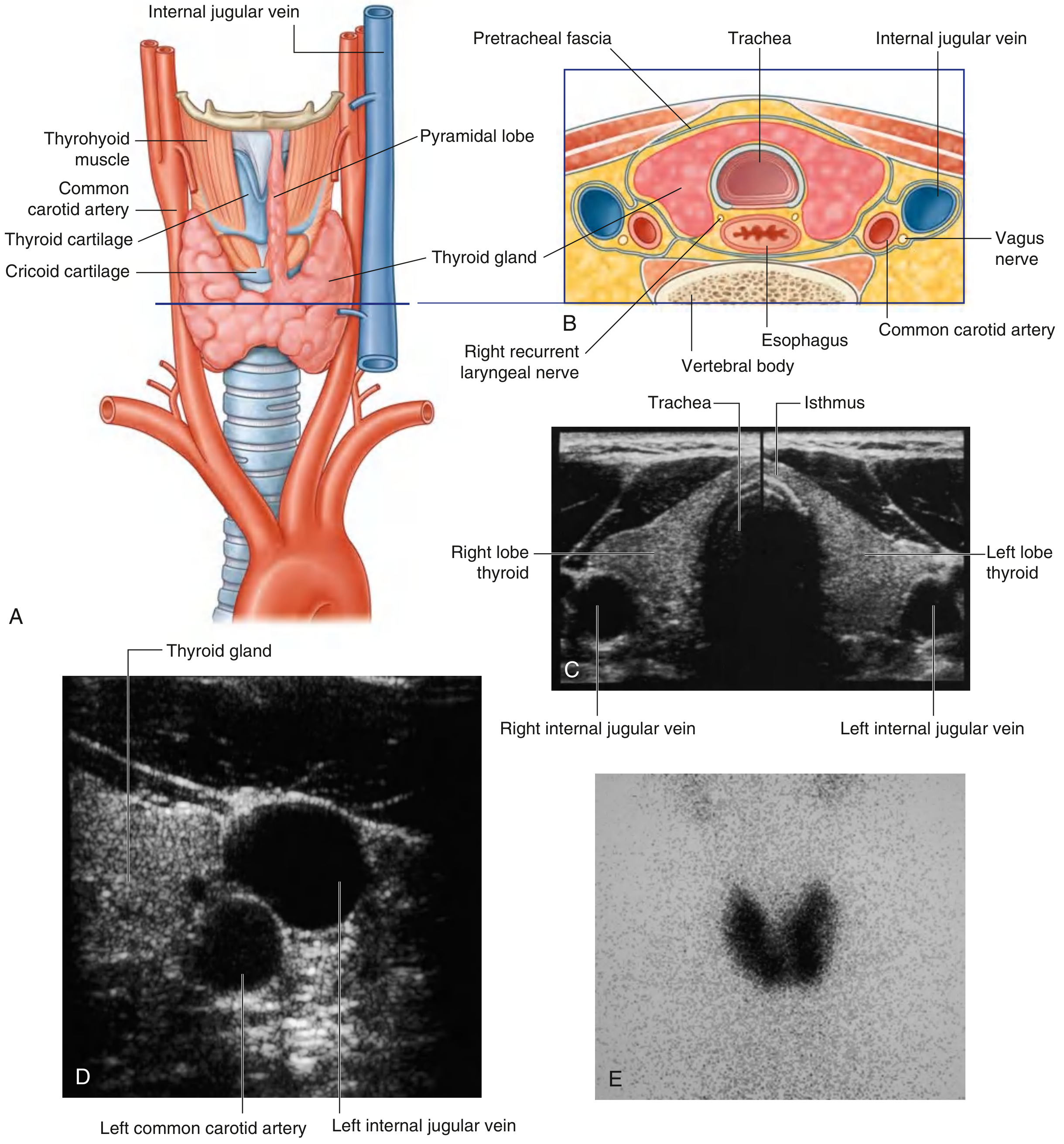
Fig. 8.181 - Thyroid Gland in the Anterior Triangle of Neck (Gray's Anatomy for Students)
Arterial Supply
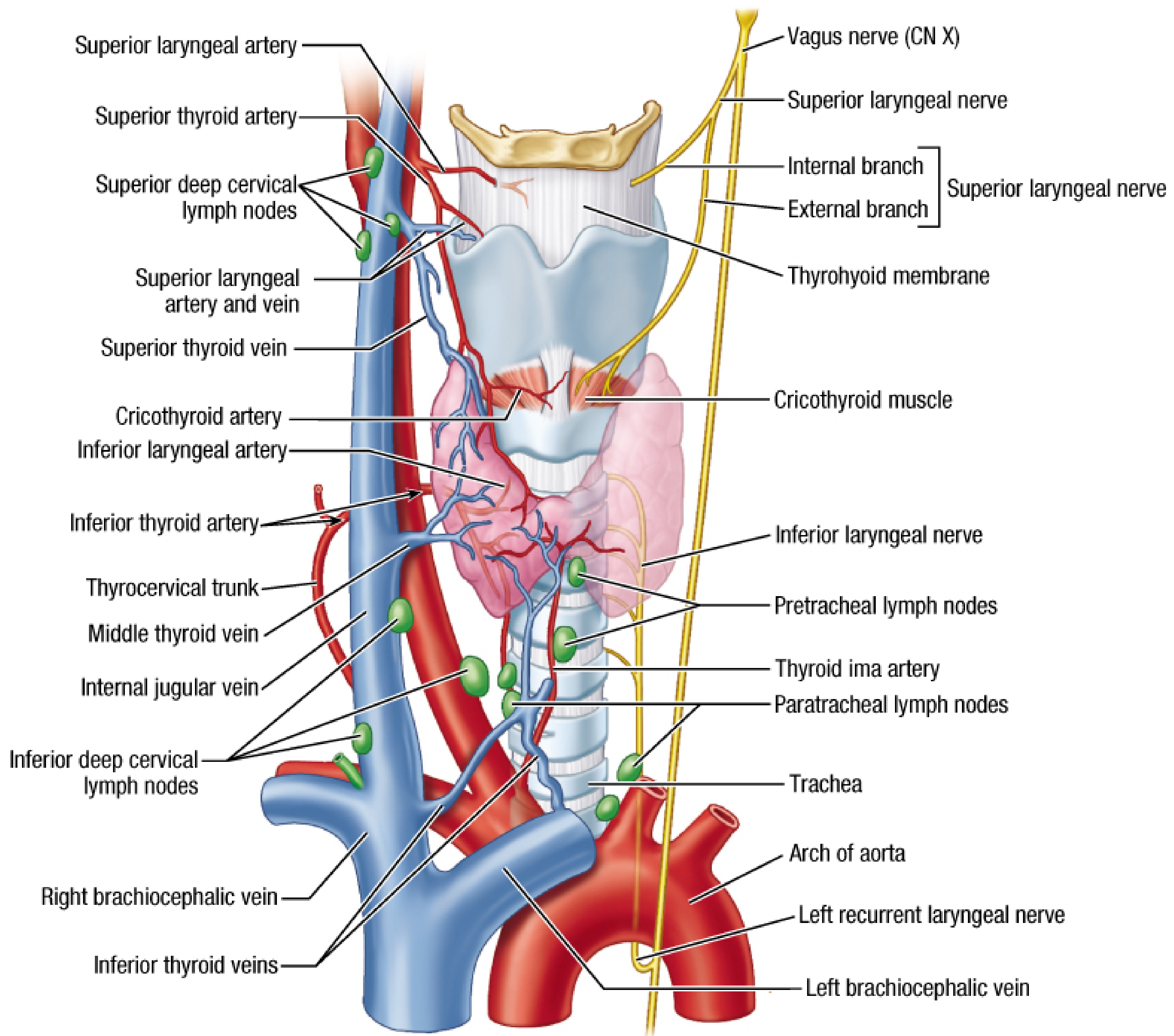
Neurovascular structures of the thyroid - showing superior thyroid artery, inferior thyroid artery, thyroid ima artery, thyrocervical trunk, and related nerves (Fischer's Mastery of Surgery)
1. Superior Thyroid Artery
- Origin: First branch of the external carotid artery
- Course: Descends along the lateral margin of the thyrohyoid muscle to reach the superior pole of the lateral lobe
- Branches at the gland:
- Anterior glandular branch - runs along the superior border of the thyroid gland and anastomoses across the isthmus with its twin from the opposite side
- Posterior glandular branch - passes to the posterior surface of the gland and may anastomose with the inferior thyroid artery
- Other branches: Gives off a superior laryngeal branch (which pierces the thyrohyoid membrane alongside the internal branch of the superior laryngeal nerve), and a cricothyroid branch
2. Inferior Thyroid Artery
- Origin: Branch of the thyrocervical trunk, which itself arises from the first part of the subclavian artery
- Course: Ascends along the medial edge of the anterior scalene (scalenus anterior) muscle, then arches medially and passes posteriorly to the carotid sheath to reach the posterior aspect of the thyroid gland
- Key surgical relationship: As it passes medially toward the gland, it crosses the recurrent laryngeal nerve (RLN) - the RLN may pass anterior, posterior, or between branches of the inferior thyroid artery. This is one of the most important surgical considerations during thyroidectomy
- Branches at the gland:
- Inferior branch - supplies the lower part of the thyroid and anastomoses with the posterior branch of the superior thyroid artery
- Ascending branch - supplies the parathyroid glands
- Parathyroid supply: The inferior thyroid artery is the primary blood supply to both superior and inferior parathyroid glands (the superior parathyroid may also receive branches from the superior thyroid artery, but the inferior thyroid is dominant for both)
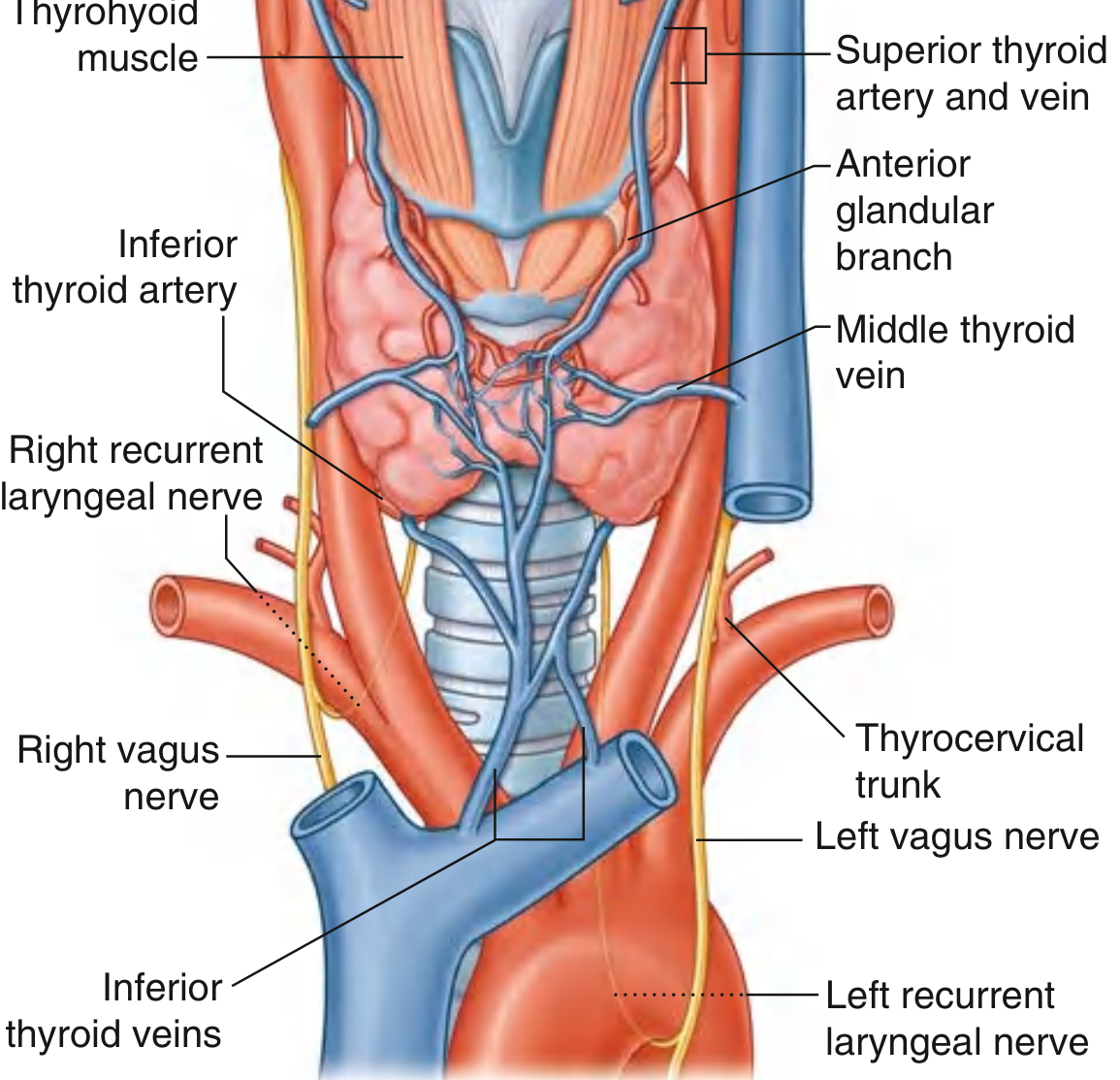
Fig. 8.182 - Vasculature of the Thyroid: Anterior View (Gray's Anatomy for Students)
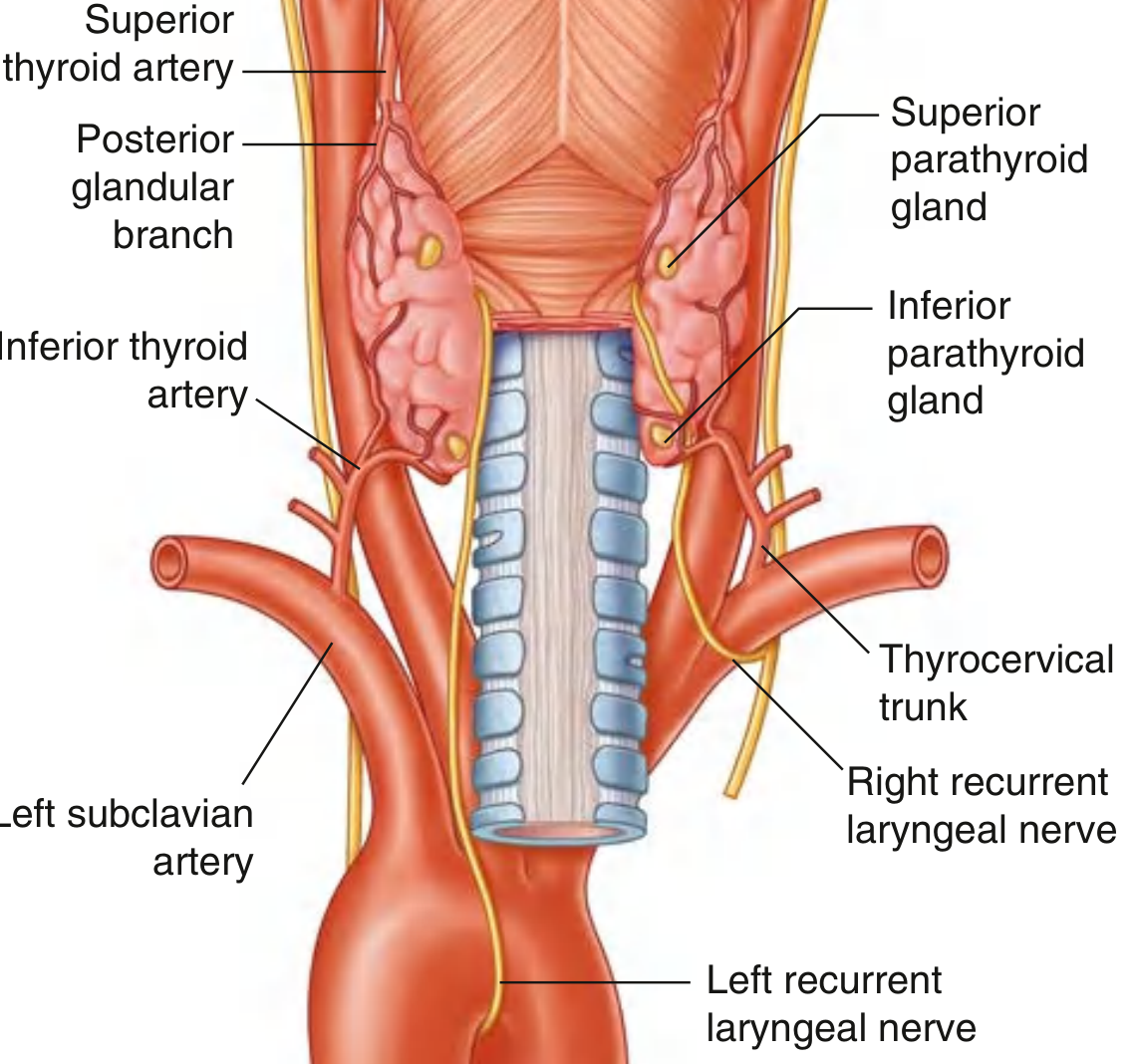
Fig. 8.183 - Superior and Inferior Thyroid Arteries, Parathyroid Glands, and Recurrent Laryngeal Nerves - Posterior View (Gray's Anatomy for Students)
3. Thyroid Ima Artery (Thyroidea Ima)
- An unpaired, inconstant vessel - present in approximately 1.5% to 12% of individuals (varies by source; about 2-10% cited across texts)
- Origin (variable): Can arise from:
- Brachiocephalic (innominate) artery - most common
- Arch of the aorta
- Right common carotid artery
- Internal thoracic artery
- Subclavian artery
- Course: Follows a midline path ascending on the anterior surface of the trachea to enter the thyroid isthmus or inferior poles of the lobes
- Surgical significance: Must be identified before midline procedures such as tracheotomy or thyroidectomy to avoid unexpected hemorrhage
Summary Table
| Artery | Origin | Territory |
|---|---|---|
| Superior thyroid artery | External carotid artery | Superior pole; anterior and posterior glandular branches |
| Inferior thyroid artery | Thyrocervical trunk (subclavian a.) | Lower pole; parathyroid glands (primary supply) |
| Thyroid ima artery | Brachiocephalic trunk / aortic arch | Isthmus/inferior poles (when present) |
Venous Drainage (for completeness)
Three veins drain the thyroid:
- Superior thyroid vein - travels with the superior thyroid artery; drains into the internal jugular vein
- Middle thyroid vein - travels without an arterial companion; drains into the internal jugular vein
- Inferior thyroid veins - unpaired vessels draining into the right and left brachiocephalic veins respectively
Clinical / Surgical Relevance
-
RLN and inferior thyroid artery relationship - The recurrent laryngeal nerve crosses the inferior thyroid artery in a variable pattern (nerve can be anterior, posterior, or between arterial branches). Surgeons must identify the RLN before ligating the inferior thyroid artery during thyroidectomy to avoid voice-damaging nerve injury.
-
Parathyroid preservation - Because the inferior thyroid artery is the dominant blood supply to all four parathyroid glands, inadvertent ligation of this artery close to its origin (rather than at the capsule) can devascularize the parathyroids and cause permanent hypoparathyroidism.
-
Thyroid ima artery - Absence or presence must be noted during tracheotomy. An unrecognized ima artery crossing the trachea can cause severe bleeding.
-
Highly vascular organ - The thyroid has a rich capsular arterial network connecting all named vessels, which is why thyroid surgery can be accompanied by significant blood loss if vessels are not carefully controlled.
Sources: Gray's Anatomy for Students (9780323934237), Fischer's Mastery of Surgery 8th ed. (9781975176433), KJ Lee's Essential Otolaryngology (9781260122237), Sabiston Textbook of Surgery (9780443124341)
This is a shared conversation. Sign in to Orris to start your own chat.