
Gluconeogenesis 10 marks from rm prasad biochem
Reading File
Now let me search the library for the RM Prasad biochemistry book and gluconeogenesis content.
Finding Sources
Finding Sources
The library doesn't have RM Prasad specifically - but it has Lippincott Biochemistry and Basic Medical Biochemistry which cover gluconeogenesis comprehensively. Let me read the gluconeogenesis sections from both biochemistry books in parallel.
Reading File
Reading File
Excellent content. Let me read more from both books - the unique steps and regulation sections.
Reading File
Reading File
I have comprehensive content. Let me also fetch the key gluconeogenesis pathway diagram and read the regulation section.
Reading File
Loading Image
I now have all the content needed for a comprehensive 10-mark answer on Gluconeogenesis. Let me compile the full answer.
Gluconeogenesis
Definition
Gluconeogenesis is the metabolic process by which glucose is synthesized de novo from non-carbohydrate precursors. It occurs primarily in the liver (responsible for ~90% after an overnight fast) and to a lesser extent in the kidney cortex (increases to ~40% during prolonged starvation of 48 hours or more). The small intestine can also contribute.
- Lippincott Illustrated Reviews: Biochemistry, 8th ed., p. 353
Why Gluconeogenesis is Needed
Glucose-dependent tissues - the brain, RBCs, kidney medulla, lens and cornea of the eye, testes, and exercising skeletal muscle - require a continuous glucose supply. Hepatic glycogen stores can meet these needs for less than 24 hours. Beyond that, gluconeogenesis from non-carbohydrate sources sustains blood glucose. Gluconeogenesis is NOT a simple reversal of glycolysis; it uses distinct enzymes to bypass the three irreversible steps of glycolysis.
Precursors (Substrates) of Gluconeogenesis
| Precursor | Source | Entry Point |
|---|---|---|
| Lactate | RBCs, exercising skeletal muscle (anaerobic glycolysis) | Converted to pyruvate (LDH), then to OAA |
| Glycerol | Hydrolysis of TAGs in adipose tissue | Glycerol → glycerol-3-P → DHAP |
| Glucogenic amino acids (all except Leu & Lys) | Muscle protein breakdown | Enter as pyruvate, OAA, or TCA intermediates |
| Alanine | Muscle (glucose-alanine cycle) | Transaminated to pyruvate |
| Propionate | Odd-chain FA oxidation | Enters as succinyl-CoA → OAA |
Note: Leucine and lysine are purely ketogenic - they CANNOT contribute to gluconeogenesis.
Key Pathway - The Three Bypass Reactions
Gluconeogenesis uses 7 reversible glycolytic enzymes in reverse, but bypasses the 3 irreversible steps of glycolysis using dedicated gluconeogenic enzymes:
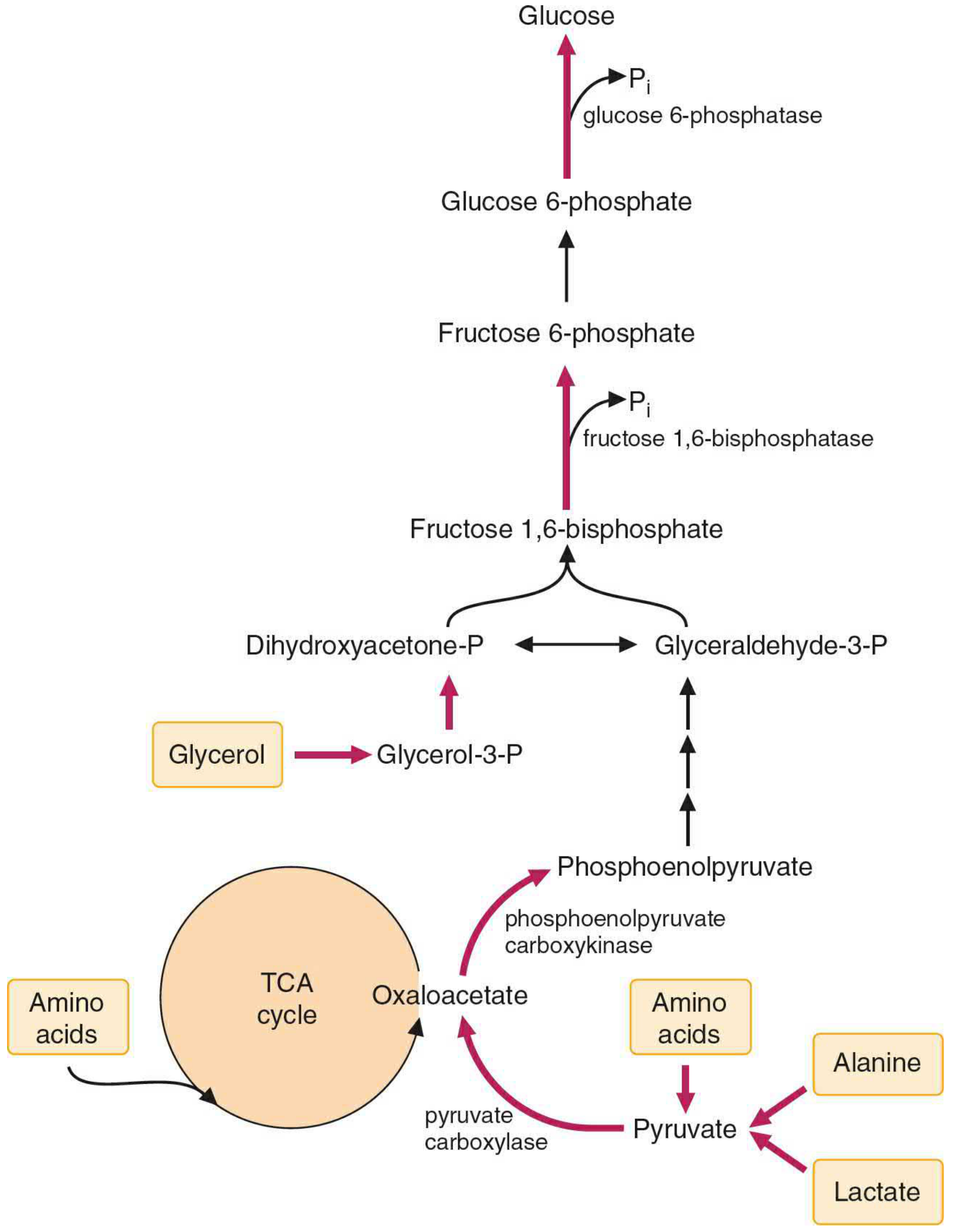
Bypass 1: Pyruvate → PEP (bypasses Pyruvate Kinase)
This is the most complex bypass and occurs in two steps via the mitochondria:
Step 1a - Pyruvate Carboxylase (PC):
Pyruvate + CO₂ + ATP → Oxaloacetate (OAA) + ADP + Pi
- Location: Mitochondrial matrix
- Cofactor: Biotin (CO₂ carrier)
- Allosteric activator: Acetyl CoA (key regulatory signal)
- OAA cannot cross the inner mitochondrial membrane directly
Step 1b - PEPCK (Phosphoenolpyruvate Carboxykinase):
OAA + GTP → PEP + CO₂ + GDP
- OAA is either transaminated to aspartate (exits as aspartate, re-converted in cytosol) or reduced to malate to exit mitochondria
- PEPCK is present in both mitochondria and cytosol
- Induced by glucagon and cortisol; suppressed by insulin
Bypass 2: Fructose-1,6-bisphosphate → Fructose-6-phosphate (bypasses PFK-1)
Enzyme: Fructose-1,6-bisphosphatase (FBPase-1)
Fructose-1,6-bisphosphate + H₂O → Fructose-6-phosphate + Pi
- Location: Cytosol
- Inhibited by AMP and fructose-2,6-bisphosphate
- Activated when fructose-2,6-bisphosphate levels are low (fasting/glucagon state)
Bypass 3: Glucose-6-phosphate → Glucose (bypasses Hexokinase/Glucokinase)
Enzyme: Glucose-6-phosphatase
Glucose-6-phosphate + H₂O → Glucose + Pi
- Location: Endoplasmic reticulum (requires glucose-6-phosphate translocase to bring substrate into ER lumen)
- Present only in gluconeogenic tissues: liver, kidney cortex, intestine (NOT muscle or brain - explains why muscle cannot release free glucose)
- Deficiency causes Glycogen Storage Disease Type Ia (Von Gierke's disease) - severe fasting hypoglycemia
Energy Cost of Gluconeogenesis
Synthesis of 1 glucose from 2 pyruvate requires:
- 4 ATP + 2 GTP + 2 NADH
- Total: 6 high-energy phosphate bonds consumed
This is energetically expensive (glycolysis yields only 2 ATP per glucose). The energy is supplied primarily by beta-oxidation of fatty acids.
The Cori Cycle (Lactic Acid Cycle)
Lactate produced by RBCs and exercising muscle is transported to the liver, reconverted to glucose by gluconeogenesis, and returned to the periphery. This shuttles carbon (not energy) from muscle to liver - the liver bears the energy cost.
Regulation of Gluconeogenesis
1. Hormonal Regulation
| Hormone | Effect | Mechanism |
|---|---|---|
| Glucagon (fasting) | Stimulates | ↓ Fructose-2,6-bisphosphate → activates FBPase-1; phosphorylates (inactivates) PK; induces PEPCK gene via CREB |
| Cortisol | Stimulates | Induces PEPCK gene expression |
| Insulin (fed state) | Inhibits | ↑ Fructose-2,6-bisphosphate → inhibits FBPase-1; suppresses PEPCK transcription |
| Epinephrine | Stimulates | Similar to glucagon (cAMP pathway) |
2. Allosteric Regulation
| Effector | Target | Effect |
|---|---|---|
| Acetyl CoA | Pyruvate Carboxylase | Activates (signals excess fatty acid oxidation → redirect pyruvate) |
| Acetyl CoA | Pyruvate Dehydrogenase | Inhibits (prevents loss of pyruvate into TCA) |
| AMP | FBPase-1 | Inhibits gluconeogenesis |
| Fructose-2,6-bisphosphate | FBPase-1 | Inhibits |
| High ATP/ADP ratio | Favors gluconeogenesis over glycolysis |
3. Substrate Availability
Increased availability of gluconeogenic precursors (amino acids, lactate, glycerol) drives the pathway forward. Decreased insulin during fasting promotes muscle protein breakdown, releasing amino acids as substrates.
Clinical Correlations
- Metformin (anti-diabetic drug): inhibits gluconeogenesis by inhibiting mitochondrial complex I, reducing NADH availability and lowering hepatic glucose output
- Von Gierke's Disease (GSD Ia): glucose-6-phosphatase deficiency - fasting hypoglycemia, hepatomegaly, lactic acidosis
- Biotin deficiency: impairs pyruvate carboxylase - hypoglycemia
- Alcohol intoxication: excess NADH (from ethanol metabolism) shifts OAA → malate and pyruvate → lactate, depleting gluconeogenic substrates - causes fasting hypoglycemia
- Diabetic Ketoacidosis: excess acetyl CoA from FA oxidation activates PC, driving gluconeogenesis and worsening hyperglycemia
Summary Table: Glycolysis vs. Gluconeogenesis
| Feature | Glycolysis | Gluconeogenesis |
|---|---|---|
| Net direction | Glucose → Pyruvate | Pyruvate → Glucose |
| Key tissue | All tissues | Liver, kidney cortex |
| Energy yield/cost | +2 ATP (net) | -6 ATP equivalents |
| Unique enzymes | HK/GK, PFK-1, PK | PC, PEPCK, FBPase-1, G6Pase |
| Stimulated by | Insulin, AMP, F-2,6-BP | Glucagon, cortisol, acetyl CoA |
| Inhibited by | -- | AMP, F-2,6-BP, insulin |
Sources: Lippincott Illustrated Reviews: Biochemistry, 8th ed., pp. 353-365; Basic Medical Biochemistry: A Clinical Approach, 6th ed., pp. 1007-1015
This is a shared conversation. Sign in to Orris to start your own chat.