
Lacunar stroke
Reading File
Reading File
Finding Sources
Searching PubMed
"Lacunar Stroke"[MeSH Terms]
Searching the Web
AHA lacunar stroke small vessel disease patient information diagnosis treatment
Searching Images
MRI lacunar infarct small deep subcortical stroke
Finding Sources
Reading Web Page
https://pmc.ncbi.nlm.nih.gov/articles/PMC10916806
Reading File
Reading File
Reading File
Lacunar stroke
A lacunar stroke is a small ischemic stroke caused by blockage of a deep penetrating brain artery. It is a manifestation of cerebral small-vessel disease, usually due to long-standing hypertension causing lipohyalinosis or small-vessel atherosclerotic change.
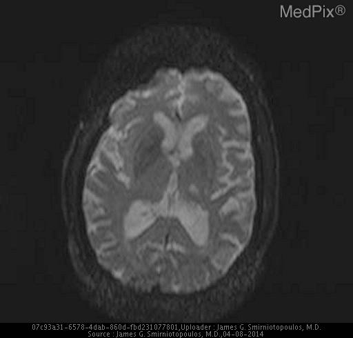
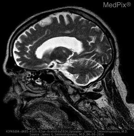
These infarcts are usually small, about 3 to 15 mm, and occur in deep structures supplied by penetrating arteries: internal capsule, basal ganglia, thalamus, pons, corona radiata, and deep white matter. Frameworks for Internal Medicine, p. 586
Major risk factors
- Chronic hypertension: strongest and most typical risk factor
- Diabetes mellitus
- Dyslipidemia
- Smoking
- Older age
Typical clinical syndromes
Lacunar strokes usually cause focal motor or sensory deficits without cortical signs.
| Syndrome | Typical features | Common location |
|---|---|---|
| Pure motor hemiparesis | Contralateral weakness of face, arm, and leg | Internal capsule or pons |
| Pure sensory stroke | Contralateral numbness or altered sensation | Thalamus |
| Sensorimotor stroke | Weakness plus sensory loss | Thalamocapsular area |
| Ataxic hemiparesis | Ipsilateral limb ataxia with contralateral weakness | Pons or internal capsule |
| Dysarthria-clumsy hand syndrome | Slurred speech and clumsy/weak hand | Pons or genu of internal capsule |
Harrison’s Principles of Internal Medicine, 22e describes these as the common small-vessel stroke syndromes.
Key clinical point
Because the cerebral cortex is generally spared, classic cortical deficits are often absent:
- No aphasia
- No neglect
- No apraxia
- No visual field defect
- Usually no seizure at onset
Their presence should prompt evaluation for another stroke mechanism, such as a cortical embolic or large-artery stroke.
Diagnosis
- Non-contrast CT head first in acute stroke, mainly to exclude hemorrhage.
- MRI brain with diffusion-weighted imaging is more sensitive for detecting a small acute infarct.
- The work-up still assesses alternative causes, including atrial fibrillation, carotid/intracranial atherosclerosis, and cardiac embolism.
Acute management
A suspected lacunar stroke is treated as an acute ischemic stroke:
- Urgent brain imaging and stroke-team assessment.
- Consider IV thrombolysis if eligible and within the treatment window.
- Mechanical thrombectomy is usually not relevant unless imaging identifies a large-vessel occlusion.
- Do not routinely lower blood pressure immediately in patients not receiving thrombolysis unless there is another indication or BP is markedly elevated.
The ESO lacunar stroke guideline recommends following standard acute ischemic-stroke pathways, including alteplase when eligible.
Secondary prevention
- Single antiplatelet therapy long term for non-cardioembolic symptomatic lacunar infarction, unless another indication changes treatment.
- Careful blood-pressure control, commonly targeting below 130/80 mmHg when appropriate.
- Statin/lipid-lowering therapy according to overall vascular-risk guidelines.
- Diabetes management, smoking cessation, regular physical activity, weight management, and a healthy diet.
- Long-term dual antiplatelet therapy is generally avoided in lacunar stroke because it increases bleeding risk without clear net benefit.
Recent guidance supports long-term single antiplatelet treatment plus risk-factor management, while the 2024 ESO guideline notes that direct lacunar-specific evidence remains limited (PMID 38380638).
Emergency warning: new facial droop, arm or leg weakness/numbness, speech difficulty, loss of balance, or sudden visual symptoms should be treated as a possible stroke. Seek emergency services immediately, even if symptoms improve.
This is a shared conversation. Sign in to Orris to start your own chat.