
Why GFR dec in neohritic syndrome and what is azotemia
Why GFR Decreases in Nephritic Syndrome
1. Inflammatory cell infiltration and glomerular hypercellularity
2. Damage to the glomerular capillary wall
3. Reduced glomerular capillary perfusion pressure
4. Net result: Decreased GFR
- Reduced filtration surface area (capillary compression)
- Damaged filtration barrier
- Reduced intraglomerular blood flow
"Glomerular diseases that present with the nephritic syndrome are usually associated with significant inflammation in the glomeruli, which damages capillary walls, leading to leakage of red cells (hematuria), and decreased GFR, which results in oliguria and azotemia."
- Robbins & Kumar Basic Pathology, p. 2297
What is Azotemia?
Elevation of blood urea nitrogen (BUN) and serum creatinine levels, due largely to a decreased GFR.
- Robbins, Cotran & Kumar - Pathologic Basis of Disease, p. 2688
Types of Azotemia
| Type | Cause |
|---|---|
| Prerenal | Hypoperfusion of kidneys (hypotension, shock, dehydration, heart failure) - no parenchymal damage |
| Renal (intrinsic) | Direct glomerular, tubular, interstitial, or vascular damage |
| Postrenal | Obstruction of urine outflow distal to the kidney; corrects when obstruction is relieved |
Azotemia vs. Uremia - Key Distinction
| Term | Definition |
|---|---|
| Azotemia | Purely a lab finding - elevated BUN and creatinine |
| Uremia | Azotemia that is severe enough to cause clinical symptoms |
- GI involvement: uremic gastroenteritis, nausea, vomiting
- Neurological: peripheral neuropathy, encephalopathy
- Cardiac: uremic fibrinous pericarditis
In Nephritic Syndrome Specifically
So why azotemia and proteinuria occurs
Why Azotemia and Proteinuria Occur in Nephritic Syndrome
1. Azotemia - Why it Occurs
Normal state:
In nephritic syndrome:
- Glomerular inflammation causes endothelial and mesangial cell proliferation + neutrophil infiltration, massively increasing cellularity inside the glomerular tuft
- This compresses and occludes the glomerular capillary lumens
- GFR falls - less blood is filtered per minute
- Urea and creatinine accumulate in the blood because the kidney cannot filter them fast enough
- Result: elevated BUN and serum creatinine = Azotemia
"Azotemia is a biochemical abnormality that refers to an elevation of blood urea nitrogen (BUN) and creatinine levels, related largely to a decreased GFR."
- Robbins, Cotran & Kumar - Pathologic Basis of Disease
2. Proteinuria - Why it Occurs
The normal filtration barrier has 3 layers:
| Layer | Role in keeping proteins out |
|---|---|
| Fenestrated endothelial cells (covered by glycocalyx) | Glycocalyx is negatively charged - electrically repels albumin (also negatively charged), preventing it from even reaching the GBM |
| Glomerular Basement Membrane (GBM) | Contains heparan sulfate proteoglycans (negative charge) + collagen type IV mesh - acts as a size and charge barrier |
| Podocyte foot processes + slit diaphragm (nephrin protein) | Final size-selective barrier; slit diaphragm is only 20-30 nm wide - too narrow for albumin (radius ~3.6 nm) to pass in significant amounts |
"The normal glomerulus is highly permeable to water and small solutes... and is impermeable to proteins of the size of albumin (~3.6-nm radius; 70 kDa) or larger."
- Robbins, Cotran & Kumar - Pathologic Basis of Disease
In nephritic syndrome:
-
Endothelial injury - disrupts the glycocalyx, removing the electrostatic repulsion of albumin. Albumin can now reach the GBM in higher concentrations.
-
GBM disruption - inflammatory mediators and neutrophil enzymes (proteases, reactive oxygen species) degrade the GBM collagen and heparan sulfate, losing both size and charge selectivity.
-
Podocyte/slit diaphragm injury - foot process effacement and nephrin loss opens up "large pores" in the slit diaphragm, allowing proteins to spill through.
"Glomerular diseases are characterized by breaks in the glomerular capillary wall, thereby leading to hematuria with proteinuria because of loss of the selective barrier to particles based on size and charge."
- Goldman-Cecil Medicine
Why is proteinuria SUBNEPHROTIC (not massive) in nephritic syndrome?
- In nephritic syndrome, the barrier disruption is partial and patchy - caused by inflammation
- In nephrotic syndrome (e.g., minimal change disease), there is diffuse, severe podocyte injury causing massive protein leak (>3.5 g/day)
- In nephritic syndrome, proteinuria is typically <3.5 g/day (subnephrotic range) because the primary injury is to the mesangium and endothelium, with less severe podocyte damage
Summary Table
| Feature | Mechanism | Result |
|---|---|---|
| Azotemia | Glomerular inflammation → ↓GFR → urea & creatinine not filtered | Elevated BUN + creatinine in blood |
| Proteinuria | Capillary wall damage → loss of size & charge selectivity of filtration barrier | Protein leaks into urine (subnephrotic range) |
| Hematuria | Same capillary wall damage → RBCs squeeze through | RBCs and RBC casts in urine |
| Hypertension | ↓GFR → Na & water retention → RAAS activation | Raised BP |
| Oliguria | ↓GFR → less filtrate formed | Reduced urine output |

I cant get 3rd immunoflorrnce of IgA nephropathy
IgA Nephropathy - Immunofluorescence (IF) Pattern
What you see on IF:
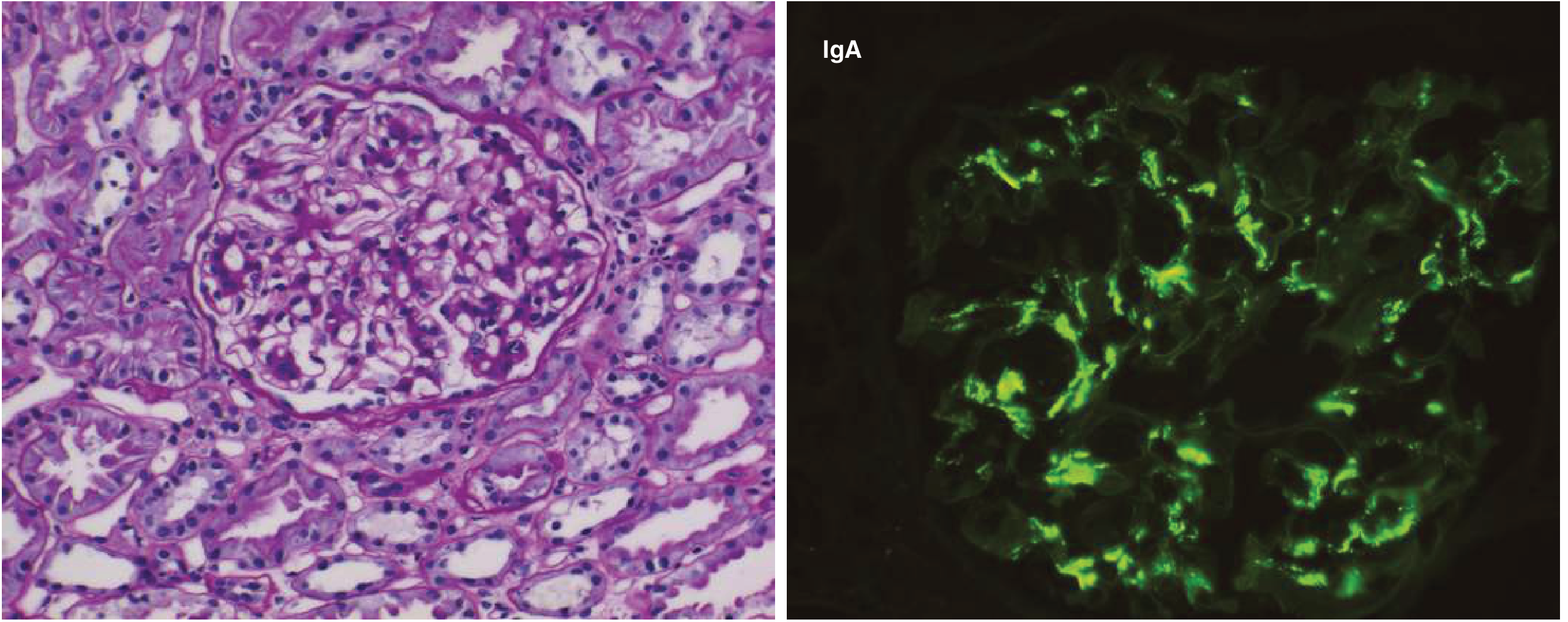
Full IF Composition:
| Immunoreactant | Presence |
|---|---|
| IgA | Dominant / codominant (MUST be present for diagnosis) |
| C3 | Often present (alternative pathway activation) |
| Properdin | Often present (confirms alternative pathway) |
| IgG or IgM | Smaller amounts, may be present |
| C1q / C4 (classical pathway) | Usually ABSENT |
"The characteristic immunofluorescence picture is of mesangial deposition of IgA, often with C3 and the alternative pathway protein properdin and smaller amounts of IgG or IgM. Early components of the classical complement pathway are usually absent."
- Robbins & Kumar Basic Pathology, p. 2405
The 3 Key IF Points to Remember (what your note probably cut off):
- Pattern: Mesangial/paramesangial (granular) - NOT linear, NOT subepithelial
- Dominant immunoglobulin: IgA (this is what makes the diagnosis - "dominant or codominant IgA")
- Complement pathway: Alternative pathway (C3 + properdin present; C1q ABSENT - no classical pathway activation)
Why Alternative Pathway?
Comparing the 3 Granular IF Patterns (exam favourite):
| Disease | IF Pattern | Location of deposits |
|---|---|---|
| IgA nephropathy | IgA dominant + C3 (alternative pathway) | Mesangium |
| PSGN | IgG + C3 (granular "starry sky") | Subepithelial + mesangial |
| Lupus nephritis | "Full house" (IgG, IgA, IgM, C3, C1q) | Mesangial + subendothelial + subepithelial |
I cant get anything
What exactly are you struggling to understand about IgA nephropathy?
IgA Nephropathy - IF Pattern, Explained Simply
First, understand what IF actually IS
What glows in IgA nephropathy?
- Left panel = Light microscopy (H&E stain) - shows mesangial cell proliferation
- Right panel = Immunofluorescence - the bright green spots = IgA deposits
The 3 things to know about where it lights up:
Normal glomerulus cross-section:

        Capillary wall (GBM)
       /
[Bowman's space] -- [Capillary loops] -- [Mesangium in the middle]
                                              ↑
                              THIS is where IgA deposits sit
- Green fluorescence in the CENTER of the glomerular tuft
- NOT along the outer capillary walls (that would be GBM/subepithelial pattern like in PSGN or membranous)
- NOT a linear line (that is Goodpasture's)
- Patchy/granular clusters in the mesangial zones
Visual comparison - WHERE the glow sits:
| Disease | Where it glows on IF | Pattern |
|---|---|---|
| IgA nephropathy | Mesangium (center of tuft) | Granular, central |
| PSGN | Capillary walls + mesangium | "Starry sky" all over |
| Goodpasture | Along entire GBM | Smooth linear line |
| Membranous GN | Outer capillary wall (subepithelial) | Granular, peripheral |
Simple memory trick:
IgA = "I go to the mesAngium"
What exactly lights up on the stain panel:
| Stain | Result |
|---|---|
| Anti-IgA | Bright green (dominant) |
| Anti-C3 | Often positive |
| Anti-IgG | Faint / minor |
| Anti-C1q | NEGATIVE (important!) |
Test me using nephrotic and nephritic syndrome from FA according to uworld and nbme
USMLE UWorld NBME high yield nephrotic nephritic syndrome tested concepts
🧪 USMLE-Style Test: Nephrotic & Nephritic Syndrome
(UWorld/NBME style - one best answer)
- A) Subepithelial "spike and dome" deposits
- B) Subendothelial deposits with "tram-track" appearance
- C) Effacement of podocyte foot processes
- D) Mesangial IgA deposits
- E) Linear IgG along GBM
- A) Mesangial cell
- B) Endothelial cell
- C) Parietal epithelial cell
- D) Podocyte
- E) Tubular epithelial cell
- A) Minimal change disease
- B) Membranous nephropathy
- C) FSGS
- D) Post-streptococcal GN
- E) Membranoproliferative GN
- A) Linear IgG along GBM
- B) Granular IgG and C3 in mesangium and capillary walls ("starry sky")
- C) Mesangial IgA deposits
- D) No deposits (pauci-immune)
- E) Subepithelial IgG deposits only
- A) Classical pathway (C1q → C4 → C2 → C3)
- B) Alternative pathway (C3 → properdin → C3b)
- C) Both classical and alternative equally
- D) Mannose-binding lectin pathway only
- E) No complement activation
- A) Anti-dsDNA
- B) Anti-GBM (type IV collagen)
- C) Anti-PLA2R (phospholipase A2 receptor)
- D) Anti-ANCA (MPO)
- E) Anti-streptolysin O
- A) Membranous nephropathy (responds to steroids)
- B) FSGS (steroid-sensitive variant)
- C) Minimal change disease (80-90% steroid responsive)
- D) IgA nephropathy (steroid responsive)
- E) Post-streptococcal GN (spontaneous resolution)
- A) Nephrotic syndrome - podocyte injury
- B) Nephritic syndrome - glomerular inflammation → decreased GFR
- C) Nephrotic syndrome - loss of charge selectivity
- D) Nephritic syndrome - thromboembolism
- E) AKI - tubular necrosis
- A) Granulomatous inflammation with necrosis
- B) Linear IgG along alveolar basement membrane
- C) Eosinophilic infiltrate
- D) Caseous necrosis
- E) Pauci-immune (no deposits)
- A) Increased IgG loss → susceptibility to encapsulated organisms
- B) Loss of antithrombin III in urine → hypercoagulable state → renal vein thrombosis
- C) Hypoalbuminemia → decreased oncotic pressure → edema
- D) Loss of thyroxine-binding globulin → hypothyroidism
- E) Increased LDL due to hepatic lipoprotein overproduction
How to respond:
C D B B A B C B B B
📊 Your Score: 7/10 — Good! Let's fix the 3 you missed.
✅ Full Breakdown
Q1 — Your answer: C ✅ CORRECT
Key: Normal LM + Normal IF + Abnormal EM (foot process effacement) = MCD
Q2 — Your answer: D ✅ CORRECT
Key FSGS associations to memorize:
- HIV → collapsing FSGS
- Heroin, sickle cell, obesity → secondary FSGS
- Idiopathic → most common in Black adults
Q3 — Your answer: B ✅ CORRECT
EM buzzword: "spike and dome" appearance - spikes of GBM material projecting between subepithelial deposits.
Q4 — Your answer: B ✅ CORRECT
LM: hypercellular glomeruli with neutrophils EM: subepithelial "humps" (pathognomonic)
Q5 — Your answer: A ❌ WRONG
- IgA does NOT activate the classical complement pathway
- Classical pathway requires IgG or IgM + C1q
- IgA activates the alternative pathway (and sometimes lectin pathway)
- This is why on IF: C3 and properdin are present, but C1q and C4 are ABSENT
- Serum complement levels are normal in IgA nephropathy (unlike PSGN where C3 is low)
Exam tip: Normal C3 + mesangial IgA + recurrent hematuria with URTI = IgA nephropathy. Low C3 = think PSGN, lupus, MPGN.
Q6 — Your answer: B ❌ WRONG
- Primary membranous nephropathy = anti-PLA2R (phospholipase A2 receptor) antibodies in ~70% of cases
- Secondary causes (Hep B, gold therapy, SLE) have the same histology but different antibody targets
- Anti-GBM → linear IF, crescents, pulmonary hemorrhage (Goodpasture) - not membranous pattern
The question stem gave you secondary membranous (gold therapy) but asked specifically about the idiopathic form's antibody = anti-PLA2R.
Q7 — Your answer: C ✅ CORRECT
- FSGS = steroid resistant in most cases
- Membranous = poor steroid response
- MCD = excellent steroid response
Q8 — Your answer: B ✅ CORRECT
- RBC casts + hematuria = glomerular inflammation (nephritic)
- Oliguria + azotemia = decreased GFR
- Subnephrotic proteinuria (<3.5 g/day) = partial barrier disruption
- Hypertension = Na/water retention + RAAS activation
The single most important distinguishing feature of nephritic vs nephrotic: RBC casts = nephritic. Massive proteinuria >3.5g + no hematuria = nephrotic.
Q9 — Your answer: B ✅ CORRECT
- Kidney: linear IgG on IF → crescentic GN → hematuria
- Lung: linear IgG on alveolar BM → alveolar hemorrhage → hemoptysis
This is Type II HSR (cytotoxic). The antibody is anti-GBM (anti-α3 chain of type IV collagen). Distinguish from Granulomatosis with polyangiitis (Wegener): also hemoptysis + hematuria, but pauci-immune IF (no deposits) + c-ANCA/PR3-ANCA positive.
Q10 — Your answer: B ✅ CORRECT
Full list of proteins lost in nephrotic syndrome to know:
- Albumin → edema, hypoalbuminemia
- Antithrombin III → thrombosis (renal vein, DVT, PE)
- IgG → infections (encapsulated bacteria - pneumococcus)
- Thyroxine-binding globulin → hypothyroidism (low T4 but normal TSH)
- Transferrin → microcytic anemia
- Complement (factor B/D) → infections
📈 Summary
| Q | Topic | You | Answer | Result |
|---|---|---|---|---|
| 1 | MCD - EM finding | C | C | ✅ |
| 2 | FSGS - cell injured | D | D | ✅ |
| 3 | Membranous nephropathy | B | B | ✅ |
| 4 | PSGN - IF pattern | B | B | ✅ |
| 5 | IgA - complement pathway | A | B | ❌ |
| 6 | Membranous - antibody target | B | C | ❌ |
| 7 | MCD - steroid response | C | C | ✅ |
| 8 | Nephritic vs nephrotic | B | B | ✅ |
| 9 | Goodpasture - lung finding | B | B | ✅ |
| 10 | Nephrotic complication | B | B | ✅ |
🎯 Focus Areas for Review:
- IgA nephropathy - alternative complement pathway (no C1q, normal serum C3)
- Anti-PLA2R - the antibody of primary/idiopathic membranous nephropathy
Create a PDF quick reference card comparing all glomerular diseases
mkdir -p /home/daytona/workspace/glomerular-ref && ls /home/daytona/workspace/
~/glomerular-ref/build_pdf.py
from reportlab.lib.pagesizes import A4, landscape
from reportlab.lib import colors
from reportlab.lib.units import cm, mm
from reportlab.platypus import (
    SimpleDocTemplate, Table, TableStyle, Paragraph, Spacer,
    HRFlowable, KeepTogether
)
from reportlab.lib.styles import getSampleStyleSheet, ParagraphStyle
from reportlab.lib.enums import TA_CENTER, TA_LEFT
from reportlab.platypus import PageBreak

OUTPUT = "/home/daytona/workspace/glomerular-ref/Glomerular_Diseases_Quick_Reference.pdf"

# ── Colour palette ──────────────────────────────────────────────
C_HEADER_BG   = colors.HexColor("#1a3a5c")   # dark navy
C_NEPHROTIC   = colors.HexColor("#1565C0")   # blue
C_NEPHRITIC   = colors.HexColor("#B71C1C")   # red
C_BOTH        = colors.HexColor("#4A148C")   # purple
C_ROW_ALT     = colors.HexColor("#F0F4FF")   # very light blue
C_ROW_ALT2    = colors.HexColor("#FFF3F3")   # very light red
C_ROW_WHITE   = colors.white
C_ACCENT      = colors.HexColor("#FF6F00")   # amber for key facts
C_LIGHT_NAVY  = colors.HexColor("#E8EFF8")
C_GREEN_LIGHT = colors.HexColor("#E8F5E9")
C_SUBHEADER   = colors.HexColor("#37474F")   # dark grey

styles = getSampleStyleSheet()

def style(name="Normal", size=8, color=colors.black, bold=False,
          align=TA_LEFT, leading=10):
    return ParagraphStyle(
        name, parent=styles["Normal"],
        fontSize=size, textColor=color,
        fontName="Helvetica-Bold" if bold else "Helvetica",
        alignment=align, leading=leading, spaceAfter=0
    )

TITLE_S   = style("Title",   size=18, color=colors.white, bold=True,  align=TA_CENTER, leading=22)
SUB_S     = style("Sub",     size=10, color=colors.HexColor("#CFD8DC"), align=TA_CENTER, leading=13)
H2_S      = style("H2",      size=11, color=C_HEADER_BG, bold=True, leading=14)
CELL_S    = style("Cell",    size=7.5, leading=10)
CELL_B    = style("CellB",   size=7.5, bold=True, leading=10)
CELL_SM   = style("CellSM",  size=7,  leading=9.5)
NEP_S     = style("Nep",     size=8,  color=C_NEPHROTIC, bold=True, align=TA_CENTER, leading=10)
NIT_S     = style("Nit",     size=8,  color=C_NEPHRITIC, bold=True, align=TA_CENTER, leading=10)
BOTH_S    = style("Both",    size=8,  color=C_BOTH, bold=True, align=TA_CENTER, leading=10)
KEY_S     = style("Key",     size=7,  color=C_ACCENT, bold=True, leading=9)
FOOT_S    = style("Foot",    size=6.5,color=C_SUBHEADER, leading=9)

def P(txt, s=None):
    if s is None: s = CELL_S
    return Paragraph(txt, s)

def bold(txt):
    return f"<b>{txt}</b>"

def col(txt, c):
    hex_c = c.hexval() if hasattr(c, 'hexval') else c
    return f'<font color="{hex_c}">{txt}</font>'

# ── Disease data ─────────────────────────────────────────────────
# Columns: Disease | Syndrome | Age/Pop | LM | IF | EM | Clinical | Key Fact
NEPHROTIC_COLOR = "#1565C0"
NEPHRITIC_COLOR = "#B71C1C"
BOTH_COLOR      = "#4A148C"

diseases = [
    # ── NEPHROTIC ─────────────────────────────────────────────
    {
        "name": "Minimal Change\nDisease (MCD)",
        "syndrome": ("NEPHROTIC", C_NEPHROTIC),
        "pop": "Children\n(#1 cause)\nAdults: Hodgkin\nlymphoma, NSAIDs",
        "lm": "NORMAL\n(no changes visible)",
        "IF": "NEGATIVE\n(no deposits)",
        "em": "Diffuse effacement\nof podocyte\nfoot processes",
        "clinical": "Massive proteinuria\n>3.5g/day\nEdema\nHypoalbuminemia\nNormal complement",
        "key": "★ Responds to\nsteroids (80-90%)\n★ No hematuria",
        "row_color": C_ROW_ALT,
    },
    {
        "name": "Focal Segmental\nGlomerulosclerosis\n(FSGS)",
        "syndrome": ("NEPHROTIC", C_NEPHROTIC),
        "pop": "Adults\n(#1 primary\nnephrotic in\nBlack adults)\nHIV, heroin,\nobesity, sickle cell",
        "lm": "FOCAL (some\nglomeruli) &\nSEGMENTAL\n(part of tuft)\nsclerosis/hyalinosis",
        "IF": "IgM + C3\n(nonspecific,\nin sclerotic areas)",
        "em": "Effacement of\nfoot processes\n(focal/segmental)",
        "clinical": "Nephrotic syndrome\nHypertension\nSteroid-RESISTANT\nProgresses to ESRD",
        "key": "★ HIV → Collapsing\nvariant (worst)\n★ Most common\nnephrotic in adults",
        "row_color": C_ROW_WHITE,
    },
    {
        "name": "Membranous\nNephropathy",
        "syndrome": ("NEPHROTIC", C_NEPHROTIC),
        "pop": "Adults 30-50y\n(#1 primary\nnephrotic in\nwhite adults)\nHep B/C, SLE,\nmalignancy, gold",
        "lm": "Thickened GBM\n'Spike & dome'\non silver stain\n(GBM spikes between\nsubepithelial deposits)",
        "IF": "Granular IgG + C3\nalong GBM\n(peripheral/\nsubepithelial)",
        "em": "Subepithelial\nelectron-dense\ndeposits",
        "clinical": "Nephrotic syndrome\nRenal vein thrombosis\n(highest risk of all\nnephrotic diseases)",
        "key": "★ Anti-PLA2R Ab\n(idiopathic form)\n★ 'Spike & dome'\non EM/silver stain",
        "row_color": C_ROW_ALT,
    },
    {
        "name": "Membrano-\nproliferative GN\n(MPGN)",
        "syndrome": ("NEPHROTIC\nor NEPHRITIC", C_BOTH),
        "pop": "Young adults\nHep C (Type I)\nC3 glomerulopathy\n(Type II)",
        "lm": "'Tram-track'\n(double contour\nof GBM)\nMesangial\nproliferation",
        "IF": "Type I: IgG+C3\n(subendothelial)\nType II: C3 only\n(intramembranous)",
        "em": "Type I: Subendothelial\ndeposits\nType II: Dense\ndeposits (ribbon-like\nin GBM)",
        "clinical": "Nephrotic OR\nnephritic features\nLow C3 (both types)\nLow C4 (Type I only)",
        "key": "★ 'Tram-track' = MPGN\n★ Type II = C3\nNephritic Factor\n(stabilizes C3\nconvertase)",
        "row_color": C_ROW_WHITE,
    },
    # ── NEPHRITIC ─────────────────────────────────────────────
    {
        "name": "Post-Streptococcal\nGN (PSGN)",
        "syndrome": ("NEPHRITIC", C_NEPHRITIC),
        "pop": "Children 2-14y\n2-3 wks after\nthroat infection\n(6 wks after skin)",
        "lm": "Diffuse hypercellular\nglomeruli\nNeutrophil\ninfiltration\n'Lumpy bumpy'",
        "IF": "Granular IgG + C3\n'Starry sky'\nsubepithelial +\nmesangial",
        "em": "Subepithelial\n'HUMPS'\n(pathognomonic)",
        "clinical": "Hematuria (tea-colored)\nRBC casts\nOliguria, azotemia\nHTN\nLOW C3, LOW C4\nElevated ASO titer",
        "key": "★ Subepithelial\n'humps' on EM\n★ Self-limiting\nin children\n★ Low C3",
        "row_color": C_ROW_ALT2,
    },
    {
        "name": "IgA Nephropathy\n(Berger Disease)",
        "syndrome": ("NEPHRITIC", C_NEPHRITIC),
        "pop": "Young adults\n(#1 GN worldwide)\nAsian > others\nRecurrent after\nURTI",
        "lm": "Mesangial\nproliferation and\nmatrix increase\n(focal or diffuse)",
        "IF": "Mesangial IgA\n(dominant)\n± C3, IgG, IgM\nNO C1q\n(alternative pathway)",
        "em": "Mesangial electron-\ndense deposits",
        "clinical": "Gross hematuria\ncoinciding with URTI\n(synpharyngitic)\nNORMAL complement\nSubnephrotic proteinuria",
        "key": "★ Normal C3\n(unlike PSGN)\n★ NO C1q on IF\n★ Alternative\ncomplement pathway",
        "row_color": C_ROW_WHITE,
    },
    {
        "name": "Goodpasture\nSyndrome",
        "syndrome": ("NEPHRITIC\n(RPGN)", C_NEPHRITIC),
        "pop": "Young men\n20-30y (HLA-DR2)\nOlder men 60-70y\nSmoking triggers\npulmonary disease",
        "lm": "Crescentic GN\n(crescents in\nBowman's space)\nFibrinoid necrosis",
        "IF": "LINEAR IgG\nalong GBM\n(smooth, ribbon-like)\n± C3",
        "em": "No immune deposits\n(antibody-mediated,\nnot immune complex)",
        "clinical": "Hemoptysis +\nHematuria\nRapid GFR decline\nAnti-GBM antibodies\n(anti-α3 collagen IV)",
        "key": "★ LINEAR IF\n(only disease)\n★ Anti-GBM Ab\n★ Pulmonary\nhemorrhage\n★ Type II HSR",
        "row_color": C_ROW_ALT2,
    },
    {
        "name": "Pauci-Immune\nRPGN (ANCA-GN)",
        "syndrome": ("NEPHRITIC\n(RPGN)", C_NEPHRITIC),
        "pop": "Adults 50-70y\nGPA (Wegener's)\nMPA\nEosinophilic GPA\n(Churg-Strauss)",
        "lm": "Crescentic GN\nFibrinoid necrosis\nNo immune deposits",
        "IF": "NEGATIVE / PAUCI-\nIMMUNE\n(no/scanty deposits)\nKey distinguishing\nfeature",
        "em": "No electron-dense\nimmune deposits",
        "clinical": "Rapid GFR decline\n± hemoptysis (GPA/MPA)\nGPA: c-ANCA/PR3-ANCA\nsinusitis, lung nodules\nMPA: p-ANCA/MPO-ANCA",
        "key": "★ NEGATIVE IF\n(pauci-immune)\n★ GPA: c-ANCA\n(PR3)\n★ MPA: p-ANCA\n(MPO)",
        "row_color": C_ROW_WHITE,
    },
    {
        "name": "Lupus Nephritis\n(Diffuse, Class IV)",
        "syndrome": ("NEPHRITIC or\nNEPHROTIC", C_BOTH),
        "pop": "Women 20-40y\nSLE\n(most common\ncause of death\nin SLE)",
        "lm": "'Wire-loop'\nlesions\n(subendothelial\ndeposits)\nDiffuse proliferative",
        "IF": "'FULL HOUSE'\nIgG + IgA + IgM\n+ C3 + C1q\n(all positive = SLE)",
        "em": "Subendothelial +\nmesangial +\nsubepithelial deposits\n('fingerprint' pattern)",
        "clinical": "Any nephritic/\nnephrotic features\nLow C3 AND C4\nAnti-dsDNA, ANA +\nLow platelets, rash",
        "key": "★ 'Full house' IF\n★ 'Wire-loop' LM\n★ LOW C3 + C4\n(classical pathway)\n★ Anti-dsDNA",
        "row_color": C_ROW_ALT,
    },
    {
        "name": "Alport Syndrome",
        "syndrome": ("NEPHRITIC", C_NEPHRITIC),
        "pop": "Males (X-linked)\nMutation in\nCOL4A5 gene\n(α5 chain type\nIV collagen)",
        "lm": "Variable; may be\nnormal early\nFocal GN later",
        "IF": "Negative\n(no immune deposits)\nAnti-GBM staining\nABSENT",
        "em": "GBM thinning AND\nthickening with\nsplitting/lamellation\n('basket-weave'\nappearance)",
        "clinical": "Hematuria (from\nchildhood)\nSensorineural\nhearing loss\nOcular defects\n(lens dislocation)",
        "key": "★ 'Basket-weave'\nGBM on EM\n★ Triad: hematuria\n+ deafness\n+ eye defects",
        "row_color": C_ROW_ALT2,
    },
]

def build_disease_table(doc_width):
    col_widths = [2.8*cm, 1.8*cm, 2.4*cm, 2.8*cm, 2.8*cm, 2.8*cm, 3.0*cm, 2.4*cm]

    header = [
        P(bold("Disease"), CELL_B),
        P(bold("Syndrome"), CELL_B),
        P(bold("Population /\nAssociations"), CELL_B),
        P(bold("Light Microscopy\n(LM)"), CELL_B),
        P(bold("Immunofluorescence\n(IF)"), CELL_B),
        P(bold("Electron\nMicroscopy (EM)"), CELL_B),
        P(bold("Clinical Features"), CELL_B),
        P(bold("★ Key USMLE\nFacts"), CELL_B),
    ]

    rows = [header]
    for i, d in enumerate(diseases):
        syn_text, syn_color = d["syndrome"]
        syn_style = ParagraphStyle(
            f"syn{i}", parent=styles["Normal"],
            fontSize=7.5, textColor=syn_color,
            fontName="Helvetica-Bold", alignment=TA_CENTER, leading=10
        )
        key_style = ParagraphStyle(
            f"key{i}", parent=styles["Normal"],
            fontSize=7, textColor=C_ACCENT,
            fontName="Helvetica-Bold", alignment=TA_LEFT, leading=9.5
        )
        row = [
            P(bold(d["name"].replace("\n","<br/>")), CELL_B),
            Paragraph(syn_text.replace("\n","<br/>"), syn_style),
            P(d["pop"].replace("\n","<br/>"), CELL_SM),
            P(d["lm"].replace("\n","<br/>"), CELL_SM),
            P(d["IF"].replace("\n","<br/>"), CELL_SM),
            P(d["em"].replace("\n","<br/>"), CELL_SM),
            P(d["clinical"].replace("\n","<br/>"), CELL_SM),
            Paragraph(d["key"].replace("\n","<br/>"), key_style),
        ]
        rows.append(row)

    tbl = Table(rows, colWidths=col_widths, repeatRows=1)

    ts = TableStyle([
        # Header row
        ("BACKGROUND",    (0,0), (-1,0), C_HEADER_BG),
        ("TEXTCOLOR",     (0,0), (-1,0), colors.white),
        ("FONTNAME",      (0,0), (-1,0), "Helvetica-Bold"),
        ("FONTSIZE",      (0,0), (-1,0), 8),
        ("ROWBACKGROUND", (0,0), (-1,0), C_HEADER_BG),
        # Borders
        ("GRID",          (0,0), (-1,-1), 0.4, colors.HexColor("#B0BEC5")),
        ("BOX",           (0,0), (-1,-1), 1,   C_HEADER_BG),
        # Vertical alignment
        ("VALIGN",        (0,0), (-1,-1), "TOP"),
        # Row padding
        ("TOPPADDING",    (0,0), (-1,-1), 4),
        ("BOTTOMPADDING", (0,0), (-1,-1), 4),
        ("LEFTPADDING",   (0,0), (-1,-1), 4),
        ("RIGHTPADDING",  (0,0), (-1,-1), 4),
        # Alternate row colors
        ("ROWBACKGROUND", (0,1), (-1,1),  colors.HexColor("#EBF5FB")),
        ("ROWBACKGROUND", (0,2), (-1,2),  colors.white),
        ("ROWBACKGROUND", (0,3), (-1,3),  colors.HexColor("#EBF5FB")),
        ("ROWBACKGROUND", (0,4), (-1,4),  colors.white),
        ("ROWBACKGROUND", (0,5), (-1,5),  colors.HexColor("#FDEDEC")),
        ("ROWBACKGROUND", (0,6), (-1,6),  colors.white),
        ("ROWBACKGROUND", (0,7), (-1,7),  colors.HexColor("#FDEDEC")),
        ("ROWBACKGROUND", (0,8), (-1,8),  colors.white),
        ("ROWBACKGROUND", (0,9), (-1,9),  colors.HexColor("#EDE7F6")),
        ("ROWBACKGROUND", (0,10),(-1,10), colors.HexColor("#FDEDEC")),
        # Syndrome column alignment center
        ("ALIGN",         (1,0), (1,-1), "CENTER"),
    ])
    tbl.setStyle(ts)
    return tbl


# ── Complement / IF quick-reference mini-table ───────────────────
def build_complement_table():
    headers = ["Disease", "C3", "C4", "C1q on IF", "IF Pattern", "Complement Pathway"]
    data = [
        ["MCD",              "Normal", "Normal", "–",    "Negative",          "None"],
        ["FSGS",             "Normal", "Normal", "–",    "IgM+C3 (focal)",    "None (nonspecific)"],
        ["Membranous",       "Normal", "Normal", "–",    "Granular IgG+C3",   "Alternative"],
        ["PSGN",             "LOW ↓",  "LOW ↓",  "–",    "Granular IgG+C3",   "Classical"],
        ["IgA Nephropathy",  "Normal", "Normal", "–",    "Mesangial IgA",     "Alternative/Lectin"],
        ["MPGN Type I",      "LOW ↓",  "LOW ↓",  "±",    "IgG+C3",            "Classical"],
        ["MPGN Type II",     "LOW ↓",  "Normal", "–",    "C3 only",           "Alternative"],
        ["Goodpasture",      "Normal", "Normal", "–",    "LINEAR IgG",        "None (anti-GBM Ab)"],
        ["Pauci-immune RPGN","Normal", "Normal", "–",    "NEGATIVE",          "None (ANCA)"],
        ["Lupus (Class IV)", "LOW ↓",  "LOW ↓",  "+++",  "Full house",        "Classical (C1q++)"],
    ]
    col_w = [3.5*cm, 1.5*cm, 1.5*cm, 1.8*cm, 3.5*cm, 4.5*cm]
    rows = [[P(bold(h), CELL_B) for h in headers]]
    for i, row in enumerate(data):
        styled = []
        for j, cell in enumerate(row):
            if "LOW" in cell or "↓" in cell:
                s = ParagraphStyle(f"r{i}{j}", parent=styles["Normal"],
                                   fontSize=7.5, textColor=C_NEPHRITIC,
                                   fontName="Helvetica-Bold", leading=10)
            elif cell in ("Normal",):
                s = ParagraphStyle(f"r{i}{j}", parent=styles["Normal"],
                                   fontSize=7.5, textColor=colors.HexColor("#2E7D32"),
                                   fontName="Helvetica-Bold", leading=10)
            elif "Full house" in cell or "LINEAR" in cell or "NEGATIVE" in cell:
                s = ParagraphStyle(f"r{i}{j}", parent=styles["Normal"],
                                   fontSize=7.5, textColor=C_BOTH,
                                   fontName="Helvetica-Bold", leading=10)
            else:
                s = CELL_SM
            styled.append(Paragraph(cell.replace("\n","<br/>"), s))
        rows.append(styled)

    tbl = Table(rows, colWidths=col_w, repeatRows=1)
    ts = TableStyle([
        ("BACKGROUND",  (0,0), (-1,0), C_HEADER_BG),
        ("TEXTCOLOR",   (0,0), (-1,0), colors.white),
        ("GRID",        (0,0), (-1,-1), 0.4, colors.HexColor("#B0BEC5")),
        ("BOX",         (0,0), (-1,-1), 1, C_HEADER_BG),
        ("VALIGN",      (0,0), (-1,-1), "MIDDLE"),
        ("TOPPADDING",  (0,0), (-1,-1), 3),
        ("BOTTOMPADDING",(0,0),(-1,-1), 3),
        ("LEFTPADDING", (0,0), (-1,-1), 4),
        ("RIGHTPADDING",(0,0), (-1,-1), 4),
        ("ROWBACKGROUND",(0,1),(-1,4),  colors.HexColor("#EBF5FB")),
        ("ROWBACKGROUND",(0,5),(-1,5),  colors.white),
        ("ROWBACKGROUND",(0,6),(-1,7),  colors.HexColor("#FDEDEC")),
        ("ROWBACKGROUND",(0,8),(-1,9),  colors.HexColor("#EDE7F6")),
        ("ROWBACKGROUND",(0,10),(-1,10),colors.HexColor("#FDEDEC")),
    ])
    tbl.setStyle(ts)
    return tbl


# ── Header banner ─────────────────────────────────────────────────
def header_banner(canvas, doc):
    canvas.saveState()
    w, h = landscape(A4)
    banner_h = 2.4*cm
    canvas.setFillColor(C_HEADER_BG)
    canvas.rect(0, h - banner_h, w, banner_h, fill=1, stroke=0)
    canvas.setFont("Helvetica-Bold", 20)
    canvas.setFillColor(colors.white)
    canvas.drawCentredString(w/2, h - 1.55*cm, "GLOMERULAR DISEASES — QUICK REFERENCE CARD")
    canvas.setFont("Helvetica", 9)
    canvas.setFillColor(colors.HexColor("#90CAF9"))
    canvas.drawCentredString(w/2, h - 2.1*cm,
        "Nephrotic Syndrome  |  Nephritic Syndrome  |  Light Microscopy  |  Immunofluorescence  |  Electron Microscopy  |  Key USMLE Facts")
    # Footer
    canvas.setFont("Helvetica", 7)
    canvas.setFillColor(C_SUBHEADER)
    canvas.drawString(1*cm, 0.5*cm, "Sources: Robbins Pathology | Goldman-Cecil Medicine | Comprehensive Clinical Nephrology | NKF Primer")
    canvas.drawRightString(w - 1*cm, 0.5*cm, f"Page {doc.page}")
    # Legend bar
    legend_y = 0.9*cm
    canvas.setFont("Helvetica-Bold", 7.5)
    canvas.setFillColor(C_NEPHROTIC)
    canvas.drawString(1*cm, legend_y, "■ NEPHROTIC")
    canvas.setFillColor(C_NEPHRITIC)
    canvas.drawString(4.5*cm, legend_y, "■ NEPHRITIC")
    canvas.setFillColor(C_BOTH)
    canvas.drawString(8*cm, legend_y, "■ BOTH / RPGN")
    canvas.setFillColor(C_ACCENT)
    canvas.drawString(13*cm, legend_y, "★ = High-yield USMLE/NBME fact")
    canvas.restoreState()


# ── Build document ────────────────────────────────────────────────
def build():
    doc = SimpleDocTemplate(
        OUTPUT,
        pagesize=landscape(A4),
        leftMargin=1*cm, rightMargin=1*cm,
        topMargin=2.9*cm, bottomMargin=1.5*cm,
    )
    w = landscape(A4)[0] - 2*cm
    story = []

    # Main disease table
    story.append(build_disease_table(w))
    story.append(Spacer(1, 0.5*cm))

    # Section divider
    story.append(HRFlowable(width="100%", thickness=1, color=C_HEADER_BG))
    story.append(Spacer(1, 0.15*cm))
    story.append(P(bold("COMPLEMENT LEVELS & IMMUNOFLUORESCENCE QUICK REFERENCE"), H2_S))
    story.append(Spacer(1, 0.2*cm))
    story.append(build_complement_table())
    story.append(Spacer(1, 0.3*cm))

    # Memory boxes
    tips = [
        ("LOW C3 + LOW C4 → Classical pathway → PSGN, Lupus, MPGN type I", C_NEPHRITIC),
        ("LOW C3 + NORMAL C4 → Alternative pathway → MPGN type II, C3 glomerulopathy", colors.HexColor("#E65100")),
        ("NORMAL complement → MCD, FSGS, Membranous, IgA nephropathy", colors.HexColor("#2E7D32")),
        ("LINEAR IF = Goodpasture (anti-GBM)  |  NEGATIVE IF = Pauci-immune (ANCA)  |  FULL HOUSE IF = Lupus", C_BOTH),
    ]

    tip_data = [[
        Paragraph(f'<font color="{c.hexval() if hasattr(c,"hexval") else "#333"}">{bold("▶")} {t}</font>',
                  ParagraphStyle(f"tip{i}", parent=styles["Normal"], fontSize=8,
                                 textColor=colors.black, leading=11,
                                 fontName="Helvetica", leftIndent=4))
        for i, (t, c) in enumerate(tips)
    ]]
    # Stack them vertically
    for i, (tip_text, tip_color) in enumerate(tips):
        hex_c = tip_color.hexval() if hasattr(tip_color, "hexval") else "#333333"
        s = ParagraphStyle(f"tipS{i}", parent=styles["Normal"],
                           fontSize=8, leading=11,
                           leftIndent=6, spaceAfter=2)
        story.append(Paragraph(
            f'<font color="{hex_c}"><b>▶</b></font>  {tip_text}', s))

    doc.build(story, onFirstPage=header_banner, onLaterPages=header_banner)
    print(f"PDF saved: {OUTPUT}")

build()
cd /home/daytona/workspace/glomerular-ref && python build_pdf.py 2>&1
ls -lh /home/daytona/workspace/glomerular-ref/Glomerular_Diseases_Quick_Reference.pdf
Glomerular Diseases Quick Reference
PDF Document · PDF
What's inside the PDF (A4 Landscape):
Main Table - 10 diseases, 8 columns each:
| Column | Content |
|---|---|
| Disease | Name + color-coded by syndrome |
| Syndrome | NEPHROTIC (blue) / NEPHRITIC (red) / BOTH (purple) |
| Population | Age group + classic associations |
| Light Microscopy | Key LM findings |
| Immunofluorescence | Pattern + deposits + location |
| Electron Microscopy | EM findings |
| Clinical Features | Presentation + labs |
| ★ Key USMLE Facts | Highlighted in amber |
Diseases covered:
- Nephrotic: MCD, FSGS, Membranous Nephropathy, MPGN
- Nephritic: PSGN, IgA Nephropathy, Goodpasture, Pauci-immune RPGN (ANCA)
- Both: Lupus Nephritis (Class IV), Alport Syndrome
Complement Quick Reference table:
Memory aids at bottom:
- LOW C3 + LOW C4 = Classical pathway (PSGN, Lupus, MPGN I)
- LOW C3 + Normal C4 = Alternative pathway (MPGN II)
- Normal complement = MCD, FSGS, Membranous, IgA
- LINEAR IF = Goodpasture | NEGATIVE IF = Pauci-immune | FULL HOUSE = Lupus