
Morrisons pouch 5 marks
Morrison's pouch is the hepatorenal recess - the most dependent part of the peritoneal cavity in the supine position, located between the liver and the right kidney. It is a common site for free fluid accumulation in trauma, ascites, and intraperitoneal bleeding.
Searching sources...
Reading File
Finding Sources
Finding Sources
Reading File
Reading File
Reading File
Loading Image
Finding Sources
Reading File
Reading File
Here is a complete 5-mark answer on Morrison's Pouch:
Morrison's Pouch (Hepatorenal Recess/Fossa) - 5 Marks
Definition & Anatomy
Morrison's pouch (also called the hepatorenal recess or hepatorenal fossa) is a peritoneal recess located between the visceral surface of the right lobe of the liver anteriorly and the upper pole of the right kidney (and adrenal gland) posteriorly. It is the posteromedial continuation of the right subhepatic space and is bounded superiorly by the right coronary ligament (bare area of the liver). The right kidney is separated from the liver specifically by this recess. - Fischer's Mastery of Surgery, p. 7623
Why It Is Clinically Important - Most Dependent Part of the Peritoneal Cavity
In the supine position, the hepatorenal recess is the lowest (most dependent) point of the peritoneal cavity in the posterior superior abdomen. Peritoneal fluid, blood, bile, or pus gravitates here preferentially. - Gray's Anatomy for Students
Free Fluid Accumulation
Any pathological intraperitoneal fluid (haemoperitoneum, ascites, bile, pus) collects here first in the supine patient. It is therefore:
- The most sensitive location to detect free intraperitoneal fluid in adults during FAST (Focused Assessment with Sonography in Trauma)
- Common site for subphrenic/subhepatic abscess formation (e.g., after perforated appendix, cholecystitis, post-operative contamination)
- Identified as a common site for peritoneal implants/metastases alongside the pouch of Douglas, right paracolic gutter, and right subhepatic space - Sabiston Textbook of Surgery
FAST Examination
The RUQ (right upper quadrant) Morrison's pouch view is the first and most important window in the FAST exam. On ultrasound:
- Normal: No anechoic stripe between liver and kidney
- Positive (abnormal): An anechoic (black) stripe between the liver and right kidney = free fluid/blood
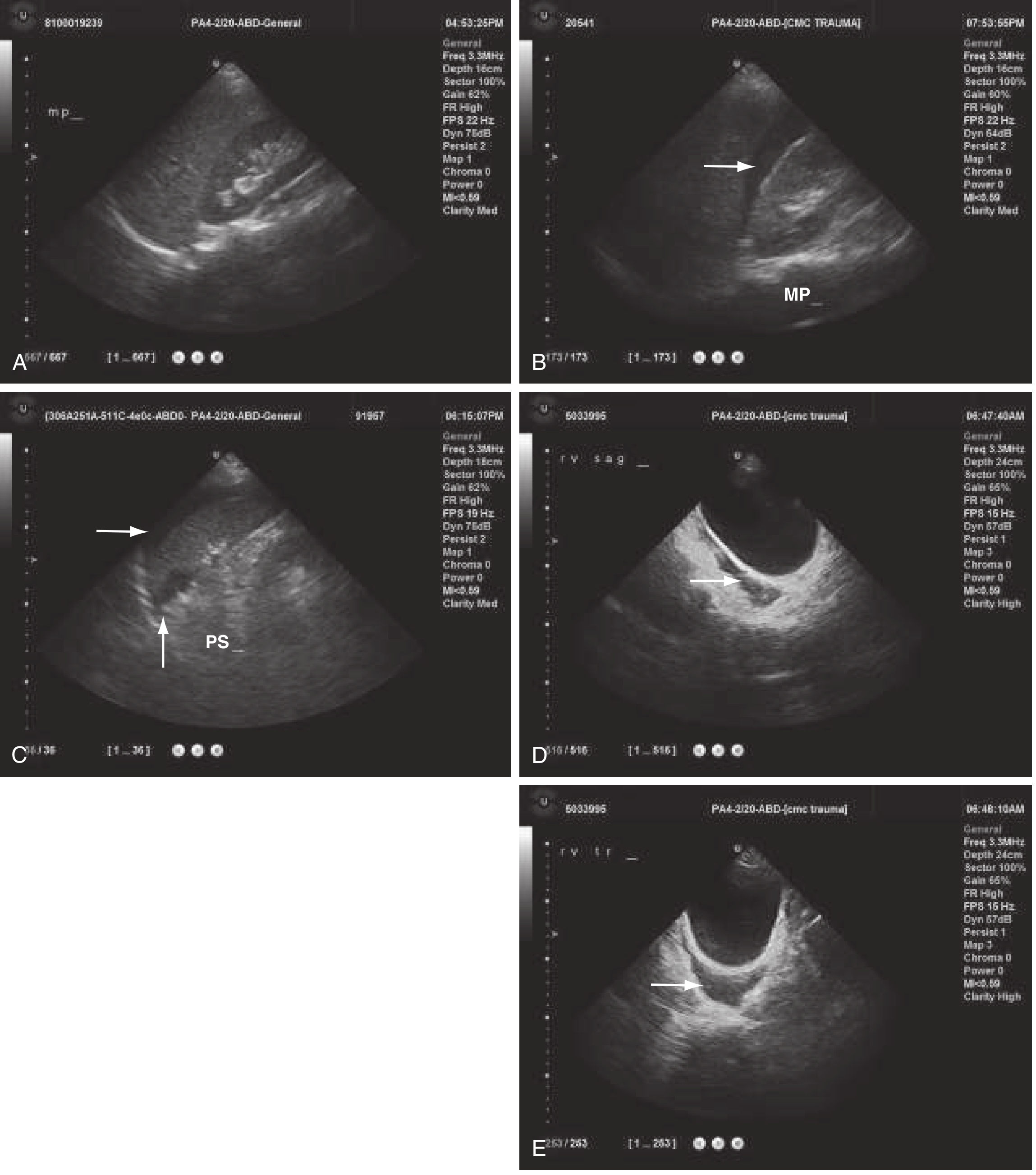
Fig. 38.1 from ROSEN's Emergency Medicine: (A) Normal Morrison's pouch - no anechoic stripe. (B) Positive Morrison's pouch - anechoic stripe (arrow) = haemoperitoneum.
Referred Pain
Pus or fluid in Morrison's pouch can track superiorly to the subphrenic space and irritate the diaphragm. Because the diaphragm is supplied by the phrenic nerve (C3-C5), irritation produces referred pain to the right shoulder tip (cutaneous distribution of C3-C5). - Gray's Anatomy for Students
Pneumoperitoneum
Free gas in the peritoneal cavity pools in Morrison's pouch on a supine plain radiograph, appearing as gas in the right upper quadrant - one of the recognised signs of pneumoperitoneum on a supine film. - Grainger & Allison's Diagnostic Radiology
Summary Table
| Feature | Detail |
|---|---|
| Location | Between right lobe of liver and right kidney |
| Also called | Hepatorenal recess / fossa, subhepatic space |
| Dependent position | Most dependent peritoneal space in supine patient |
| Clinical uses | FAST exam (trauma), abscess site, peritoneal metastasis |
| Free fluid detection | Anechoic stripe on RUQ ultrasound |
| Referred pain | Right shoulder tip (phrenic nerve C3-C5) |
| Pneumoperitoneum | Gas visible here on supine X-ray |
This is a shared conversation. Sign in to Orris to start your own chat.