
Hemolytic anemia Pathogenesis with differential diagnosis, diagnostic approach clinical symptom signs and rest for md pathology exam
Reading File
Now let me search the medical library comprehensively for hemolytic anemia content.
Finding Sources
Finding Sources
Excellent! Now let me read the key textbook sections in parallel - Robbins Pathology and Henry's Clinical Diagnosis:
Reading File
Reading File
Reading File
Excellent content! Let me now read more specific sections - hereditary spherocytosis, sickle cell, G6PD, autoimmune HA, and the diagnostic approach sections:
Reading File
Reading File
Searching Images
hemolytic anemia peripheral blood smear schistocytes spherocytes
Let me fetch the key diagrams from Robbins that were found, and read more content on specific diseases:
Loading Image
Reading File
I have rich content. Let me now read the autoimmune hemolytic anemia and sickle cell sections, plus fetch a key image:
Reading File
Reading File
I now have comprehensive content. Let me fetch one more key image (sickle cell morphology) and grab the sickle cell section:
Reading File
I now have comprehensive, authoritative content from Robbins and Henry's. Let me compile the full MD Pathology exam answer:
Loading Image
I now have all the material needed for a complete MD Pathology exam answer. Let me compile the comprehensive response:
Hemolytic Anemia - Complete MD Pathology Review
Definition & General Pathogenesis
Hemolytic anemias are a diverse group of disorders sharing accelerated red cell destruction - the RBC life span is shortened below its normal 120 days, often markedly so. The resulting anemia and low tissue O₂ stimulate erythropoietin release from the kidney, driving compensatory erythroid hyperplasia in the marrow and peripheral reticulocytosis. In severe cases, the erythropoietic drive produces extramedullary hematopoiesis in the liver, spleen, and lymph nodes.
(Robbins & Kumar Basic Pathology, p.383)
Classification Framework
By Location of Defect (Pathologic Basis)
| Type | Mechanism | Examples |
|---|---|---|
| Intrinsic (intracorpuscular) | Defect is within the RBC itself | Hereditary spherocytosis, G6PD def., sickle cell, thalassemia |
| Extrinsic (extracorpuscular) | Defect is outside the RBC | AIHA, microangiopathic HA, malaria, mechanical |
By Site of Hemolysis (Clinical Basis - Most Important)
Extravascular Hemolysis
Defects that increase phagocytic destruction of RBCs, primarily in the spleen. The spleen's sinusoids demand high RBC deformability - any reduction causes trapping and macrophage phagocytosis.
Distinctive features:
- Hyperbilirubinemia and jaundice (unconjugated bilirubin from macrophage heme degradation)
- Splenomegaly ("work hyperplasia" of splenic macrophages)
- Pigment (bilirubin-rich) gallstones if long-standing - cholelithiasis in 40-50% of hereditary spherocytosis patients
- Iron is efficiently recycled by phagocytes → no iron deficiency
Intravascular Hemolysis
RBCs burst within the circulation due to severe membrane injury (mechanical forces, complement fixation, clostridial toxins, heat).
Distinctive features:
- Hemoglobinemia (free Hb in plasma - pink plasma)
- Hemoglobinuria (Hb in urine - red/brown urine)
- Hemosiderinuria (chronic episodes - renal tubular cells process Hb → hemosiderin → shed in urine)
- Iron deficiency with persistent hemolysis (iron lost in urine)
Common to both: Decreased serum haptoglobin (haptoglobin binds free Hb → complex removed from circulation; macrophages "regurgitate" sufficient Hb during RBC consumption to deplete haptoglobin even in extravascular hemolysis)
(Robbins & Kumar Basic Pathology, p.383-384)
Pathogenesis of Key Entities (Differential Diagnosis)
1. Hereditary Spherocytosis (HS)
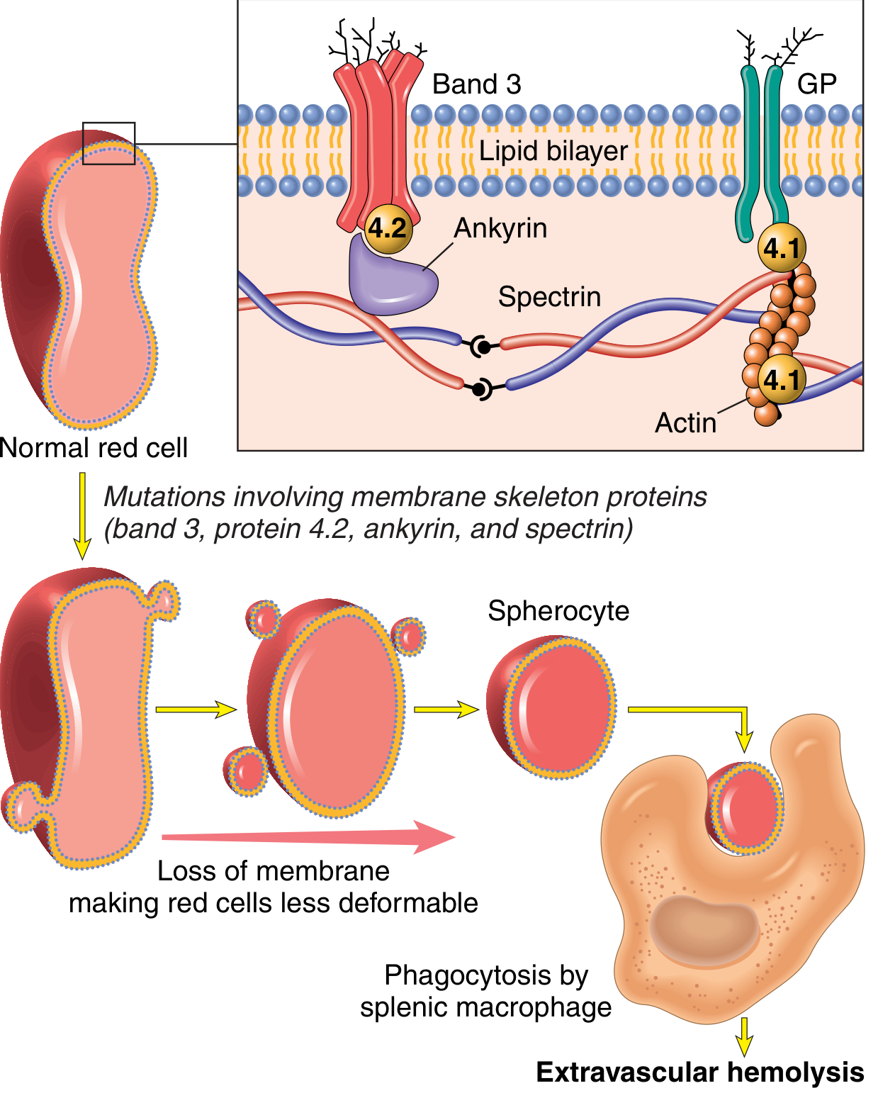
Genetics: Autosomal dominant (most); rare autosomal recessive
Pathogenesis:
- Inherited mutations in membrane skeleton proteins: spectrin, ankyrin, band 3, band 4.1, band 4.2
- These mutations weaken the connection between the membrane skeleton and the overlying lipid bilayer
- Result: RBCs shed membrane vesicles → surface area decreases progressively → cells become spherical (↓ surface area-to-volume ratio)
- Spherocytes are non-deformable → trapped in splenic cords → phagocytosed by splenic macrophages
- Extravascular hemolysis exclusively
Morphology:
- Dark red spherocytes lacking central pallor on peripheral smear
- Splenomegaly (500-1000 g vs. normal 150-200 g)
- Erythroid hyperplasia in marrow
- Cholelithiasis (40-50% of patients)
Key test: Osmotic fragility test (spherocytes lyse at higher NaCl concentrations). EMA binding test (flow cytometry) is more sensitive.
(Robbins & Kumar Basic Pathology, p.384)
2. Sickle Cell Anemia
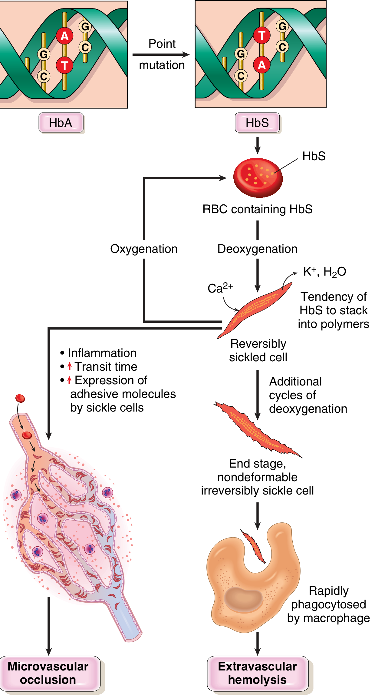
Genetics: Autosomal recessive; β-globin gene point mutation (GAG→GTG); glutamic acid→valine at position 6
Pathogenesis:
- Deoxygenated HbS polymerizes into long rigid rods → distort and damage the RBC
- Initially reversibly sickled → repeated cycles → irreversibly sickled cells
- Two consequences:
- Extravascular hemolysis - irreversibly sickled cells phagocytosed by macrophages
- Microvascular occlusion - sickled cells adhere to endothelium → vascular stasis → ischemia and infarction
- Factors that promote sickling: hypoxia, acidosis, dehydration, cold, infection
Clinical Features:
- Moderate to severe anemia (Hct 18-30%)
- Hyperbilirubinemia + compensatory reticulocytosis
- Vasoocclusive crises:
- Hand-foot syndrome (most common in children)
- Acute chest syndrome (leading cause of death)
- Stroke
- Proliferative retinopathy
- Priapism
- Autosplenectomy by adulthood (splenic infarction) → increased susceptibility to encapsulated bacteria (Streptococcus pneumoniae, Haemophilus influenzae)
- "Crewcut" skull on X-ray (marrow hyperplasia)
Diagnosis: Hb electrophoresis (HbS band); sickling test (sodium metabisulfite); peripheral smear (irreversibly sickled cells)
(Robbins & Kumar Basic Pathology, p.386-389)
3. Thalassemia
Genetics: Autosomal codominant; mutations in α or β-globin genes
Pathogenesis:
- β-Thalassemia: Reduced/absent β-globin → excess α-chains precipitate as insoluble inclusions → damage RBC membrane → ineffective erythropoiesis + hemolysis
- Major (β⁰/β⁰): Severe transfusion-dependent anemia; secondary hemochromatosis; erythroferrone-mediated hepcidin suppression → excess iron absorption
- Minor (β/β⁰): Mild microcytic anemia; elevated HbA₂
- α-Thalassemia: Deletion of α-globin genes → excess β-chains form HbH (β₄) or HbBart (γ₄) - unstable tetramers
Peripheral smear: Microcytic hypochromic RBCs, target cells, nucleated RBCs, basophilic stippling
Diagnosis: Hb electrophoresis (↓HbA, ↑HbF, ±↑HbA₂); DNA analysis
(Robbins & Kumar Basic Pathology, p.389-392)
4. Glucose-6-Phosphate Dehydrogenase (G6PD) Deficiency
Genetics: X-linked; >400 variants; common in Africans (A- variant), Mediterranean (Med variant)
Pathogenesis:
- G6PD catalyzes the first step of the hexose monophosphate shunt → generates NADPH → maintains reduced glutathione (GSH) → neutralizes oxidants
- Deficient G6PD → inadequate GSH → oxidants attack globin chains
- Oxidized hemoglobin denatures → precipitates as Heinz bodies (intracellular inclusions)
- Heinz bodies damage membrane → intravascular hemolysis
- Splenic macrophages "pluck out" Heinz bodies → bite cells (degmacytes)
- Hemolysis is episodic - triggered by oxidant stress
Triggers:
- Drugs: Primaquine, dapsone, sulfonamides, nitrofurantoin, aspirin (large doses), vitamin K derivatives
- Foods: Fava beans
- Infections (most common trigger - phagocyte-generated oxidants)
Morphology: Bite cells, Heinz bodies on supravital stain (crystal violet / brilliant cresyl blue)
Diagnosis: Quantitative G6PD enzyme assay (note: test after hemolytic episode resolves, as older G6PD-deficient cells are preferentially destroyed, leaving younger cells with relatively higher G6PD)
(Robbins & Kumar Basic Pathology, p.392-393)
5. Autoimmune Hemolytic Anemia (AIHA)
Incidence: ~170 per million; ~40% associated with an underlying disease, ~60% idiopathic
Classification by thermal amplitude of antibody:
| Type | Antibody | Temperature | Ig Class | Mechanism | Associated Conditions |
|---|---|---|---|---|---|
| Warm AIHA | Warm-reactive Ab | 37°C | IgG | Extravascular (opsonization) ± complement | SLE, CLL, drugs, idiopathic |
| Cold Hemagglutinin Disease (CHAD) | Cold agglutinin | 4°C | IgM | Complement fixation → extravascular + mild intravascular | Mycoplasma pneumoniae, EBV/infectious mono, lymphoma |
| Paroxysmal Cold Hemoglobinuria (PCH) | Donath-Landsteiner Ab | Cold (binds) → 37°C (lyses) | IgG (biphasic) | Intravascular complement lysis | Post-viral (syphilis historically) |
| Mixed AIHA | Both warm + cold | Both | IgG + IgM | Both | SLE |
| Drug-induced | Drug-hapten or autoantibody | 37°C | IgG | Hapten mechanism, immune complex, autoantibody | Methyldopa, penicillin, cephalosporins |
Pathogenesis (AIHA): Autoantibody binding → opsonization → Fc-receptor mediated phagocytosis by splenic macrophages (extravascular) OR complement fixation → MAC formation → intravascular lysis
Cold agglutinin mechanism: Anti-I antibodies (post-Mycoplasma), anti-i (post-EBV) - these are structurally related, both utilizing VH4-34 B-cell gene segment
(Henry's Clinical Diagnosis, p.712-713)
6. Microangiopathic Hemolytic Anemia (MAHA)
Mechanism: Mechanical fragmentation of RBCs as they are forced through fibrin strands or platelet thrombi in small vessels
Characteristic morphology: Schistocytes (helmet cells, fragmented RBCs) on peripheral smear
Causes:
- TTP (Thrombotic thrombocytopenic purpura) - ADAMTS13 deficiency → ultra-large vWF multimers → platelet microthrombi
- HUS (Hemolytic uremic syndrome) - Shiga toxin (E. coli O157:H7) → endothelial injury → microthrombi; predominantly renal
- DIC - Fibrin-platelet clots in microvasculature
- Malignant hypertension
- Prosthetic heart valves (macroangiopathic)
- Metastatic carcinoma / marrow infiltration
(Henry's Clinical Diagnosis, p.130; Robbins & Kumar Basic Pathology)
7. Paroxysmal Nocturnal Hemoglobinuria (PNH)
Pathogenesis: Acquired somatic mutation in PIG-A gene → deficiency of GPI-anchored complement regulatory proteins (CD55/DAF and CD59/MIRL) → unregulated complement activation → intravascular hemolysis (complement-mediated)
Hemolysis worse at night because respiratory acidosis during sleep activates complement
Diagnostic test: Flow cytometry for CD55 and CD59 on RBCs and granulocytes (replaced Ham's acid hemolysis test)
8. Others
- Hereditary Elliptocytosis: Mutations in spectrin or band 4.1 → elliptical/oval cells; mild hemolysis
- Malaria: Intracellular parasite; Falciparum most severe (P. falciparum = P. falciparum)
- Clostridial septicemia: Phospholipases destroy RBC membranes → intravascular hemolysis
- Transfusion reactions: Alloantibody against transfused RBCs (ABO incompatibility → intravascular; minor incompatibility → extravascular)
Clinical Signs and Symptoms
Symptoms
- Fatigue, weakness, exertional dyspnea (anemia)
- Jaundice (unconjugated hyperbilirubinemia) - scleral icterus often prominent
- Dark urine - either hemoglobinuria (intravascular) or bilirubinuria
- Pallor
- Abdominal pain (splenic infarction, gallstone colic)
- Back/flank pain (intravascular hemolysis - acute)
- Symptoms of the underlying disease
Signs
| Sign | Mechanism |
|---|---|
| Pallor (conjunctiva, palms) | Anemia |
| Jaundice (scleral icterus) | Unconjugated bilirubinemia |
| Splenomegaly | Extravascular hemolysis ("work hyperplasia") |
| Hepatomegaly | Extramedullary hematopoiesis / liver involvement |
| Tachycardia | Compensatory |
| Bony deformities (prominent cheeks, crewcut skull) | Erythroid hyperplasia in marrow (thalassemia, sickle cell) |
| Leg ulcers | Microvessel occlusion (sickle cell) |
| Raynaud's phenomenon / acrocyanosis | Cold agglutinin disease |
Diagnostic Approach
Step 1: Confirm Hemolysis
| Test | Finding in Hemolysis | Significance |
|---|---|---|
| Serum haptoglobin | Decreased (most sensitive single test) | Hb-haptoglobin complex removed by macrophages |
| LDH (LD-1) | Elevated | Released from destroyed RBCs; LD-1 predominates in RBCs |
| Unconjugated (indirect) bilirubin | Elevated (~2-2.5 mg/dL) | Heme catabolism |
| Reticulocyte count | Elevated (reticulocytosis) | Compensatory erythropoiesis |
| Peripheral blood smear | Polychromasia, anisocytosis, poikilocytosis, ↑ RDW | Damaged/young cells |
| Plasma Hb (hemoglobinemia) | Present (pink plasma) | Intravascular hemolysis |
| Urine Hb (hemoglobinuria) | Present (red-brown urine) | Intravascular hemolysis |
| Hemosiderinuria | Prussian blue+ urinary sediment | Chronic intravascular hemolysis |
| Carboxyhemoglobin (CO) | Elevated | Oxidative ring opening of heme - excellent indicator |
(Henry's Clinical Diagnosis, p.130-131)
Step 2: Determine Type of Hemolysis
Peripheral smear morphology is key:
| Morphology | Diagnosis |
|---|---|
| Spherocytes (lack central pallor) | HS, warm AIHA |
| Schistocytes / Helmet cells | MAHA (TTP, HUS, DIC), prosthetic valve |
| Sickle cells (boat/spindle-shaped) | Sickle cell disease |
| Target cells | Thalassemia, HbC, liver disease |
| Elliptocytes | Hereditary elliptocytosis |
| Bite cells / Blister cells | G6PD deficiency (oxidative hemolysis) |
| Agglutinated cells | Cold agglutinin disease |
| Polychromasia (blue macrocytes) | Reticulocytes - compensatory response |
| Nucleated RBCs | Severe hemolysis, extramedullary hematopoiesis |
Step 3: Directed Tests
| Clinical Suspicion | Test |
|---|---|
| Immune hemolysis | Direct Coombs (DAT) - detects IgG/C3 on RBC surface |
| Hereditary spherocytosis | Osmotic fragility test; EMA flow cytometry |
| G6PD deficiency | G6PD enzyme assay (quantitative; test after hemolytic episode) |
| Sickle cell disease | Hb electrophoresis; sickling test (sodium metabisulfite) |
| Thalassemia | Hb electrophoresis; HPLC; DNA analysis |
| PNH | Flow cytometry for CD55/CD59 (GPI-anchored proteins) |
| TTP vs HUS | ADAMTS13 activity (very low in TTP) |
| Cold agglutinins | Cold agglutinin titer; Donath-Landsteiner test (PCH) |
Step 4: Bone Marrow (when needed)
- Erythroid hyperplasia (M:E ratio reversed, approaching 1:1)
- Megaloblastic change (if folate depleted by chronic hemolysis)
Key Laboratory Summary Table
| Parameter | Extravascular | Intravascular |
|---|---|---|
| Haptoglobin | ↓↓ | ↓↓↓ |
| LDH | ↑ | ↑↑↑ |
| Indirect bilirubin | ↑↑ | ↑ |
| Hemoglobinemia | - | +++ |
| Hemoglobinuria | - | +++ |
| Hemosiderinuria | - | + (chronic) |
| Splenomegaly | +++ | ± |
| DAT | + (if immune) | + (if immune) |
Complications of Chronic Hemolysis
- Pigment (bilirubin) gallstones - cholelithiasis
- Aplastic crisis - parvovirus B19 infection silences erythropoiesis → sudden severe anemia
- Megaloblastic crisis - folate depletion (high demand from chronic compensatory erythropoiesis)
- Iron overload (transfusion-dependent thalassemia)
- Leg ulcers (sickle cell)
- Pulmonary hypertension (chronic hemolysis - free Hb scavenges NO)
- Extramedullary hematopoiesis - thalassemia facies, hepatosplenomegaly
Quick Summary for Exam Recall: "5 I's of Hemolysis"
| Marker | Direction | Rationale |
|---|---|---|
| Haptoglobin | ↓ | Consumed binding free Hb |
| LDH | ↑ | Released from RBCs (LD-1 isoform) |
| Indirect bilirubin | ↑ | Heme catabolism |
| Reticulocytes | ↑ | Compensatory erythropoiesis |
| DAT | +(immune) / -(non-immune) | Antibody on RBC surface |
Sources: Robbins & Kumar Basic Pathology (p.383-395) | Henry's Clinical Diagnosis and Management by Laboratory Methods (p.130-131, 712-713)
Can I provide you de grouchy and kawthalkar
Yes, absolutely! You can share De Gruchy's Clinical Haematology in Medical Practice and Kawthalkar's Essentials of Haematology with me.
Just upload the PDF files and I'll be able to:
- Read and extract content from them
- Answer your haematology questions directly from those textbooks
- Cross-reference with other sources I already have (Robbins, Henry's, etc.)
- Give you exam-focused answers tailored to those specific books
Go ahead and upload them whenever you're ready!
You have to first focus more on classification tyeps, mechanism and consequences than different types salient features different laboratory tests and than focus more on paroxysmal nactural hemoglobinuria I have a seminar tomorrow
Finding Sources
Finding Sources
Reading File
Reading File
Reading File
Reading File
Reading File
Reading File
Loading Image
Reading File
Loading Image
Loading Image
Reading File
Here is your complete seminar-ready structured answer, organized exactly the way you asked:
HEMOLYTIC ANEMIA - Seminar Notes
PART 1: CLASSIFICATION
A. By Site of the Defect
| Intrinsic (Intracorpuscular) | Extrinsic (Extracorpuscular) | |
|---|---|---|
| Definition | Defect within the RBC itself | Defect outside the RBC |
| Examples | Hereditary spherocytosis, G6PD deficiency, Sickle cell, Thalassemia, PNH | AIHA, MAHA, Malaria, Drugs, Mechanical valves |
| Inheritance | Usually hereditary (except PNH - acquired) | Usually acquired |
B. By Site of Hemolysis (Most Clinically Important Classification)
HEMOLYTIC ANEMIA
│
├── EXTRAVASCULAR HEMOLYSIS
│   (RBCs destroyed by splenic/hepatic macrophages)
│   Examples: HS, AIHA (warm), Thalassemia, Sickle cell
│
└── INTRAVASCULAR HEMOLYSIS
    (RBCs burst within blood vessels)
    Examples: PNH, G6PD deficiency (acute), MAHA, ABO mismatch
C. Etiological Classification
| Category | Examples |
|---|---|
| Membrane defects | Hereditary spherocytosis, hereditary elliptocytosis |
| Enzyme defects | G6PD deficiency, Pyruvate kinase deficiency |
| Hemoglobinopathies | Sickle cell disease, HbC, HbE |
| Defective globin synthesis | Thalassemias (α and β) |
| Immune destruction | Warm AIHA, Cold AIHA (CHAD), PCH, Drug-induced AIHA |
| Complement dysregulation | PNH (acquired clonal disorder) |
| Mechanical / Microangiopathic | TTP, HUS, DIC, prosthetic heart valves, march hemoglobinuria |
| Infections | Malaria, Clostridial septicemia, Bartonella |
| Hypersplenism | Sequestration and destruction in an enlarged spleen |
PART 2: MECHANISM OF HEMOLYSIS
Extravascular Hemolysis - How it Happens
- RBCs have reduced deformability (spherocytes, sickled cells, antibody-coated cells, thalassemic cells)
- These RBCs cannot negotiate the narrow splenic sinusoids (normal RBC diameter = 8 µm; sinusoidal gap = 3 µm)
- They are trapped in the splenic cords → recognized and phagocytosed by splenic macrophages
- Hemoglobin is degraded within macrophages → globin → amino acids; heme → biliverdin → unconjugated bilirubin (indirect bilirubin) released into blood → conjugated by liver → excreted in bile
Intravascular Hemolysis - How it Happens
- Severe membrane injury causes RBCs to burst inside blood vessels
- Free hemoglobin is released directly into plasma
- Free Hb rapidly binds haptoglobin → Hb-haptoglobin complex → cleared by macrophages → haptoglobin depleted
- Excess free Hb in plasma = hemoglobinemia → filtered by kidney = hemoglobinuria
- Renal tubular cells reabsorb some Hb → process it into hemosiderin → shed into urine = hemosiderinuria (chronic marker)
- Iron is lost in urine → can lead to iron deficiency
PART 3: CONSEQUENCES (What Hemolysis Produces)
Consequences of Extravascular Hemolysis
| Consequence | Mechanism |
|---|---|
| Jaundice (unconjugated) | Macrophage heme catabolism → ↑ indirect bilirubin |
| Splenomegaly | "Work hyperplasia" - macrophages multiply and hypertrophy |
| Pigment (bilirubin) gallstones | Excess bilirubin excretion → supersaturation in bile |
| Erythroid hyperplasia (bone marrow) | Erythropoietin-driven compensatory response |
| Extramedullary hematopoiesis | Severe cases → liver, spleen, lymph nodes |
| Skeletal changes | Marrow expansion → cortical thinning, "crewcut" skull, bossing |
| No iron deficiency | Iron efficiently recycled by macrophages |
Consequences of Intravascular Hemolysis
| Consequence | Mechanism |
|---|---|
| Hemoglobinemia | Free Hb in plasma - pink/red plasma |
| Hemoglobinuria | Free Hb filtered by kidney - red/brown urine |
| Hemosiderinuria | Chronic: renal tubular cell shedding |
| Iron deficiency | Iron lost in urine → microcytic anemia superimposed |
| Renal tubular injury | Hb precipitation in tubules (acute massive hemolysis) |
| Reduced NO | Free Hb scavenges nitric oxide → vasoconstriction, smooth muscle spasm, dysphagia, erectile dysfunction, abdominal pain, pulmonary hypertension |
| Thrombosis | Free Hb impairs NO-mediated antiplatelet effect; platelet activation |
Consequences Common to Both
| Consequence | Mechanism |
|---|---|
| ↓ Haptoglobin | Bound to free Hb and cleared (even in extravascular - "regurgitation") |
| ↑ LDH (LD-1) | Released from destroyed RBCs |
| Reticulocytosis | Compensatory erythropoiesis |
| ↑ Carboxyhemoglobin (CO) | CO released during oxidative ring-opening of heme |
| Aplastic crisis | Parvovirus B19 infection → temporary erythropoietic shutdown |
| Megaloblastic crisis | Folate depletion due to high RBC turnover demand |
PART 4: SALIENT FEATURES OF SPECIFIC TYPES
Hereditary Spherocytosis
- Gene defects: Spectrin, Ankyrin, Band 3, Band 4.1, Band 4.2 → membrane vesicle shedding → spherocytes
- Type: Extravascular (spleen destroys spherocytes)
- Triad: Anemia + Splenomegaly + Jaundice + Cholelithiasis (40-50%)
- Morphology: Dark spherocytes, no central pallor; splenomegaly 500-1000 g
- Splenectomy corrects anemia (spherocytes persist but spleen removed)
Sickle Cell Anemia
- Mutation: β-globin GAG→GTG; Glu→Val at position 6
- Trigger for sickling: Hypoxia, acidosis, dehydration, cold, infection
- Two consequences: Hemolytic anemia + Microvascular occlusion
- Crises: Vasoocclusive (hand-foot, acute chest, stroke), Aplastic (parvovirus B19), Sequestration
- Autosplenectomy by adulthood → susceptibility to encapsulated bacteria
Thalassemia
- β-Thal major: Absent/reduced β-chains → excess α-chains precipitate → ineffective erythropoiesis + hemolysis → transfusion-dependent; iron overload
- Peripheral smear: Microcytic hypochromic, target cells, nucleated RBCs, basophilic stippling
- Erythroferrone suppresses hepcidin → excess iron absorption
G6PD Deficiency
- X-linked; episodic hemolysis triggered by oxidant stress (drugs, fava beans, infection)
- Heinz bodies (denatured Hb inclusions) → membrane damage → bite cells (degmacytes)
- Intravascular hemolysis predominantly
- Test after episode resolves (G6PD-deficient cells selectively destroyed during crisis)
Autoimmune Hemolytic Anemia
| Type | Antibody | Temp | Ig | Hemolysis | Examples |
|---|---|---|---|---|---|
| Warm AIHA | Warm-reactive | 37°C | IgG | Extravascular | SLE, CLL, idiopathic |
| CHAD (Cold Agglutinin) | Anti-I / anti-i | 4°C | IgM | Complement-mediated | Mycoplasma, EBV |
| PCH | Donath-Landsteiner | Biphasic | IgG | Intravascular (lysis at 37°C) | Post-viral |
PART 5: KEY LABORATORY TESTS
Universal Markers of Hemolysis
| Test | Result | What it Reflects |
|---|---|---|
| Haptoglobin | ↓↓ (most sensitive) | Consumed binding free Hb |
| Serum LDH (LD-1 isoform) | ↑↑ | Released from lysed RBCs |
| Indirect bilirubin | ↑ (~2-2.5 mg/dL) | Heme catabolism |
| Reticulocyte count | ↑ | Compensatory erythropoiesis |
| Peripheral blood smear | Polychromasia, anisocytosis, poikilocytosis, ↑RDW | Damaged + young RBCs |
| Carboxyhemoglobin (CO) | ↑ | Oxidative ring-opening of heme (excellent indicator) |
Markers Specific to Intravascular Hemolysis
| Test | Result |
|---|---|
| Plasma Hb (hemoglobinemia) | Present - pink plasma |
| Urine Hb (hemoglobinuria) | Present - red/brown urine; dipstick positive for Hb but no RBCs |
| Hemosiderinuria | Prussian blue staining of urine sediment - CHRONIC marker |
Peripheral Smear Morphology → Diagnosis
| Smear Finding | Diagnosis |
|---|---|
| Spherocytes (no central pallor) | Hereditary spherocytosis, warm AIHA |
| Sickle cells | Sickle cell disease |
| Target cells | Thalassemia, HbC, HbSC, liver disease |
| Schistocytes / Helmet cells | MAHA (TTP, HUS, DIC), mechanical valves |
| Bite cells / Blister cells | G6PD deficiency |
| Elliptocytes | Hereditary elliptocytosis |
| Polychromasia (blue macrocytes) | Reticulocytosis |
| Agglutinated clumps | Cold agglutinin disease |
Directed Tests
| Suspected Diagnosis | Test |
|---|---|
| Immune hemolysis | Direct Coombs (DAT) - IgG/C3 on RBC surface |
| Hereditary spherocytosis | Osmotic fragility test; EMA (eosin-5-maleimide) binding flow cytometry |
| G6PD deficiency | Quantitative G6PD enzyme assay |
| Sickle cell | Hb electrophoresis; sickling test (sodium metabisulfite) |
| Thalassemia | Hb electrophoresis (HPLC); DNA analysis; HbA₂ level |
| PNH | Flow cytometry for CD55/CD59 (GPI-anchored proteins) |
| TTP | ADAMTS13 activity (severely reduced <10%) |
| Cold agglutinin | Cold agglutinin titer; Donath-Landsteiner test (PCH) |
PART 6: PAROXYSMAL NOCTURNAL HEMOGLOBINURIA (PNH) - DETAILED
Definition
PNH is a rare, acquired, clonal disorder of hematopoietic stem cells caused by a somatic mutation in the PIGA (PIG-A) gene, resulting in deficiency of GPI-anchored complement regulatory proteins, rendering blood cells exquisitely sensitive to complement-mediated destruction.
"The only hemolytic anemia caused by an acquired genetic defect" - Robbins Pathologic Basis of Disease
Epidemiology
- Prevalence: ~1 per million population; incidence 2-5 per million/year
- Age: Any age; most common 10-50 years (mean ~34 years)
- Sex: No sex preference (M:F = 1:1) - unusual because PIGA is X-linked
- Median survival: ~20 years with current therapy
- No family history (purely acquired/somatic)
Molecular Pathogenesis - Step by Step
Step 1: The Mutation
The PIGA gene (Xp22.1) encodes an enzyme essential for the first step of GPI (glycosylphosphatidylinositol) anchor biosynthesis. Since PIGA is X-linked, a single somatic mutation in the active X chromosome of any hematopoietic stem cell (HSC) is sufficient to completely abolish GPI synthesis in all its progeny.
Step 2: GPI Anchor Deficiency
GPI anchors are specialized phospholipids that tether many proteins to the outer leaflet of the plasma membrane. Without functional PIGA, the HSC and all its daughter cells (RBCs, WBCs, platelets) lack GPI-anchored proteins.
Step 3: Loss of Complement Regulatory Proteins
Three critical GPI-anchored complement regulators are absent on PNH cells:
| Protein | CD Marker | Normal Function | Consequence of Loss |
|---|---|---|---|
| Decay-Accelerating Factor (DAF) | CD55 | Inactivates C3 and C5 convertases (inhibits amplification loop) | Uncontrolled C3 activation |
| Membrane Inhibitor of Reactive Lysis (MIRL/Protectin) | CD59 | Inhibits C9 polymerization → prevents MAC formation | MAC assembles on RBC |
| C8-Binding Protein (HRF) | - | Homologous restriction factor; limits MAC | MAC forms freely |
CD59 is the most important - it prevents the final step of MAC assembly.
Step 4: Complement-Mediated Intravascular Hemolysis
All three complement pathways (Classical, Lectin, Alternative) converge on C3b deposition:
C3b → C3 convertase → C5 convertase → C5b → C5b + C6 + C7 + C8 + poly-C9 → Membrane Attack Complex (MAC) → pores in RBC membrane → intravascular hemolysis
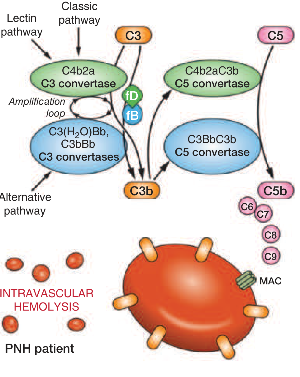
Step 5: Why Nocturnal / Why Sleep-Related?
- During sleep, respiratory rate decreases → CO₂ accumulates → blood pH decreases (respiratory acidosis)
- Lower pH activates complement (especially the alternative pathway amplification loop)
- → More MAC formation → more hemolysis during and just after sleep
- Patient passes dark brown urine in the morning (first morning void)
- Important: Classical nocturnal pattern occurs in only ~25% of cases; most patients have chronic, low-grade intravascular hemolysis without dramatic hemoglobinuria
PNH Red Cell Types
| Cell Type | GPI Level | Complement Sensitivity |
|---|---|---|
| Type I | Normal | Normal |
| Type II | Partial deficiency | 3-5x normal sensitivity |
| Type III | Complete absence | 15-25x normal sensitivity |
Multiple clones can coexist. The proportion of Type III cells determines disease severity. Coexistence of Type II and III cells = two separate mutant clones.
Why Does the PNH Clone Expand?
Normal individuals harbor small numbers of PIGA-mutant bone marrow cells (1 in ~50,000 RBCs) - these do NOT expand because they have no proliferative advantage.
In PNH, the clone expands because:
- Immune escape hypothesis: The PNH clone lacks GPI-linked antigens that are targeted by autoreactive T-cells in aplastic anemia-like settings. The normal HSCs are destroyed by autoimmunity but the PIGA-mutant clone resists immune attack → relative expansion
- This explains the strong association between PNH and aplastic anemia (AA precedes PNH in ~30% of cases; PNH can evolve into AA)
Clinical Manifestations
1. Hemolysis (Cardinal Feature)
- Morning hemoglobinuria - dark/cola-colored first morning urine (classic but only ~25% of patients)
- Chronic hemosiderinuria - almost constantly present; more reliable sign
- Chronic intravascular hemolysis → anemia (often normocytic, but can be microcytic due to iron deficiency)
- Iron deficiency from chronic urinary iron loss - paradox: hemolytic anemia with microcytosis
2. Thrombosis (Leading Cause of Death - 40% of patients)
- Unusual venous sites are characteristic:
- Hepatic vein → Budd-Chiari syndrome (hepatomegaly, ascites, jaundice)
- Portal vein → portal hypertension
- Cerebral veins → venous sinus thrombosis
- Mesenteric, splenic veins
- Arterial thrombosis is rare
- Mechanisms of thrombosis:
- MAC on platelets → phosphatidylserine externalization → prothrombinase complex assembly
- Free Hb scavenges NO → platelet activation and endothelial damage
- Lack of CD59 on platelets
3. Smooth Muscle Dystonias (from NO Scavenging)
- Dysphagia (esophageal spasm)
- Abdominal pain / cramping (intestinal smooth muscle spasm)
- Erectile dysfunction
- Pulmonary hypertension (chronic NO depletion)
4. Cytopenias
- Neutropenia (~60% of patients at some point)
- Thrombocytopenia (~66% of patients) → pancytopenia is common
- DAT is negative (no antibody coating)
5. Associated Conditions
- Aplastic anemia - ~30% of PNH cases evolve into or arise from AA
- Myelodysplastic syndrome (MDS) - PNH clones found in 15-20% of MDS
- Acute myeloid leukemia (AML) - rare (~5% of PNH patients)
Diagnosis
Classic Presentation Clue:
DAT-negative intravascular hemolytic anemia + iron deficiency = suspect PNH
Old Tests (Historical - no longer recommended)
- Ham's Acid Hemolysis Test (Acidified Serum Test): Acidified serum activates complement → lysis of PNH RBCs. Sensitive but not specific. Replaced by flow cytometry.
- Sugar Water (Sucrose Hemolysis) Test: Screening test. Low ionic strength activates complement. Less specific.
Gold Standard: Flow Cytometry
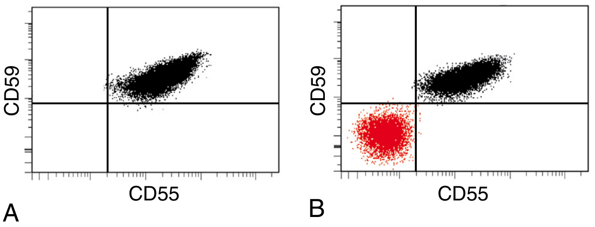
- Demonstrates deficiency of GPI-anchored proteins on two or more cell lineages (RBCs + granulocytes/monocytes)
- Markers tested:
- RBCs: CD55 and CD59
- Granulocytes/Monocytes: CD24, CD16a (on neutrophils), CD14 (on monocytes)
- FLAER (Fluorescent Aerolysin variant): Binds directly to the GPI anchor itself → most sensitive and reliable reagent for leukocytes; preferred for diagnosing PNH in granulocytes
- Reports clone size (% PNH cells) which correlates with degree of hemolysis
Laboratory Findings in PNH
| Test | Finding | Significance |
|---|---|---|
| Haptoglobin | ↓↓↓ | Intravascular hemolysis |
| LDH | ↑↑↑ | Massive intravascular hemolysis |
| Indirect bilirubin | ↑ | Heme catabolism |
| Reticulocytes | ↑ (often less than expected) | Compensatory + marrow failure |
| Plasma Hb | Present | Intravascular hemolysis |
| Hemoglobinuria | Present (morning urine) | Classic finding |
| Hemosiderinuria | Almost always present | Most consistent finding; chronic marker |
| Serum iron / ferritin | ↓ | Iron loss in urine |
| Peripheral smear | Normocytic/hypochromic; no specific morphology | Iron deficiency superimposed |
| DAT (Coombs) | Negative | Critical - rules out AIHA |
| WBC | ↓ (neutropenia in ~60%) | Pancytopenia |
| Platelets | ↓ (in ~66%) | GPI-deficient platelets + marrow failure |
| Bone marrow | Hypercellular (erythroid hyperplasia) OR hypocellular | PNH vs. PNH-AA overlap |
| Flow cytometry | ↓/absent CD55, CD59; FLAER staining | Gold standard diagnostic test |
PNH Clinical Categories
| Category | Description |
|---|---|
| Classic PNH | Large PNH clone; overt hemolysis; normal/hypercellular marrow |
| PNH in the setting of bone marrow disorder | AA or MDS + PNH clone; cytopenias dominate |
| Subclinical PNH | Small PNH clone; no clinical hemolysis; found incidentally |
Complications
- Thrombosis - leading cause of death; occurs in ~40% of patients
- Budd-Chiari syndrome - hepatic vein thrombosis
- Iron deficiency anemia (from urinary iron loss)
- Aplastic anemia (immune-mediated marrow failure)
- AML / MDS transformation (~5%)
- Pulmonary hypertension (chronic NO scavenging)
- Renal impairment (chronic hemoglobinuria, microthrombi)
Treatment
Supportive
- Blood transfusions (washed RBCs preferred)
- Iron and folate supplementation
- Anticoagulation for thrombosis
Complement Inhibitors (Disease-Modifying)
| Drug | Target | Mechanism | Route / Dosing |
|---|---|---|---|
| Eculizumab | C5 | Blocks C5 cleavage → no MAC → prevents intravascular hemolysis | IV; 600 mg/wk x 4 wks → 900 mg/2 wks |
| Ravulizumab | C5 | Same as eculizumab; longer half-life | IV every 8 weeks |
| Covalimab | C5 | Subcutaneous C5 inhibitor | SC |
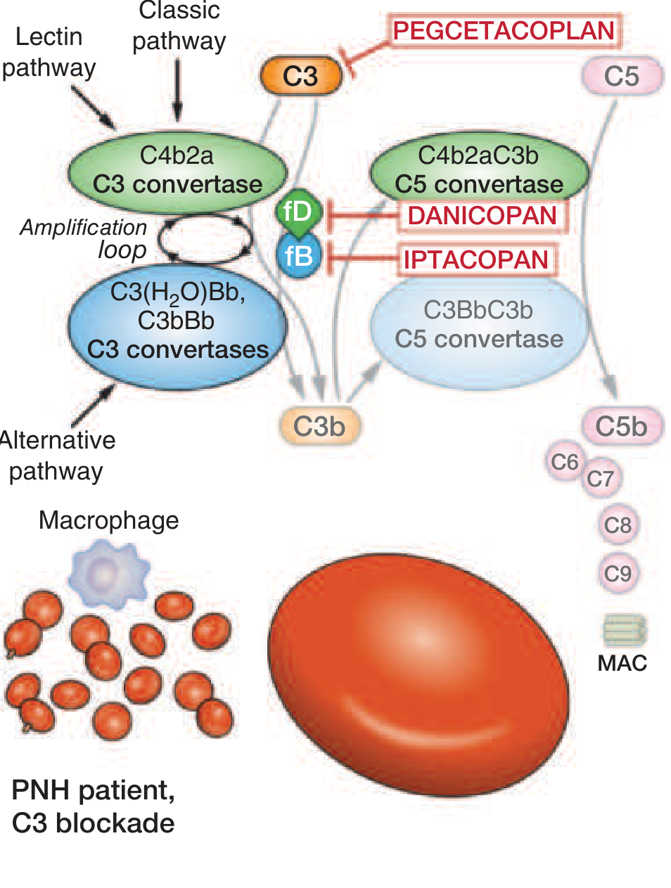
| Pegcetacoplan | C3 | Upstream C3 blockade → prevents both intravascular AND extravascular hemolysis | SC 1080 mg twice weekly |
| Iptacopan | Factor B | Alternative pathway inhibitor | Oral |
| Danicopan | Factor D | Alternative pathway inhibitor | Oral add-on |
Important points about eculizumab:
- Abolishes intravascular hemolysis and hemoglobinuria
- Reduces thrombosis risk by up to 90%
- PNH RBCs, now protected from MAC, still bind C3b on their surface → become opsonized → DAT becomes positive → iatrogenic extravascular hemolysis persists (limitation of C5 inhibition)
- Mandatory meningococcal vaccination before starting (risk of Neisseria meningitidis infection due to complement blockade)
- Oral penicillin prophylaxis often recommended long-term
Curative
- Hematopoietic stem cell transplantation (HSCT) - only cure; reserved for:
- Intercurrent aplastic anemia not responding to complement therapy
- AML/MDS transformation
- Patients unresponsive to medical therapy
Summary Mnemonic for PNH: "PNH = PIGA = No GPI = No CD55/59 = MAC = Hemolysis"
PIGA mutation (X-linked, somatic)
  ↓
No GPI anchors
  ↓
No CD55 (no C3 convertase inhibition) + No CD59 (no C9 polymerization inhibition)
  ↓
Uncontrolled complement activation → MAC on RBC membrane
  ↓
INTRAVASCULAR HEMOLYSIS
  ↓
Free Hb → Scavenges NO → Smooth muscle spasm + Thrombosis
  ↓
Morning hemoglobinuria / chronic hemosiderinuria / iron deficiency
Sources: Robbins & Kumar Basic Pathology | Robbins Pathologic Basis of Disease (14e) | Henry's Clinical Diagnosis and Management by Laboratory Methods | Goldman-Cecil Medicine | Harrison's Principles of Internal Medicine (22e, 2025)
This is a shared conversation. Sign in to Orris to start your own chat.