
Endoscopic dacryoxystorhinostomy in detail, anatominof lacrimal gland, sac syringing perfectly explained and whole surgical steps along with complications, according to standard ENT textbook in simplified manner
endoscopic dacryocystorhinostomy DCR lacrimal sac surgical anatomy
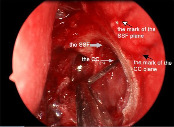
This endoscopic clinical photograph depicts a surgical field during an endoscopic dacryocystorhinostomy (DCR). The circular frame shows a view of the lateral nasal wall where the lacrimal sac has been exposed through marsupialization. Key anatomical structures identified include the sac superior fundus (SSF), marked with a gray arrow at the superior aspect of the sac, and the common canaliculus (CC) opening, indicated by a second gray arrow. To ensure anatomical accuracy, two preoperative reference markings are visible on the surrounding nasal mucosa as shallow horizontal incisions: a white arrow with a black edge points to the SSF plane mark, and a solid black arrow points to the CC plane mark. These markings are oriented at right angles to the long axis of the lacrimal sac. A surgical probe or instrument is seen entering from the inferior aspect of the frame to assist in localizing and measuring these structures. This visual demonstrates the verification of preoperative surface landmarks against actual intranasal anatomy for effective lacrimal system drainage.
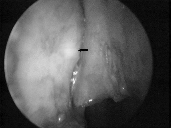
This endoscopic clinical photograph displays the internal anatomy of the lateral nasal cavity during a dacryocystorhinostomy (DCR) procedure. The image focuses on the bony lateral nasal wall, where the overlying mucosa has been surgically reflected or removed to expose the underlying bone. A black arrow indicates a distinct 'transillumination point,' a localized area of increased brightness. This point represents light from an optical fiber probe inserted through the lacrimal canaliculus, used to precisely locate the lacrimal sac through the lacrimal bone. The surrounding structures include the vertical orientation of the bony wall and adjacent mucosal folds. This diagnostic and surgical imaging technique is essential for ophthalmological and ENT surgeons to identify the correct site for creating an osteotomy, ensuring direct access to the lacrimal sac for bypass of a nasolacrimal duct obstruction. The visual provides a clear example of intraoperative navigation and anatomical localization within the nasal meatus.
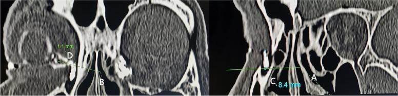
This diagnostic image consists of two parasagittal computed tomographic dacryocystography (CT-DCG) views of the lacrimal drainage system and nasal cavity. The imaging demonstrates the spatial anatomy of a small lacrimal sac relative to key surgical landmarks used in endoscopic dacryocystorhinostomy (DCR). In the left panel, the common canaliculus (CC) is identified, with a horizontal reference line (B) drawn at a right angle to the sac's long axis; measurement 'D' indicates the height of the sac fundus above the CC (1.1 mm). The right panel focuses on the middle turbinate axilla (MTA), with a reference line (A) drawn perpendicular to the sac; measurement 'C' shows the height of the sac fundus above the MTA (8.4 mm). Visible structures include the contrast-filled lacrimal sac, the maxillary frontal process, ethmoid air cells, and the middle turbinate. This imaging approach provides precise preoperative localization of the lacrimal sac and fundus in relation to intranasal landmarks, essential for planning surgical osteotomy and mucosal incisions.
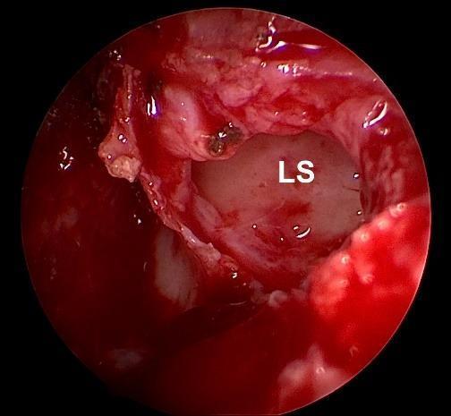
This endoscopic clinical photograph depicts an intraoperative view during an endonasal dacryocystorhinostomy (DCR). The focal point is a wide, circular marsupialization of the lacrimal sac (LS), indicated by the label. The interior mucosa of the lacrimal sac appears pale, smooth, and whitish, contrasting with the surrounding hyperemic and vascularized nasal mucosa. The surgical field shows active but controlled bleeding and moist tissues, typical of an endonasal procedure. The anatomical region shown is the lateral nasal wall where the lacrimal bone and frontal process of the maxilla were removed to expose the sac. This image demonstrates a successful surgical opening that is sufficiently large to maintain patency without the need for silicone stenting. Key educational concepts include lacrimal system anatomy, surgical management of nasolacrimal duct obstruction, and endoscopic visualization techniques in otolaryngology and ophthalmology.
lacrimal drainage system anatomy punctum canaliculus nasolacrimal duct diagram
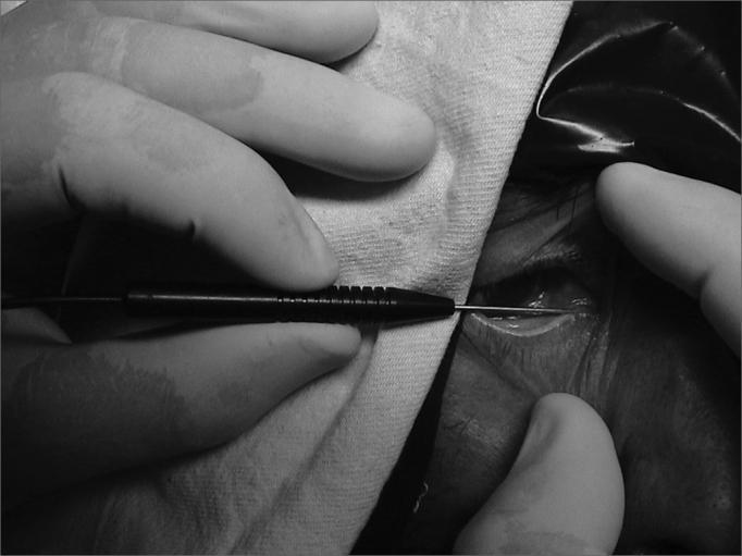
This monochrome intraoperative clinical photograph demonstrates the insertion of a thin metallic optic fiber into the inferior lacrimal canaliculus of a patient. The procedure is part of a dacryocystorhinostomy (DCR) or similar nasolacrimal duct intervention where transillumination is required. A medical professional, wearing sterile surgical gloves, is seen manipulating the instrument. The optic fiber is attached to a black cylindrical handle with textured grip markings for precise control. The fiber enters through the lacrimal punctum of the lower eyelid, following the anatomical path of the canaliculus toward the lacrimal sac. Visible anatomical landmarks include the lower eyelid margin, periorbital skin folds, and the medial canthal region. The surgical field is partially draped with a sterile cloth, focusing the view on the lacrimal drainage system. This image serves as an educational guide for ophthalmology and otolaryngology trainees on the technique of canalicular probing and the use of transillumination to identify the lacrimal fossa from a nasal perspective.
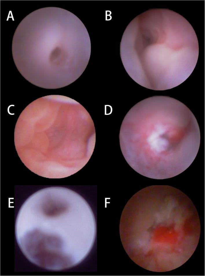
A series of six dacryoendoscopic images (A-F) illustrating the clinical appearance of the lacrimal drainage system in pediatric patients with membranous congenital nasolacrimal duct obstruction (CNLDO). Image A shows a normal, smooth lacrimal canaliculus for baseline comparison. Image B depicts the common canalicula area including the Valve of Rosenmüller. Images C-F highlight various pathological findings associated with failed prior probing and persistent obstruction: (C) shows the primary obstruction point at the distal end of the nasolacrimal duct (NLD); (D) demonstrates an irregular, white-colored scar between the lacrimal sac and the canaliculus; (E) reveals a false passage near the common canaliculus, a common complication of blind probing; and (F) displays mucosal inflammation with associated purulent secretions within the duct. These images provide critical diagnostic visual markers for ophthalmologists identifying reasons for probing failure, such as scarring, false passages, or persistent distal membranes (Hasner's valve), and emphasize the utility of direct endoscopic visualization for precise surgical management.
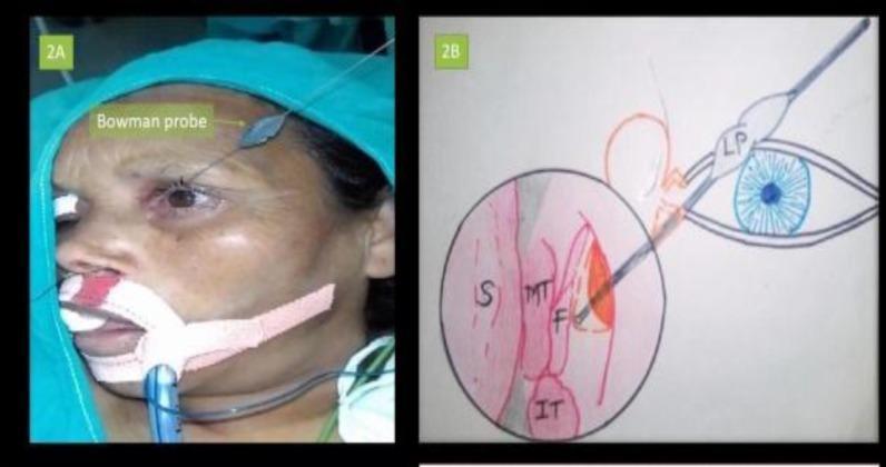
A multi-panel image illustrating the Bowman’s technique for lacrimal probing during endonasal dacryocystorhinostomy (DCR). Panel 2A is a clinical procedural photograph of a patient under general anesthesia, prepped with surgical drapes and an endotracheal tube. A Bowman lacrimal probe is visible, inserted through the superior punctum of the left eye to navigate the lacrimal drainage system. Panel 2B is a corresponding anatomical diagram and endoscopic view illustration. It depicts the lacrimal probe (LP) passing through the canalicular system and emerging into the nasal cavity. Key endonasal landmarks are labeled: Nasal Septum (S), Middle Turbinate (MT), Inferior Turbinate (IT), and the surgically created Mucosal Flap (F). This visual aids in understanding the spatial relationship between external ophthalmic structures and endonasal anatomy during surgical management of nasolacrimal duct obstruction. The content is designed for intermediate to advanced medical education in ophthalmology and otolaryngology.
Endoscopic Dacryocystorhinostomy (En-DCR): Complete Guide
Part 1: Anatomy of the Lacrimal Drainage System
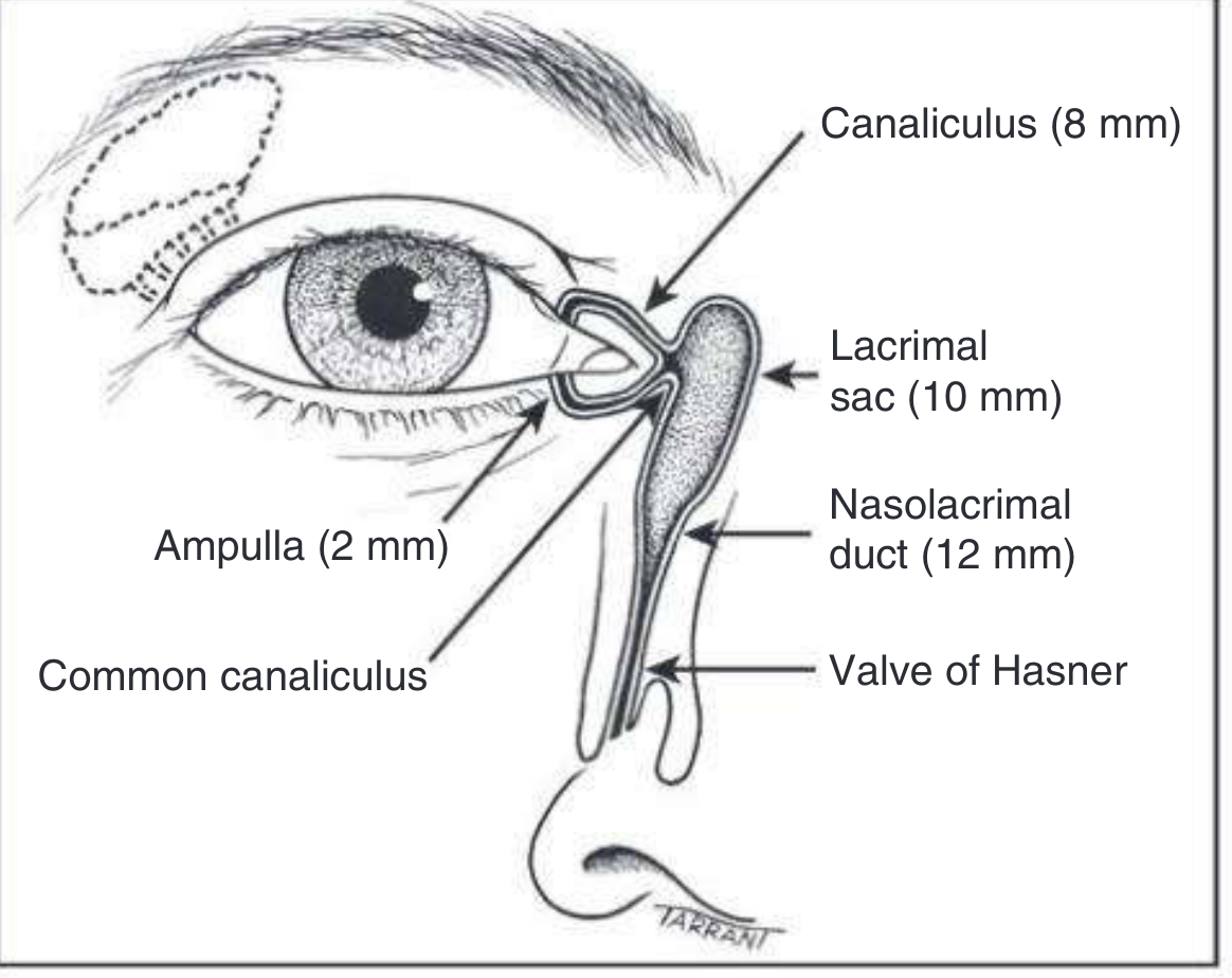
Puncta
- Located at the posterior edge of the lid margin, at the junction of the lash-bearing lateral 5/6 (pars ciliaris) and the medial non-ciliated 1/6 (pars lacrimalis).
- Normally face slightly posteriorly and can be seen by everting the medial aspect of the lids.
Canaliculi
- Each canaliculus first runs vertically for ~2 mm from the lid margin - this vertical segment is called the ampulla.
- It then turns medially and runs horizontally for ~8 mm to reach the lacrimal sac.
- The horizontal canaliculus is surrounded by the Horner muscle (palpebral part of orbicularis oculi).
- The superior and inferior canaliculi unite in >90% of cases to form the common canaliculus, which opens into the lateral wall of the lacrimal sac.
- A mucosal fold called the valve of Rosenmüller guards the junction of the common canaliculus and the sac, preventing reflux.
Lacrimal Sac
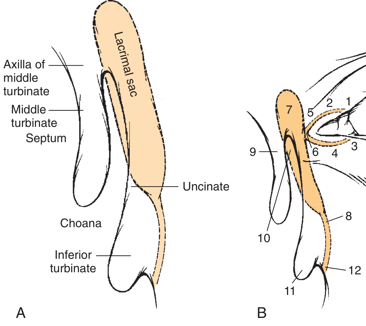
- 10-12 mm long, lies in the lacrimal fossa between the anterior and posterior lacrimal crests.
- Bounded laterally by the frontal process of the maxilla and medially by the lacrimal bone, which separates it from the middle meatus of the nasal cavity.
- The upper 1/3 of the sac lies just superior to the anterior insertion of the middle turbinate.
- The remaining inferior 2/3 is oriented vertically, just under the anterior maxillary line (lacrimomaxillary suture).
- The axilla of the middle turbinate is the most reliable endoscopic landmark to locate the lacrimal sac.
- The common canaliculus (common internal punctum) opens into the sac approximately 3 mm above the axilla of the middle turbinate.
Nasolacrimal Duct
- 12-18 mm long - the inferior continuation of the lacrimal sac.
- Descends slightly laterally and posteriorly to open into the inferior nasal meatus, lateral to and below the inferior turbinate.
- Opening is partially covered by a mucosal fold: the valve of Hasner.
Part 2: Physiology of Tear Drainage (Why the Pump Matters)
- Tears flow along the upper and lower marginal strips, pooling in the lacus lacrimalis medial to the lower puncta.
- With each blink: orbicularis oculi (pretarsal part) compresses the ampullae, shortens the horizontal canaliculi, closes the puncta medially, and creates positive pressure forcing tears down the nasolacrimal duct - mediated by helically arranged connective tissue fibers around the sac.
- When the eyes open: canaliculi and sac expand, creating negative pressure that draws tears from canaliculi into the sac.
Part 3: Sac Syringing (Lacrimal Irrigation) - Step by Step
Equipment
- Lacrimal dilator (Nettleship)
- 25-gauge blunt lacrimal cannula on a 2 mL syringe filled with saline
- Optional: fluorescein dye
Technique
- Dilate the punctum with a Nettleship dilator using a rotating motion, first vertically then horizontally.
- Insert the 25-gauge blunt cannula into the inferior (or superior) punctum - initially vertical for 2 mm, then horizontal for 8 mm following the canalicular anatomy.
- Gently irrigate with saline.
Interpreting Results
| Finding | Meaning |
|---|---|
| Patient tastes saline / fluid passes freely into nasopharynx | Patent system - no complete obstruction |
| Reflux from same punctum being syringed | Canalicular obstruction (pre-sac) |
| Reflux from opposite punctum with no nasal flow | Nasolacrimal duct obstruction (sac or duct level) - DCR indicated |
| Reflux of mucus/pus | Active dacryocystitis with NLD obstruction |
| Hard stop felt on probing | Probe hits medial wall of sac/bone - canaliculi are patent |
| Soft stop felt on probing | Impeded progress before entering sac - canalicular stenosis |
Jones Tests
- Jones I test: Fluorescein drops in conjunctival fornix. Normal = complete disappearance within 5 min with dye recoverable from nose/throat on cotton swab (positive Jones I). Abnormal = obstruction present.
- Jones II test: After a negative Jones I, the conjunctival sac is irrigated with saline after placing fluorescein. Recovery of fluorescein-stained saline from the nose = obstruction is distal (duct level). Recovery of unstained saline = functional obstruction. No recovery = complete obstruction.
Part 4: What Is Endoscopic DCR?
Epiphora vs Watery Eye
- Epiphora: tears overflow the lid margin and run down the face - caused by obstruction, treated surgically.
- Watery eye: abnormally thick tear film alters vision but does not drain down the skin - caused by tear film composition problems or reflex tearing, treated medically.
Types of Obstruction
| Type | Description | Surgery Success |
|---|---|---|
| Anatomic (70%) | Complete physical blockage between sac and nasal cavity | Better outcomes |
| Functional (30%) | Critical narrowing or failure of the lacrimal pump | Inferior outcomes |
Advantages of Endoscopic over External DCR
- No external facial scar
- Similar success rates to external DCR
- Better visualization of the agger nasi region
- Ideal for revision cases
- Preserves the medial canthal tendon and lacrimal pump mechanism
Part 5: Pre-operative Workup and Diagnostics
Clinical Examination
- Exclude lid laxity, malposition, punctal anomalies, blepharitis.
- Palpate over the lacrimal sac - reflux of mucopurulence = dacryocystitis (obstruction amenable to DCR).
- Dye disappearance test (Jones I) as described above.
- Probing with Bowman probe to differentiate hard stop (sac entry - patent canaliculi) from soft stop (canalicular stenosis).
- Syringing (Jones II) to confirm site of obstruction.
Imaging
- Dacryocystogram (DCG): Contrast injected into lacrimal system. Anatomic obstruction shows dye failing to pass beyond the sac-duct junction. Functional obstruction shows dye penetrating the nasal cavity but with delayed scintigraphy.
- Lacrimal scintigraphy: More physiologic - uses a drop of radiolabeled isotope in the conjunctival sac to measure transit time.
- CT-DCG: Best for pre-operative planning in endoscopic DCR - localizes the fundus of the sac relative to the middle turbinate axilla.
Part 6: Surgical Steps of Endoscopic DCR (Powered Technique)
Setup and Anesthesia
- General or local anesthesia with sedation.
- Patient positioned with head raised ~30 degrees to reduce venous congestion.
- 1% lidocaine with 1:100,000 epinephrine injected into the anterior lateral nasal wall for vasoconstriction and hydrodissection.
- Neuropatties containing 1:1000 epinephrine packed into the nasal cavity.
- 30-degree nasal endoscope used throughout, aimed in a superolateral direction.
Step 1: Septoplasty (if needed - ~50% of cases)
- A high endoscopic septoplasty is performed if the axilla of the middle turbinate cannot be completely visualized due to a deviated nasal septum.
- This creates adequate working space in the anterior-superior nasal cavity.
Step 2: Mucosal Flap Elevation
- A posteriorly pedicled mucoperiosteal flap is outlined with a No. 15 blade:
- Vertical incision from the axilla of the middle turbinate downward
- Horizontal incision along the inferior border
- The flap dimensions are approximately 10 mm wide x 15 mm tall
- A sharp-suction Freer elevator achieves the elevation.
- The flap must be aggressively elevated off the entire lacrimal bone so that it remains attached only inferiorly at the uncinate process and superiorly at the axilla.
- Hemostasis is regained with topical vasoconstrictors before proceeding.
Step 3: Bone Removal
- A 4-mm Hajek-Koeffler punch (forward-biting) removes the frontal process of the maxilla covering the anterior lacrimal sac.
- Critical technique: Release the punch after each bite - failure to do so may inadvertently tear the sac wall.
- The thin lacrimal bone over the posterior sac is easily flaked off with a round knife.
- A DCR drill (diamond bur) then "saucerizes" the remaining bone to the edges of the original mucosal incisions.
- Drilling is complete when a hemisphere of the lacrimal sac sits "proud" on top of the saucerized bone - this allows flaps to lie open during healing rather than curl inward.
- The agger nasi cell is opened in almost all DCRs (incorporated into the marsupialized sac in ~55%).
Step 4: Cannulation (Probe Insertion)
- Superior and inferior puncta are dilated with a lacrimal dilator.
- A Bowman probe (00 size) is passed through the inferior canaliculus:
- Initially directed horizontally and slightly posteriorly
- As the sac is approached, tip is directed superiorly through the common canaliculus into the lacrimal sac
- Confirmation is essential: The tip of the probe must be clearly visible tenting the medial sac wall before cutting. If the whole sac moves with the probe but the tip is not visible, the probe is still in the common canaliculus - cutting here will damage the common internal punctum and cause surgical failure.
- An optical fiber light guide can alternatively be used for transillumination to identify the sac through the nasal wall.
Step 5: Marsupialization of the Lacrimal Sac
- Once the probe tip is clearly tenting the sac, a spear knife (sickle knife) opens the sac vertically from top to bottom.
- A ball probe is then passed through the medial sac wall incision to check adequacy of bone removal - any remaining bone is removed at this point.
- The common internal punctum (opening of common canaliculus) should now be visible approximately 3 mm above where the axilla was located.
- Anterior and posterior mucosal flaps of the sac are trimmed to create a wide opening (anterior flap retained, posterior flap trimmed or folded back against the septum/middle turbinate).
- The mucosal flap elevated from the lateral nasal wall is folded back over the middle turbinate to oppose the posterior sac flap and promote mucosalization rather than granulation.
Step 6: Silicone Stent Placement (Optional)
- Bicanalicular silicone intubation (Crawford tubes) may be placed through both canaliculi, through the new ostium, and out via the nasal cavity where they are clipped or tied.
- Stents maintain the ostium during early healing and prevent scarring.
- Note: Current evidence shows silicone tubing is not always necessary after primary En-DCR when the ostium is adequately large.
- Tubes are left in place for 1-6 months and removed via the nose (not pulled from the eye) to prevent aspiration.
Step 7: Post-operative Care
- Nasal saline irrigations begin the following day.
- Topical nasal steroid sprays reduce mucosal inflammation.
- Antibiotic-steroid eye drops for 4-6 weeks.
- Endoscopic review at 4-6 weeks to assess the ostium and remove any granulations.
Part 7: Complications of Endoscopic DCR
Intraoperative Complications
| Complication | Cause / Note |
|---|---|
| Orbital penetration | Excessive lateral dissection; can damage ocular muscles (especially medial rectus) |
| Damage to the medial canthal tendon | Aggressive superior dissection |
| Damage to common canaliculus | Cutting down onto probe while tip is still in common canaliculus, not in sac - most common cause of immediate failure |
| Excessive bleeding | Inadequate vasoconstriction; injury to angular vessels |
Post-operative Complications
| Complication | Details |
|---|---|
| Surgical failure / recurrent epiphora (most common) | Due to inadequate marsupialization, granulation tissue occluding the ostium, or "cheese-wiring" |
| Granuloma formation | Found in 47% of ostia at the edge; most respond to topical steroids; persistent ones need excision or intralesional triamcinolone |
| Cheese-wiring | Silicone stent placed under too much tension tears through the lacrimal puncta; causes iatrogenic epiphora; reported in 0.1%; prevented by pulling a loop of tubing before intranasal clipping |
| Dacryolith / remaining bone | Any unopened portion of the lacrimal sac develops a fluid meniscus, increases resistance, and causes pump failure |
| Frontal or maxillary sinusitis | From inadvertent damage to sinus drainage pathways; rare and associated with inexperience |
| CSF leak | Rare but reported; due to disorientation at the skull base |
| CPAP-related air regurgitation | 80% of patients on CPAP machines may experience post-op symptoms; half may need to stop using CPAP |
| Lacrimal sac tumor discovered late | Present in ~1/3 of delayed failures; signs: bloody nasal drainage, medial canthal swelling, proptosis; squamous cell and transitional cell carcinoma are most common malignant types |
Part 8: Causes of Surgical Failure and How to Prevent Them
- Inadequate bone removal (most common cause overall): Any remaining bone creates a fluid meniscus. Prevention: Complete saucerization until the sac sits proud; use a ball probe to palpate the opened sac for hidden bony ledges.
- Sac not fully opened: Not all of the sac mucosa is marsupialized. Prevention: Identify and center the common internal punctum in the ostium.
- Granuloma at the ostium: Prevention: Mucosa-to-mucosa apposition of sac flap to nasal mucosal flap; topical steroids post-operatively.
- Cheese-wiring by stent: Prevention: Loop technique when securing tube in the nose.
- Proximal obstruction: DCR cannot succeed if the obstruction is at the level of the canaliculi (common canalicular stenosis) - must be diagnosed pre-operatively.
Quick Summary Table
| Step | Action | Key Point |
|---|---|---|
| 1 | Septoplasty if needed | Creates space in anterior superior nasal cavity |
| 2 | Mucosal flap elevation | Posteriorly pedicled; expose frontal process of maxilla |
| 3 | Bone removal | Punch + drill (saucerize); sac should sit "proud" |
| 4 | Probe insertion | Confirm tip tents medial sac wall before cutting |
| 5 | Marsupialization | Open sac top to bottom; anterior flap retained; identify common internal punctum |
| 6 | Silicone stent | Optional; 1-6 months; remove via nose |
| 7 | Post-op care | Saline irrigation + steroid spray + eye drops |