
Stroke no short forms
"stroke"[MeSH Terms] AND "ischemic stroke"
- Definition and types: ischemic stroke, intracerebral hemorrhage, subarachnoid hemorrhage, transient ischemic attack
- Causes and risk factors: atherosclerosis, cardioembolism, small vessel disease, atrial fibrillation, hypertension, diabetes, smoking
- Vascular anatomy and syndromes: the middle cerebral artery, anterior cerebral artery, posterior circulation, and lacunar syndromes
- Recognition and emergency assessment: symptoms, scoring scales, time windows
- Imaging and diagnostic workup: non-contrast computed tomography, magnetic resonance imaging, vessel imaging, cardiac evaluation
- Acute treatment: intravenous thrombolysis, mechanical thrombectomy, blood pressure management, hemorrhage management
- Secondary prevention: antiplatelet drugs, anticoagulants, statins, carotid revascularization
- Rehabilitation and complications
Which part of stroke should I cover in detail (all terms written out in full)?
- Definition and types: ischemic stroke, intracerebral hemorrhage, subarachnoid hemorrhage, transient ischemic attack
- Causes and risk factors: atherosclerosis, cardioembolism, small vessel disease, atrial fibrillation, hypertension, diabetes, smoking
- Vascular anatomy and syndromes: the middle cerebral artery, anterior cerebral artery, posterior circulation, and lacunar syndromes
- Recognition and emergency assessment: symptoms, scoring scales, time windows
- Imaging and diagnostic workup: non-contrast computed tomography, magnetic resonance imaging, vessel imaging, cardiac evaluation
- Acute treatment: intravenous thrombolysis, mechanical thrombectomy, blood pressure management, hemorrhage management
- Secondary prevention: antiplatelet drugs, anticoagulants, statins, carotid revascularization
- Rehabilitation and complications
Stroke
ischemic stroke brain imaging infarct diffusion-weighted
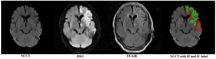
A multi-modal comparison of brain imaging from a patient with an acute ischemic stroke, featuring four axial views: Non-Contrast Computed Tomography (NCCT) with skull removal, Diffusion-Weighted Imaging (DWI), Fluid-Attenuated Inversion Recovery (FLAIR), and a labeled NCCT. The image demonstrates a clear clinicoradiological mismatch. The DWI modality reveals a distinct area of hyperintensity in the left hemisphere, signifying cytotoxic edema and the restricted diffusion characteristic of an acute infarct. In contrast, the FLAIR sequence shows minimal signal changes in the corresponding area, suggesting a 'DWI-FLAIR mismatch' indicative of hyperacute stroke within the thrombolysis window. The final panel displays the NCCT with manual segmentations: the 'Infarct Core' (IC) is highlighted in green, while the surrounding 'Infarct Penumbra' (IP) is highlighted in red. This comparison highlights the difficulty of visualizing early ischemic changes on NCCT compared to the sensitivity of MRI sequences, emphasizing the role of automated segmentation in identifying salvageable brain tissue.
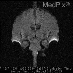
**Imaging Modality:** Magnetic Resonance Imaging (MRI); Diffusion-Weighted Imaging (DWI).
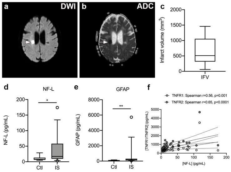
This composite image illustrates diagnostic findings and biomarker data from ischemic stroke (IS) patients. (a, b) Axial brain MRI scans showing a subcortical lacunar infarct in the right hemisphere. Panel (a) is a Diffusion-Weighted Imaging (DWI) sequence where the acute infarct appears as a bright hyperintense lesion (white arrow). Panel (b) is the corresponding Apparent Diffusion Coefficient (ADC) map, showing the same lesion as a dark hypointense area (white arrow), confirming restricted diffusion. (c) A box plot representing the infarct volume distribution (mm³) among IS patients. (d, e) Comparative box plots showing significantly elevated serum levels of Neurofilament light chain (NF-L) and Glial Fibrillary Acidic Protein (GFAP) in IS patients versus healthy controls (Ctl), indicating neuro-axonal and glial damage. (f) A correlation plot with linear regression and 95% confidence intervals demonstrating positive associations between serum NF-L and plasma TNFR1 and TNFR2 levels. These data illustrate the relationship between structural brain injury, inflammatory signaling, and biomarker release post-ischemic stroke.
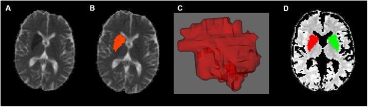
This composite diagnostic image illustrates the process of defining a Volume of Interest (VOI) for an acute ischemic stroke using neuroimaging. Panel A shows an axial slice of a diffusion-weighted MRI (DWI) displaying a hyperintense lesion in the region of the basal ganglia, consistent with an acute infarct. Panel B demonstrates the manual segmentation of this infarct, highlighted as a red Region of Interest (ROI). Panel C presents a 3D reconstruction of the segmented infarct volume, providing a volumetric representation of the lesion. Panel D shows an axial map (likely an Apparent Diffusion Coefficient or Fractional Anisotropy map) with two mirrored ROIs: the primary infarct site in the left hemisphere (red) and its anatomically mirrored counterpart in the unaffected right hemisphere (green). This methodology is used in neuroradiology research to compare diffusion parameters between ischemic and healthy brain tissue, excluding cerebrospinal fluid (CSF) to ensure data accuracy. The image is intended for advanced medical education regarding stroke imaging analysis and neuroimaging post-processing techniques.
Stroke
1. Types
| Type | Mechanism | Approximate share |
|---|---|---|
| Ischemic stroke | Blocked artery | About 85 percent |
| Intracerebral hemorrhage | Bleeding into brain tissue, usually from hypertension | About 10 percent |
| Subarachnoid hemorrhage | Bleeding around the brain, often from a ruptured aneurysm | About 5 percent |
| Transient ischemic attack | Temporary blockage with full recovery and no dead tissue on imaging | Warning event |
2. Causes of ischemic stroke
- Large artery thrombosis accounts for about one third of ischemic strokes. A clot forms on an ulcerated atherosclerotic plaque, typically where an artery branches, such as the internal carotid artery.
- Small vessel (lacunar) stroke produces infarcts from a few millimetres to 2 centimetres. It affects the basal ganglia, thalamus, pons, and internal capsule. About 80 to 90 percent of these patients have hypertension, and diabetes is also common.
- Cardioembolic stroke accounts for about one quarter. Atrial fibrillation is the most common source and raises stroke risk about fivefold.
- Artery-to-artery embolism comes from a carotid plaque. One example is amaurosis fugax, which is temporary loss of vision in one eye.
- In the young (3 to 4 percent of strokes occur at ages 15 to 45), carotid or vertebral artery dissection is the leading identified cause. Other causes are pregnancy, oral contraceptives, antiphospholipid antibodies, protein C or S deficiency, sickle cell anemia, polycythemia, and cocaine or amphetamine use.
- More than one third of first strokes have no clear cause identified.
3. Recognition
- facial droop
- weakness or numbness of one arm or leg, usually on one side
- slurred or lost speech, or trouble understanding speech
- loss of vision in one eye or half of the visual field
- loss of balance, vertigo, or double vision (suggesting the back of the brain)
- a sudden severe headache (suggesting hemorrhage)
4. Emergency assessment and imaging
- Check airway, breathing, circulation, and blood glucose. Low blood sugar can mimic stroke.
- Perform urgent non-contrast computed tomography of the head to exclude hemorrhage. It is often normal in the first hours of an ischemic stroke.
- Image the blood vessels (computed tomography angiography or magnetic resonance angiography) to find a large vessel occlusion.
- Use magnetic resonance imaging with diffusion-weighted sequences or perfusion imaging to show the dead core and the salvageable surrounding tissue, called the penumbra.
- Later workup includes a heart rhythm recording, an echocardiogram, a lipid profile, and glycated hemoglobin.
5. Acute treatment of ischemic stroke
- It was first approved for a 3-hour window, then extended to 3 to 4.5 hours after onset.
- Selected patients can benefit up to 9 hours from last seen well when advanced imaging shows salvageable tissue.
- Its limits are the narrow time window, a weak effect on large proximal occlusions, and the need for advanced imaging at later times.
- Fewer than 5 percent of eligible patients received it in the textbook's cited data (Bradley and Daroff's Neurology in Clinical Practice, p. 1131).
- Blood pressure must be below about 185 over 110 millimetres of mercury before treatment, and there must be no hemorrhage on imaging.
- Tenecteplase is used as an alternative in many centres. I did not verify current local guidance, so check it for your setting.
- Five randomized trials in 2015 showed benefit for large vessel occlusion in the early window.
- The DAWN and DEFUSE 3 trials extended benefit to as late as 24 hours from last seen well in patients selected by imaging (Bradley and Daroff's Neurology in Clinical Practice, p. 1131).
- Patient selection matters to avoid both overtreatment and undertreatment.
- Admit to a dedicated stroke unit.
- Correct low oxygen levels.
- Avoid aggressive blood pressure lowering unless the patient is receiving thrombolysis or has very high pressure.
- Treat fever and high or low glucose.
- Screen swallowing before giving food or drink.
- Give aspirin within 24 to 48 hours if thrombolysis was not given.
- Lower the blood pressure.
- Reverse any anticoagulant medicine.
- Involve neurosurgery for large cerebellar bleeds, hydrocephalus, or ruptured aneurysm.
- For subarachnoid hemorrhage, secure the aneurysm by clipping or coiling and give nimodipine to reduce delayed ischemia.
6. Secondary prevention
- Antiplatelet therapy: aspirin or clopidogrel. After a minor stroke or high-risk transient ischemic attack, short-term (about 21 days) combined aspirin and clopidogrel is used.
- Anticoagulation for atrial fibrillation, with a direct oral anticoagulant preferred over warfarin in most patients.
- Blood pressure control, a high-intensity statin, and diabetes control.
- Carotid endarterectomy or stenting for symptomatic narrowing of the carotid artery, mainly 70 to 99 percent.
- Lifestyle: stop smoking, exercise, adopt a Mediterranean-style diet, limit alcohol, and manage weight.
7. Recovery and complications
- Start rehabilitation early, with physical, occupational, and speech therapy.
- Watch for aspiration pneumonia, deep vein thrombosis, pressure sores, depression, seizures, spasticity, shoulder pain, and swelling of the brain after a large infarct.
Recent evidence check
- a review of telling ischemic from hemorrhagic stroke by presenting symptoms (PubMed identifier 40759192)
- a review of stroke despite oral anticoagulation in atrial fibrillation (41427999)
- a review of prognosis in ischemic stroke with a negative diffusion-weighted scan (38737347)