
Left bundle branch block ECG symptoms and management
Reading File
Finding Sources
Finding Sources
Searching PubMed
"left bundle branch block" AND management
Reading File
Reading File
Reading File
Reading File
Finding Sources
Finding Sources
Loading Image
Reading File
Loading Image
I now have everything needed for a comprehensive, well-illustrated answer. Here it is:
Left Bundle Branch Block (LBBB): ECG Features, Symptoms & Management
What is LBBB?
Left bundle branch block is an intraventricular conduction disturbance caused by failure of the left bundle branch to conduct impulses normally. This forces the left ventricle to depolarize late, via slow cell-to-cell spread from the right ventricle - producing characteristic wide QRS complexes and secondary ST-T changes. - Harrison's Principles of Internal Medicine 22E, p.1915
ECG Criteria for LBBB
Diagnostic criteria (complete LBBB):
| Feature | Finding |
|---|---|
| QRS duration | ≥ 120 ms (complete); 110-120 ms = incomplete |
| Lead V1 | Wide, predominantly negative (broad QS or rS complex) |
| Lead V5/V6 | Entirely positive, broad, monophasic R wave (no q wave, no S wave) |
| Lateral leads (I, aVL) | Broad R wave, no septal q wave |
| T wave | Opposite in polarity to the last QRS deflection (discordant) |
| Axis | Usually leftward |
Key mechanism: LBBB alters both early and later phases of ventricular depolarization. The major QRS vector is directed left and posteriorly. The normal left-to-right septal activation is disrupted - septal depolarization now proceeds right to left, generating the characteristic QS in V1 and broad positive R in V6. - Harrison's 22E
Visual comparison of RBBB vs LBBB waveforms:
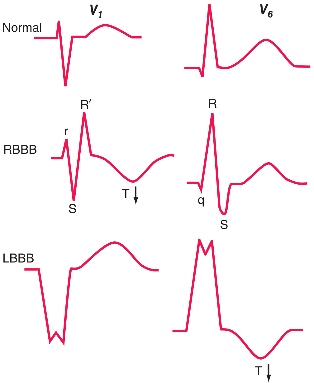
Figure: Normal, RBBB, and LBBB waveform comparison in V1 (left column) and V5/V6 (right column). Note the broad QS in V1 and wide R in V6 with LBBB, with discordant T waves (arrows). - Harrison's 22E, Fig. 247-10
Associated Conditions (Causes)
LBBB is most commonly a marker of underlying structural heart disease. The four major associated conditions are: - Harrison's 22E
- Coronary artery disease (ischemic heart disease, frequently with impaired LV function)
- Hypertensive heart disease
- Aortic valve disease (including after TAVR - transcatheter aortic valve replacement)
- Cardiomyopathy (dilated, infiltrative)
LBBB may also be rate-related (appearing above a critical heart rate), intermittent, or occasionally idiopathic.
Clinical Symptoms
Intraventricular conduction delays are themselves typically asymptomatic. However, LBBB causes ventricular dyssynchrony - the right ventricle contracts before the left ventricle. In some patients this dyssynchrony leads to mitral regurgitation. The result can be the precipitation or worsening of heart failure. - Goldman-Cecil Medicine
Symptoms, when present, are usually those of the underlying disease:
- Dyspnoea, fatigue, reduced exercise tolerance (from heart failure or LV dysfunction)
- Syncope or presyncope (suggesting associated high-degree AV block)
- Angina (if ischemic heart disease is the cause)
- Palpitations (if rate-related or alternating block)
A rare entity called Painful LBBB Syndrome (Sympathetic Crural Syndrome) is characterized by chest pain occurring at higher heart rates that induces rate-dependent LBBB - a 2025 systematic review (PMID 39913085) reviewed treatment strategies in this condition.
LBBB in Acute MI: Sgarbossa Criteria
LBBB is a major diagnostic challenge in suspected acute MI because it obscures ST-segment analysis. A new or presumed new LBBB with clinical/lab findings suggesting acute MI is associated with high mortality and should be treated the same as STEMI (immediate reperfusion). - Goldman-Cecil Medicine, p.3412
Modified Sgarbossa Criteria (to detect AMI in LBBB):
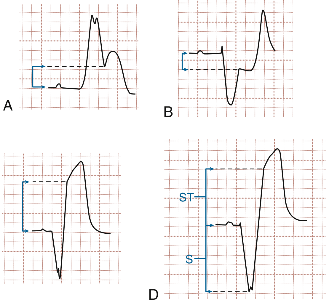
Figure: Sgarbossa criteria. A = concordant ST elevation ≥1 mm; B = concordant ST depression ≥1 mm in V1-V3; C = discordant ST elevation ≥5 mm; D = modified criteria using ST/S ratio >0.25 for discordant leads. - Rosen's Emergency Medicine, Fig. 64.21
| Criterion | Threshold | Points |
|---|---|---|
| Concordant ST elevation (same direction as QRS) | ≥ 1 mm | Strongly suggestive |
| Concordant ST depression in V1, V2, or V3 | ≥ 1 mm | Suggestive |
| Discordant ST elevation (opposite to QRS) | ≥ 5 mm (original) or ST/S ratio > 0.25 (modified) | Suggestive |
The modified Sgarbossa criteria replace the absolute 5 mm cut-off with a ratio of ST elevation to the depth of the S wave > 0.25, which improves sensitivity for detecting acute coronary occlusion. - Rosen's Emergency Medicine
Evaluation
When LBBB is found, the evaluation focuses on: - Goldman-Cecil Medicine
- Review prior ECGs - is this new or old? New LBBB = urgent rule-out of ACS
- Echocardiography - assess LV function and structure
- Stress test (echo or nuclear) - to evaluate for underlying ischemic heart disease
- Cardiac MRI - if infiltrative cardiomyopathy is suspected
- Rhythm monitoring - indicated if LBBB is associated with prolonged PR interval, alternating bundle branch block, or symptoms of lightheadedness/presyncope
Management
Treatment is targeted at the underlying cause. Rhythm-specific interventions include: - Goldman-Cecil Medicine
1. Cardiac Resynchronization Therapy (CRT / Biventricular Pacing)
- Indicated in patients with heart failure with reduced ejection fraction (HFrEF) and LBBB with QRS ≥ 130 ms
- CRT counteracts the electrical and mechanical dyssynchrony of LBBB, improving contractility, exercise tolerance, and quality of life - Miller's Anesthesia 10e; Fuster and Hurst's The Heart 15e
- A 2026 systematic review (PMID 42152765) evaluated conduction system pacing (His-bundle pacing, left bundle branch area pacing) as alternatives to biventricular pacing for CRT
2. Pacemaker Implantation
- Bifascicular block + syncope: Pacemaker is preferred over implantable cardiac monitor, regardless of QRS morphology or PR interval
- New bifascicular block (LBBB or RBBB + fascicular block) + PR prolongation: Prophylactic pacemaker is often placed due to high risk for progression to complete heart block, especially in acute MI
3. Acute MI with New LBBB
- Treat as STEMI equivalent - urgent triage for reperfusion therapy (PCI or thrombolysis)
4. Post-TAVR LBBB
- A 2025 meta-analysis (PMID 40620053) investigated predictors of post-TAVR LBBB, which occurs in a significant proportion of patients and may require pacemaker implantation
Prognosis
- LBBB increases all-cause mortality approximately 2-3 fold overall
- The risk is approximately 6-fold higher in patients with underlying structural heart disease
- Risk is minimally increased (if at all) when exercise testing and cardiac MRI show no underlying heart disease
- The combination of RBBB + left anterior or posterior fascicular block (bifascicular block) carries high risk of needing a permanent pacemaker, but prophylactic pacing is not indicated in asymptomatic patients - Goldman-Cecil Medicine
Key takeaway: LBBB is rarely a benign finding - it almost always warrants workup for structural heart disease. New LBBB in the setting of chest pain must be treated as STEMI until proven otherwise, using the Sgarbossa criteria to assess for ischemic changes. Management is dictated by the underlying cause, with CRT being the major cardiac-specific intervention in heart failure.
This is a shared conversation. Sign in to Orris to start your own chat.