
Foot hand mouth disease
hand foot mouth disease rash vesicles skin lesions
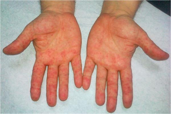
This clinical photograph displays the bilateral palmar surfaces of an adult's hands, demonstrating a widespread, symmetrical vesicular rash. The dermatological findings include numerous small, discrete, erythematous macules and papulovesicles. The lesions are distributed across the entire palmar aspect, extending onto the volar surfaces of the fingers and thumbs. The skin exhibits generalized erythema and mild swelling, particularly around the concentrated areas of vesicles. The morphology and characteristic distribution on the palms are hallmark clinical signs of Hand, Foot, and Mouth Disease (HFMD), typically caused by Coxsackievirus A16 or Enterovirus 71. This visual serves as a primary educational example for diagnosing viral exanthems in a clinical setting, highlighting the transition from macular redness to distinct vesicular lesions on acral surfaces.
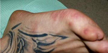
This clinical photograph displays the dorsal and lateral aspect of a human foot, illustrating dermatological manifestations characteristic of Hand, Foot, and Mouth Disease (HFMD) in an adult. The skin features multiple discrete, erythematous maculopapular lesions and small vesicles. These lesions are notably concentrated along the dorsal surface of the toes and the lateral border of the foot. Some lesions appear as slightly raised, circular reddish papules, while others show signs of early vesiculation or central erosion surrounded by an inflammatory erythematous halo. The distribution is asymmetric and non-confluent. The surrounding skin exhibits mild localized erythema but otherwise maintains normal texture, aside from the presence of a decorative tattoo on the dorsal midfoot. This image serves as an educational example of the peripheral exanthem associated with Coxsackievirus or Enterovirus infections, highlighting that while primarily a pediatric condition, HFMD can present with symptomatic cutaneous involvement in adults.
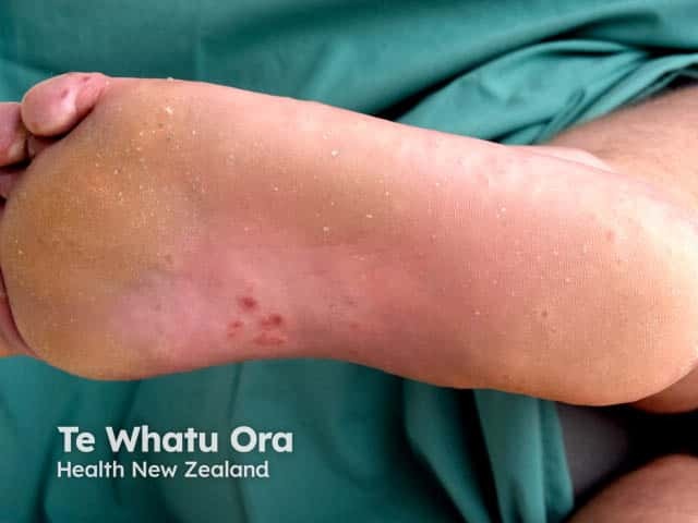
Clinical photography of a pediatric foot plantar surface demonstrates vesicular lesions consistent with hand-foot-mouth disease (HFMD) involving the instep and toes. The image captures small, 1–3 mm vesicles and papules with clear vesicular roofs, some with adjacent erythema and mild edema. Several lesions appear isolated while others are clustered along pressure-bearing areas of the instep and toe pads. In early stages, vesicles are translucent; as they evolve, superficial erosions and crusts may form. The surrounding skin remains largely intact, and there is no evidence of purulence. No mucosal involvement is evident in this view, though HFMD commonly involves oral mucosa and presents with fever and malaise in children. Etiology is enteroviral, predominantly Coxsackievirus A16 or enterovirus 71. The image is valuable for dermatologic morphology education, aiding recognition of acral vesiculopapular eruptions and differentiation from varicella, pompholyx, eczema, or contact dermatitis. It supports clinical correlation with systemic symptoms and oral findings, guides triage, infection control, and patient counseling. For researchers, it illustrates lesion topography on the plantar surface and helps quantify lesion density and progression over time. This photograph emphasizes the characteristic distribution on the feet with toe involvement and instep involvement, reinforcing a HFMD diagnosis in pediatric patients.
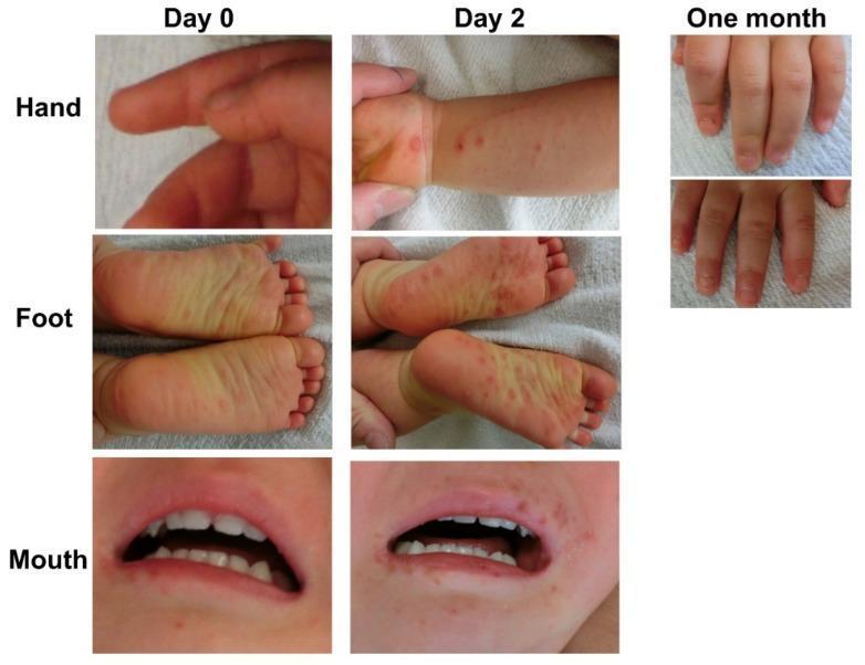
This clinical photographic montage illustrates the progression and sequelae of Hand, Foot, and Mouth Disease (HFMD) in a pediatric patient over a one-month period. The visual data is organized by anatomical site (hand, foot, and mouth) and time point (Day 0, Day 2, and one month). At Day 0, early manifestations include mild erythema and a few subtle red macules. By Day 2, the lesions progress into more distinct, numerous erythematous papules and vesicles: the hand shows clusters on the wrist and palm, the soles of the feet exhibit a widespread maculopapular rash, and the perioral and intraoral regions demonstrate pronounced papular eruptions and early ulceration. The far-right images document a late-stage complication at one month: while the primary rash has resolved, the fingernails show clear evidence of onycholysis (separation of the nail plate from the nail bed). This progression is characteristic of atypical HFMD caused by Coxsackievirus A6 (CV-A6), which is frequently associated with more extensive skin involvement and subsequent nail changes such as onychomadesis or onycholysis.
"hand foot mouth disease" enterovirus
Hand, Foot, and Mouth Disease (HFMD)
Overview
Epidemiology
- Most common in children 6 months to 10 years; infants and toddlers are especially vulnerable
- Seasonal pattern: primarily spring and summer months
- Spreads via fecal-oral route, respiratory droplets, and direct contact with vesicle fluid
- Exposed adults can develop disease (particularly with CVA6, to which most adults lack immunity)
- High secondary attack rates within households
Pathogen Breakdown
| Strain | Clinical Feature |
|---|---|
| CVA16 | Classic/typical HFMD - most common |
| CVA6 | "Atypical HFMD" - more widespread eruption, perioral lesions, higher hospitalization rate |
| EV-A71 | Severe disease; associated with life-threatening rhombencephalitis/CNS complications |
Clinical Features
Prodrome
Oral Lesions (Enanthem)
- Present in ~90% of patients
- Appear 1-2 days after prodrome
- Start as erythematous macules on buccal mucosa, tongue, soft palate, gingiva, uvula
- Rapidly vesiculate and ulcerate into painful 4-8 mm ulcers with a red areola
- Pain often causes reduced oral intake and mild dehydration
Skin Lesions (Exanthem)
- Asymptomatic red papules progressing to small 3-7 mm gray vesicles with a red halo
- Oval, linear, or crescentic - run parallel to skin lines on fingers and toes
- Distribution: palms, soles, dorsa of fingers and toes, and buttocks (especially in diaper-wearing children)
- Lesions resolve over 4-7 days
Atypical HFMD (CVA6)
- Wider distribution with numerous lesions on the trunk
- Perioral lesions that can mimic severe impetigo or Stevens-Johnson syndrome
- In adults: widespread purpuric lesions simulating vasculitis or erythema multiforme
- In children with atopic dermatitis (AD): vesicular/erosive eruption in areas of dermatitis = "eczema coxsackium" (similar to eczema herpeticum)
- Higher hospitalization rate due to dehydration and pain
- Child-to-adult transmission more common
Complications
- Onychomadesis (nail loss/separation) - occurs ~1 month after acute illness, especially with CVA6; also seen as onycholysis
- Viral meningitis / meningoencephalitis - rare
- Myocarditis - rare
- Rhombencephalitis - life-threatening, associated with EV-A71 (notably in the Taiwan EV-71 outbreak, 80% of CNS cases had HFMD skin lesions)
- Dehydration from poor oral intake
Diagnosis
- Primarily clinical based on characteristic distribution and appearance
- PCR: virus can be recovered from vesicle swabs or stool specimens
- Viral culture: from vesicle fluid or stool
- Histopathology (rarely needed): intraepidermal blister with vacuolar and reticular degeneration of keratinocytes; no inclusion bodies or multinucleated giant cells (distinguishes from HSV)
Differential Diagnosis
| Condition | Distinguishing Feature |
|---|---|
| Herpangina | Oral lesions only (posterior pharynx/tonsillar pillars), no skin lesions |
| Herpes gingivostomatitis | High fever, friable bleeding gums, tender cervical adenopathy, multinucleated giant cells on Tzanck smear |
| Varicella | Centripetal distribution, all stages of lesions simultaneously, crusting |
| Pompholyx / dyshidrotic eczema | Chronic/recurrent, no oral involvement, not contagious |
| Stevens-Johnson syndrome | Severe systemic illness, mucosal sloughing, target lesions |
Treatment
- Antipyretics: paracetamol/ibuprofen for fever and pain
- Oral analgesics / topical agents: for painful mouth ulcers (e.g., viscous lidocaine gel with caution in young children)
- Oral rehydration: mainstay for dehydration
- IV fluids: for significant dehydration or inability to tolerate oral intake
- In CVA6 HFMD with AD ("eczema coxsackium"): treat underlying AD to reduce pruritus; monitor for secondary bacterial infection
- Disease typically resolves in less than 1 week
Prevention
- Handwashing is the most important preventive measure, especially for children and caregivers
- Avoid contact with vesicle fluid, respiratory secretions, and stool of infected individuals
- No licensed vaccine is available in most countries (though EV-A71 vaccines have been developed and used in China)