
Management of parapharyngeal abscess
I'll invoke the medical library skill first before answering.
Reading File
Now I'll run parallel searches across the library and PubMed simultaneously.
Finding Sources
Finding Sources
Searching PubMed
parapharyngeal abscess management treatment
Excellent. Now let me read the key textbook sections in parallel.
Reading File
Reading File
Reading File
Let me now read the comprehensive Scott-Brown section on deep neck space infections (which covers parapharyngeal space in detail) and also Rosen's emergency medicine section.
Reading File
Reading File
Let me read the remaining Rosen's section for full clinical detail, and also read the Cummings otolaryngology section on parapharyngeal space.
Reading File
Finding Sources
Now let me fetch the anatomical diagram and also get Rosen's antibiotic table and the Cummings section on deep neck infections.
Loading Image
Finding Sources
Let me now read the antibiotics table from Rosen's and also get more detail on the deep neck space infection surgical treatment from Scott-Brown's.
Finding Sources
Reading File
Now let me read the KJ Lee antibiotic table and also check the Scott-Brown section on specific parapharyngeal space details.
Reading File
Reading File
I now have comprehensive information from multiple authoritative sources. Let me compile the complete answer.
Management of Parapharyngeal Abscess
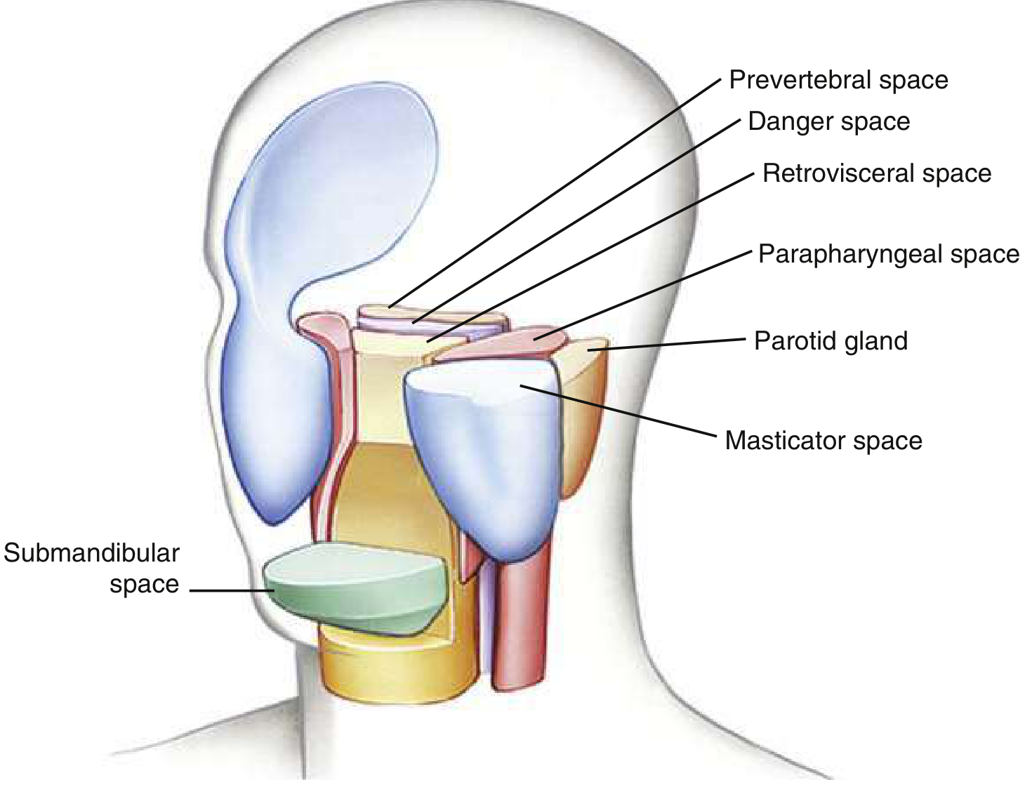
Anatomy and Significance
The parapharyngeal space (PPS) lies on either side of the neck, extending from the skull base to the styloglossus muscle at the angle of the mandible. It has two important sub-compartments:
- Anterior PPS (prestyloid): Contains fat - prone to liquefaction and abscess formation; requires aggressive surgical drainage
- Posterior PPS (poststyloid/retrostyloid): Contains the carotid sheath (carotid artery, internal jugular vein, sympathetic chain, CN IX-XII), lymph nodes; more often causes lymphadenitis and may be managed more conservatively
The posterior aspect is separated from the retropharyngeal and "danger space" by a fascial plane, creating a direct pathway to the mediastinum.
- Scott-Brown's Otorhinolaryngology, p. 671
- Rosen's Emergency Medicine, p. 965
Aetiology and Microbiology
Common sources include:
- Peritonsillar or retropharyngeal abscess (direct spread)
- Dental infection / mandibular molar periapical abscess
- Suppurative cervical lymphadenitis
- Parotitis
- Penetrating trauma, nerve blocks, or surgical procedures (iatrogenic)
- IV drug abuse
Organisms are typically polymicrobial - gram-positive cocci (streptococci, staphylococci), gram-negative rods, and anaerobes (Fusobacterium, Bacteroides, Peptostreptococcus).
Clinical Features
| Feature | Details |
|---|---|
| Neck pain and swelling | Lateral neck mass, often obscured by SCM |
| Trismus | Involvement of pterygoid muscles - key distinguishing sign vs. peritonsillar abscess |
| Odynophagia / dysphagia | Edema and pharyngeal displacement |
| Torticollis | If SCM is involved |
| Fever and systemic sepsis | Tachycardia, rigors |
| Medial tonsillar displacement | In anterior compartment infections |
| Voice change / muffled voice | Pharyngeal edema |
| Airway compromise | Dyspnea, stridor, inability to manage secretions - emergency |
Note: Unlike peritonsillar abscess, the maximal swelling is behind the posterior faucial pillar with little or no oedema of the soft palate. - Bailey & Love's Surgery, p. 480
Complications (Life-threatening - must anticipate)
- Airway obstruction - edema, abscess mass effect
- Descending mediastinitis - via retropharyngeal/danger space communication (crude mortality up to 2%)
- Lemierre syndrome - septic thrombophlebitis of the internal jugular vein; suspect when sore throat + sepsis + multisystem involvement
- Carotid artery erosion, aneurysm, or rupture - any signs of bleeding warrant CT/MR angiography urgently
- Horner syndrome - sympathetic chain involvement (CN IX-XII neuropathies)
- Cavernous sinus thrombosis - proptosis, impaired EOM, pupillary changes
- Aspiration pneumonia / lung abscess / empyema - from abscess rupture into airway
- Retroperitoneal sepsis - rare but described
Investigations
Imaging is mandatory (unless airway is immediately at risk - secure airway first):
- Contrast-enhanced CT of neck + chest - gold standard; demonstrates rim-enhancing abscess, extent of infection, and any mediastinal involvement. Always include the mediastinum in the scan field. Cellulitis appears as fat stranding along fascial planes; abscesses show characteristic ring enhancement
- MRI - comparable to CT for delineating extent; useful when radiation is to be minimized
- Ultrasound - may assess whether abscess has sufficiently liquefied and can guide needle aspiration, but limited in deep spaces
- CT or MR angiography - when vascular complications suspected
- Orthopantomogram (OPG) - if dental origin suspected
- Fibreoptic nasendoscopy - assess upper aerodigestive tract and airway patency simultaneously (performed cautiously to avoid triggering gag reflex)
Scott-Brown's ORL, p. 671; Rosen's Emergency Medicine, p. 965
Management: Step-by-Step
Step 1 - Airway Assessment and Securing (Priority Zero)
This is the pre-eminent step before any other intervention.
- Assess for: dyspnea, tachypnea, stridor, inability to manage secretions, agitation, drooling
- Conventional laryngoscopy is often impossible due to trismus, reduced neck extension, laryngopharyngeal edema, and friable mucosa
- Do NOT paralyze before securing airway
- Preferred method: awake fibreoptic nasotracheal or orotracheal intubation with adequate topicalization
- Have emergency tracheostomy prepared and set up before commencing anaesthesia; pre-infiltrate with 2% lignocaine + 1:80,000 adrenaline at tracheostomy site
- Transfer to operating theatre with anaesthetic and surgical teams experienced in difficult airway management
- Supine positioning for CT is only safe once airway is deemed stable
Scott-Brown's ORL, p. 673; Rosen's Emergency Medicine, p. 965
Step 2 - Intravenous Antibiotics (Immediate, Do Not Delay for Cultures)
Start broad-spectrum empiric IV antibiotics without delay:
| Scenario | Regimen |
|---|---|
| Community-acquired (first-line) | Ampicillin-sulbactam 1.5-3.0 g IV every 6 hours |
| Penicillin allergy | Clindamycin 600-900 mg IV every 8 hours |
| Eikenella suspected (dental origin) | Add moxifloxacin 400 mg QD |
| MRSA suspected | Clindamycin + vancomycin 1 g IV every 12 hours |
| Immunocompromised / nosocomial | Piperacillin-tazobactam 3 g IV every 6 h OR imipenem-cilastatin 500 mg IV every 6 h |
| Necrotizing fasciitis | Ceftriaxone 2 g + clindamycin 600-900 mg + metronidazole 500 mg IV every 6 h |
| Actinomyces | Penicillin G 10-20 MU/day for 4 weeks, then oral penicillin V for 4-6 months |
- Alternatively: amoxicillin-clavulanate, or 2nd/3rd generation cephalosporins with metronidazole
- Review all antibiotic choices once culture and sensitivity results are available
- Note rising resistance: clindamycin 11-18%, erythromycin 14%, penicillins 7-8%
- In children under 2: clindamycin preferred as first line due to MRSA rates
KJ Lee's Essential Otolaryngology, p. 710; Scott-Brown's ORL, p. 671
Step 3 - Corticosteroids (Adjunctive)
- Dexamethasone 0.6 mg/kg IV/IM (maximum 10 mg) given with antibiotics
- Associated with decreased need for surgical drainage in select cases
- Reduces pharyngeal edema and systemic inflammation
Rosen's Emergency Medicine, p. 965
Step 4 - Surgical Drainage (Decision)
Indications for surgery:
- Confirmed abscess on CT with liquefaction
- Failure to respond to conservative treatment after 24-48 hours (persistent pyrexia, worsening pain)
- Airway compromise from the abscess
- Vascular complications
Conservative (non-surgical) management may be appropriate if:
- Intranodal abscess or phlegmon (cellulitis without pus collection)
- Abscess size <2.5 cm contained within a single neck space
- Most pediatric cases (especially posterior PPS / phlegmon)
- Patient is responding clinically to antibiotics within 24-48 hours
Approximately 80% of patients ultimately require surgical drainage, though this varies widely by center (range 10-100%). - Scott-Brown's ORL, p. 672
Step 5 - Surgical Approach Options
A. Transcervical (external) approach - standard for most parapharyngeal abscesses
- Incision anterior to SCM, dissecting along the great vessels
- Approach anterior and medial to the carotid sheath
- Curette abscess wall; break down loculations
- Insert drain (usually corrugated or suction drain)
B. Intraoral drainage - only if abscess clearly points into the oropharynx
- Blunt instrument drainage through the oropharynx (medial to the pterygomandibular raphe)
- Risk of inadequate drainage; limited visualization
C. Ultrasound-guided needle aspiration
-
Shown in a prospective RCT to reduce hospital stay from 5.2 to 3.1 days and reduce cost by 41%, with no difference in efficacy or complications
-
Better for microbiological sampling (increases anaerobe isolation)
-
Useful when abscess is superficially accessible
-
Scott-Brown's ORL, p. 672; Bailey & Love's Surgery, p. 480
Repeat imaging and/or surgical re-intervention is indicated if there is no improvement after 48-72 hours of therapy.
Step 6 - ICU Admission and Monitoring
- Admit to ICU if airway or vascular compromise is present
- Monitor for septic complications, mediastinal spread, vascular involvement
- Patients with stable airways may be admitted to a high-dependency floor bed
- IV fluid resuscitation; nutritional support (NG or parenteral if swallowing compromised)
- Serial clinical and radiological reassessment
Special Considerations
Tuberculosis / fungal infection: In immunosuppressed patients or endemic regions with insidious onset, consider TB. Seek infectious disease + ENT consultation. Dedicated cervical spine imaging if ligamentous/osseous destruction is suspected.
Lemierre syndrome: If sore throat followed by sepsis + multisystem involvement - antibiotics for 6 weeks (penicillin + metronidazole or co-amoxiclav). Consider anticoagulation if spreading thrombophlebitis. - Scott-Brown's ORL, p. 480
Children: The majority of cases; often younger children; posterior PPS infections (lymphadenitis) more common and frequently managed conservatively with antibiotics alone. Use clindamycin as first-line in children under 2.
Recent Evidence
A 2023 systematic review on deep neck space infections (Sheikh Z et al., Clin Otolaryngol 2023) is the most current high-level evidence on assessment and management, supporting the multi-modal approach described above. A 2024 review (Wang X et al., PMID 38029535) specifically examined adjuvant corticosteroids in pediatric retropharyngeal and parapharyngeal abscess, supporting dexamethasone use to reduce the need for surgical drainage.
Summary Algorithm
Parapharyngeal Abscess
        |
        v
Airway Compromise? ──YES──> Awake fiberoptic intubation
        |                   (+ prepare emergency tracheostomy)
        NO
        |
        v
Contrast-enhanced CT Neck + Chest
        |
   _____|_____
  |           |
Cellulitis/  Abscess
Phlegmon     confirmed
  |           |
IV Abx       IV Abx + Dexamethasone
+ Dex        + Otolaryngology consult
  |           |
Reassess     Failure to respond  ──> Surgical drainage
at 24-48h    at 24-48 hours         (transcervical / US-guided)
References:
- Rosen's Emergency Medicine - Concepts and Clinical Practice, pp. 964-966
- Scott-Brown's Otorhinolaryngology Head & Neck Surgery (Vol 2), pp. 671-674
- Bailey & Love's Short Practice of Surgery (28th ed.), p. 480
- KJ Lee's Essential Otolaryngology, pp. 709-711
- Sheikh Z et al. (2023). Systematic review of deep neck space infections. Clin Otolaryngol. PMID 37147934
Management of chronic retropharyngeal abscess ( k/a prevertebral abscess )
Reading File
Quick answer: Chronic retropharyngeal abscess (prevertebral abscess) is almost always tuberculous in origin (Pott's disease of the cervical spine). Management involves anti-tubercular therapy (ATT) as the cornerstone, with surgical drainage reserved for large collections or neurological compromise. The abscess is drained by a lateral cervical approach - never by incision through the oropharynx (risk of rupture + aspiration).
Searching sources...
Finding Sources
Finding Sources
Searching PubMed
retropharyngeal abscess tuberculosis prevertebral management
Good - Bailey & Love has a dedicated section. Let me read it fully and run parallel searches for related content.
Reading File
Finding Sources
Searching PubMed
Pott disease cervical spine abscess treatment
Excellent. Now let me read the Cummings prevertebral section, the Scott-Brown section, and the Rheumatology/Radiology sections in parallel.
Reading File
Reading File
Reading File
Now let me get the anti-tubercular therapy regimen details and the Rheumatology TB treatment section, plus fetch the CT image.
Finding Sources
Loading Image
Reading File
Now let me get the ATT regimen from the Rheumatology textbook and also look for any ENT-specific ATT dosing details.
Reading File
Finding Sources
Let me now search specifically for the ATT regimen for spinal/bone TB and also the Grainger radiology section on prevertebral space.
Finding Sources
Reading File
Let me look specifically for the ATT treatment regimen (standard WHO regimen for osteoarticular/spinal TB) and the Rheumatology treatment section.
Finding Sources
Finding Sources
Let me get the Rheumatology treatment section and fetch the spinal TB X-ray image, plus the recent systematic review abstract.
Reading File
Reading Article
PMID: 41319734
I now have comprehensive information from all sources. Let me compile the full, detailed answer.
Management of Chronic Retropharyngeal Abscess (Prevertebral Abscess)
Definition and Key Distinction
Chronic retropharyngeal abscess - also termed prevertebral abscess or cold abscess of the neck - is fundamentally different from acute retropharyngeal abscess in its aetiology, age group, pathological process, and management approach. The single most critical clinical rule:
A chronic retropharyngeal abscess must NEVER be opened into the mouth - unlike an acute abscess. Intraoral drainage risks secondary pyogenic contamination of a sterile tuberculous collection and aspiration.
- Bailey & Love's Surgery, 28th ed., p. 809
Aetiology
Virtually always tuberculous in origin - specifically Pott's disease of the cervical spine (TB spondylitis), which spreads through the following pathway:
Haematogenous seeding of cervical vertebral body
           |
   Bone destruction + caseation
           |
Extension beneath the Anterior Longitudinal Ligament
           |
Spread into the Prevertebral Space (behind prevertebral muscles)
           |
Cold abscess tracks anteriorly → Retropharyngeal swelling
           |
Presents as bulge in posterior pharyngeal wall
In the cervical spine specifically, the pus may:
- Spread to the posterior cervical triangle
- Bulge into the retropharyngeal space - visible as a posterior pharyngeal wall swelling
- Track down into the mediastinum (along prevertebral fascia)
A particular variant, subligamentous TB, involves spread up and down the spine beneath the anterior longitudinal ligament, scalloping multiple anterior vertebral bodies without disc involvement - more common in the cervical spine.
- Firestein & Kelley's Textbook of Rheumatology, p. 2503
- Bailey & Love's Surgery, p. 808
Clinical Features
| Feature | Details |
|---|---|
| Age | Almost exclusively adults (vs. acute retropharyngeal abscess which is predominantly in children) |
| Onset | Insidious - weeks to months |
| Cervical pain and stiffness | Most common presenting symptom (69.5% in systematic review) |
| Posterior pharyngeal wall swelling | Visible on oropharyngeal inspection - often the presenting sign to ENT |
| Neck fullness | Fullness behind the sternocleidomastoid on one side |
| Dysphagia / odynophagia | From pharyngeal displacement |
| Hoarseness | Laryngeal edema or compression |
| Torticollis | Cervical instability or muscle spasm |
| Constitutional symptoms | Low-grade fever, weight loss, night sweats, anorexia |
| Neurological deficits | Motor-sensory deficits in 46.2%; myelopathy from cord compression - serious |
| Radicular pain | In 31% of cases |
| Absence of acute inflammatory signs | No trismus, no acute toxicity (hence "cold" abscess) |
The cervical spine may be frankly unstable - undue manipulation risks precipitating a neurological event (atlantoaxial dislocation, quadriplegia). - Bailey & Love's Surgery, p. 809
Investigations
Imaging
MRI is the modality of choice for suspected discitis and prevertebral abscess, especially when TB is the concern. In practice, it may not be possible immediately if the patient is too unwell.
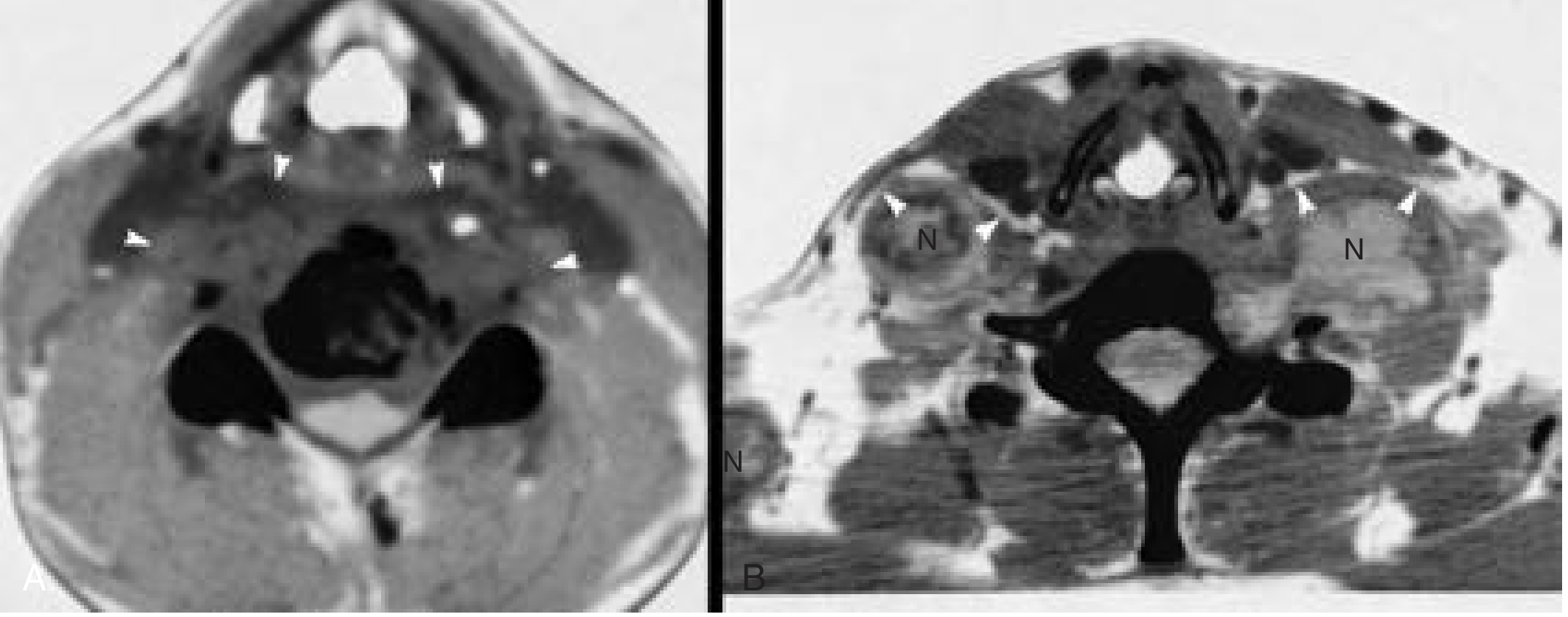
Contrast-enhanced CT is used when MRI is not available or when the patient is acutely unwell. Key CT findings:
- Rim-enhancing low-density collection anterior to the prevertebral muscles (the abscess is posterior to pharyngeal mucosa and prevertebral muscles displace anteriorly - distinguishing it from a retropharyngeal space mass where the prevertebral muscles are displaced posteriorly)
- Vertebral body destruction - loss of vertebral height, osteolysis of anterior vertebral body
- Disc space narrowing and intervertebral disc involvement
- Loss of normal cervical lordosis / kyphotic deformity
- Gibbus deformity in advanced cases
- Occasional calcification within the abscess (pathognomonic of TB)
- Epidural extension and cord compression in severe cases
Plain X-ray cervical spine (AP + lateral):
- Osteolysis of anterior vertebral bodies
- Disc involvement (unlike metastatic disease which spares disc)
- Retropharyngeal soft tissue shadow widening
- Gibbus / kyphotic deformity
- Partial calcification of paravertebral mass
- Loss of normal curvature
Technetium-99 bone scan - more sensitive than plain films for early disease but may give false-negatives in highly destructive or early indolent disease.
Ultrasound - high sensitivity (96%) and specificity (82%) for pus collections; can show whether collection has liquefied and may guide aspiration; less useful for deep prevertebral collections.
Microbiological / Histological Confirmation
This is mandatory before committing to long-term ATT:
-
CT-guided / US-guided percutaneous needle aspiration of the abscess - send fluid for:
- AFB smear (only 40% positive from psoas/paravertebral abscesses)
- Mycobacterial culture (gold standard but takes 6-8 weeks; positive in 80-95% of biopsy specimens with TB histology)
- GeneXpert/NAAT (Nucleic Acid Amplification Test) - sensitivity 85-95%, specificity 96-100%, result in 48 hours - now the preferred rapid test
- Standard bacterial culture (to exclude pyogenic co-infection)
- Drug sensitivity testing (DST) to exclude MDR-TB
-
CT-guided needle biopsy of the vertebral body/disc - send for:
- Histology: granulomatous inflammation with central caseation, Langhans giant cells
- AFB culture
-
Blood tests: ESR (elevated), CRP, FBC (anaemia of chronic disease), liver function (pre-ATT baseline), renal function
-
Mantoux / tuberculin skin test - positive in >80%
-
IGRA (Interferon-Gamma Release Assay) - QuantiFERON-TB Gold - more specific in BCG-vaccinated populations
-
Chest X-ray - screen for concurrent pulmonary TB (present in 52% of cases)
-
HIV testing (alters treatment regimen and duration)
-
Firestein & Kelley's Rheumatology, pp. 2503-2504
Management
Step 1 - Cervical Spine Precautions (Immediate Priority)
Before any other intervention:
- Avoid neck manipulation - the spine may be unstable; undue force can precipitate atlantoaxial dislocation, subluxation, or acute cord compression
- Cervical immobilization with a rigid collar if instability is demonstrated or suspected on imaging
- Senior clinician involvement essential for examination
Step 2 - Anti-Tubercular Therapy (ATT) - The Cornerstone of Treatment
ATT is the primary treatment. Drainage of the abscess may not be necessary at all if appropriate ATT is instituted and the patient responds.
Standard WHO regimen for osteoarticular / spinal TB:
| Phase | Duration | Drugs | Notes |
|---|---|---|---|
| Intensive phase | 2 months | HRZE (Isoniazid + Rifampicin + Pyrazinamide + Ethambutol) | Daily, supervised (DOT preferred) |
| Continuation phase | 10 months (total 12 months) | HR (Isoniazid + Rifampicin) | Extended vs. pulmonary TB due to poor drug penetration into avascular bone/abscess |
Standard doses (adult, weight-based):
| Drug | Abbreviation | Daily dose |
|---|---|---|
| Isoniazid | H | 5 mg/kg (max 300 mg/day) |
| Rifampicin | R | 10 mg/kg (max 600 mg/day) |
| Pyrazinamide | Z | 25 mg/kg (max 2 g/day) |
| Ethambutol | E | 15-20 mg/kg (max 1.6 g/day) |
Duration: Most guidelines recommend 12 months total for spinal TB (vs. 6 months for pulmonary TB), given the avascular nature of bone/abscess cavities and the risk of relapse.
MDR-TB: If GeneXpert detects rifampicin resistance, extended regimens with second-line agents (fluoroquinolones, injectables) under infectious disease guidance are required.
Monitoring on ATT:
- Baseline LFTs, uric acid, urea/creatinine, visual acuity (for ethambutol)
- Monthly clinical assessment
- Monitor for hepatotoxicity (commonest toxicity - isoniazid, rifampicin, pyrazinamide)
- Ethambutol optic neuritis - check visual acuity monthly
- Pyridoxine (vitamin B6) 10 mg/day with isoniazid to prevent peripheral neuropathy
Step 3 - Drainage of the Abscess (Surgical Decision)
Drainage may not be required if ATT produces clinical response. However, drainage is indicated when:
- Large collection causing significant airway compromise or dysphagia
- Failure to respond to ATT after 6-8 weeks (persistent abscess, worsening symptoms)
- Need for microbiological confirmation if CT-guided aspiration is not feasible or has failed
- Neurological compromise (see Step 4 below)
- Abscess pointing superficially and at risk of spontaneous rupture
HOW TO DRAIN - The Critical Rule:
| Approach | Rule |
|---|---|
| ❌ Intraoral / transoral drainage | ABSOLUTELY CONTRAINDICATED - risk of secondary pyogenic infection converting a sterile cold abscess into a mixed infection, and aspiration |
| ✅ External cervical approach | The correct route |
Surgical technique (external approach):
- Incision anterior to the sternocleidomastoid muscle
- Dissect medially, approaching anterior and medial to the carotid sheath
- Enter the retropharyngeal/prevertebral space
- Open the cavity, suction dry thoroughly
- Send biopsy material for histology, AFB culture, and GeneXpert
- Cavity not primarily closed - drain insertion if needed
- No dependent drainage (unlike pyogenic abscess) - prevertebral abscesses are often under no tension
- Bailey & Love's Surgery, p. 809
Step 4 - Surgery for Spinal Decompression / Stabilization
Indicated when:
- Progressive neurological deficit (myelopathy, cord compression, paraplegia / quadriplegia)
- Spinal instability with risk of cord injury
- Significant kyphotic deformity (gibbus) requiring correction
- Large epidural abscess compressing the cord
- Failure of conservative management with ATT
Surgical options for cervical Pott's disease:
| Approach | Indication |
|---|---|
| Anterior approach (anterior cervical discectomy / corpectomy + fusion) | Most common (used in 60.4% of cases); for anterior column disease, vertebral body destruction, and abscess |
| Posterior approach (laminectomy + posterior instrumented fusion) | For posterior element involvement, epidural abscess, or if anterior access is not feasible |
| Combined anterior + posterior | For severe kyphosis, multilevel disease, or cases requiring both decompression and long-segment stabilization |
Key points from the 2026 systematic review (PMID 41319734):
- Surgical intervention was performed in 71.9% of cervical spine TB patients
- Bone fusion was achieved at a mean of 4.1 months
- Surgery-related mortality: 1.0%
- A combination of medical and surgical treatment produces favorable neurological outcomes
- MRI findings showed epidural/paravertebral abscesses with cord involvement in 72.9% of cases
Corticosteroids - adjunctive dexamethasone is sometimes used when there is significant cord edema or spinal cord compression, though evidence specific to TB spondylitis is limited.
Step 5 - General Supportive Measures
- Nutritional support - TB is a catabolic disease; high-protein diet, nutritional supplementation
- Analgesia - NSAIDs (avoid if hepatotoxicity is a concern on ATT)
- Immobilization - cervical orthosis / collar for pain relief and stability during the healing phase
- Physiotherapy - after spinal fusion is achieved; graduated mobilization and strengthening
- Notification to public health authorities (TB is a notifiable disease)
- Contact tracing and screening of household contacts
- HIV management if co-infected (adjust ATT timing/regimen; avoid rifampicin interactions with some antiretrovirals)
Differential Diagnosis of Prevertebral/Retropharyngeal Mass
| Condition | Distinguishing features |
|---|---|
| Acute retropharyngeal abscess | Children, acute onset, fever, trismus, NO bone destruction |
| Metastatic disease (vertebral) | Disc space usually preserved, known primary, no calcification |
| Primary vertebral tumour | MRI/biopsy differentiates |
| Pyogenic vertebral osteomyelitis | More acute, higher fever, CRP markedly elevated, CT/MRI differentiates |
| Retropharyngeal lymph node abscess (non-TB) | Younger patients, responds to antibiotics, no vertebral destruction |
| Fungal spondylitis (Aspergillus, Candida) | Immunocompromised, culture-based diagnosis |
| Sarcoidosis | Non-caseating granulomas, ACE level elevated |
Summary Algorithm
Posterior pharyngeal wall swelling / prevertebral mass in an adult
                    |
          Cervical spine precautions
          (Rigid collar; avoid manipulation)
                    |
       MRI / Contrast-enhanced CT neck + cervical spine
                    |
   Vertebral destruction + paravertebral cold abscess?
                    |
         YES → Suspect TB spondylitis
                    |
    ┌───────────────┼──────────────────┐
    |               |                  |
 CT/US-guided    Chest X-ray      Blood tests:
 aspiration/    HIV testing    ESR, CRP, FBC,
  biopsy        Mantoux/IGRA   LFT, GeneXpert
    |
 Confirm Mycobacterium tuberculosis
    |
 Start ATT: 2HRZE + 10HR (12 months total)
    |
 Reassess at 6-8 weeks
    |
 Responding?                Not responding / Large collection /
    |                       Neurological deficit / Instability
    |                                  |
Continue ATT                   External cervical drainage
                               (anterior to SCM, medial to
                               carotid sheath) + Spinal surgery
                               (decompression/fusion) if needed
Key Teaching Points
- Cold abscess = TB until proven otherwise - insidious onset, adults, no acute toxicity
- Never drain intraorally - secondary infection risk; always external cervical approach
- Drainage may not be needed - ATT alone can resolve the abscess in many cases
- Always get histology + GeneXpert before starting ATT wherever possible
- Cervical spine instability is a real danger - handle the neck with care
- 12 months of ATT for spinal/osteoarticular TB (not 6 months as in pulmonary TB)
- Neurological deficit = surgical emergency - do not delay decompression
Sources:
- Bailey & Love's Short Practice of Surgery, 28th ed., pp. 808-809
- Firestein & Kelley's Textbook of Rheumatology, pp. 2502-2505
- Cummings Otolaryngology Head & Neck Surgery (imaging section)
- Scott-Brown's Otorhinolaryngology Head & Neck Surgery, p. 456
- Grainger & Allison's Diagnostic Radiology (perivertebral space section)
- Murhega RB et al. (2026). Cervical spine tuberculosis: systematic review and meta-analysis. World Neurosurg. PMID 41319734
LUDWIG'S ANGINA
Finding Sources
Finding Sources
Searching PubMed
Ludwig angina management treatment
Excellent coverage. Now reading all key textbook sections in parallel.
Reading File
Reading File
Reading File
Reading File
Now reading the remaining rich sections - Rosen's management details, Miller's Anesthesia airway section, Tintinalli, and Scott-Brown surgical treatment.
Reading File
Reading File
Reading File
Reading File
Now let me get the antibiotic table from Rosen's and the Scott-Brown surgical drainage details.
Reading File
Reading File
Loading Image
Now let me get the antibiotic table from Rosen's and the Ludwig's angina anterior neck erythema image, plus the clinical image.
Finding Sources
Loading Image
I now have comprehensive material from 6 authoritative sources. Let me write the complete answer.
Ludwig's Angina
Definition
Ludwig's angina is a rapidly progressive, bilateral, gangrenous cellulitis involving all three submandibular spaces simultaneously: the bilateral sublingual spaces and the submandibular (submaxillary) space, separated by the mylohyoid muscle, plus the submental space. It is NOT primarily an abscess - it is a woody, brawny cellulitis that spreads through fascial planes (not lymphatics), making palpable fluctuation characteristically absent.
"A rapidly progressive, bilateral, gangrenous cellulitis of all submandibular spaces that can rapidly lead to death within hours"
- Rosen's Emergency Medicine, p. 961
Anatomy of the Submandibular Space
The submandibular space is subdivided by the mylohyoid muscle into:
- Sublingual space (above mylohyoid) - contains tongue, sublingual glands, deep lingual vessels
- Submylohyoid / submaxillary space (below mylohyoid) - contains submandibular gland, facial vessels
The bilateral open posterior communication between these spaces allows infection to spread rapidly across both sides and then posteriorly via the styloglossus muscle into the parapharyngeal space and onward to the retropharyngeal space and ultimately the mediastinum.
Aetiology and Predisposing Factors
Source:
- Dental origin (80-90%) - particularly mandibular molar periapical abscesses
- The roots of mandibular 2nd and 3rd molars insert below the mylohyoid line on the mandible - when infected, pus naturally tracks into the submylohyoid space
- The lingual cortex of the mandible at this level is very thin, facilitating spread
- Mandibular fracture
- Oral trauma: tongue piercing, lingual laceration, iatrogenic (intubation lacerations)
- Secondary infection of oral malignancy
- Suppurative parotitis
- Adjacent deep neck infections spreading in
Predisposing conditions:
- Diabetes mellitus (most important - commonest comorbidity)
- Immunocompromised states (HIV, malignancy, chemotherapy)
- Malnutrition
- IV drug abuse
- Poor dental hygiene / neglected dentition
Rosen's EM, p. 961; Tintinalli's EM, p. 839
Microbiology
Infection is polymicrobial, reflecting normal oral flora:
- Streptococcus species (especially viridans group) - most common; virulent streptococcal infections are classic
- Gram-negative rods (Klebsiella, Escherichia coli, Haemophilus)
- Anaerobes (Bacteroides, Peptostreptococcus, Fusobacterium) - essential to cover
- Staphylococcus aureus including MRSA in at-risk populations
- Eikenella corrodens (bite-related cases)
- Putrid halitosis is characteristic of anaerobic infection
Bailey & Love's Surgery, p. 808; Scott-Brown's Vol 2, p. 480
Clinical Features
| Feature | Description |
|---|---|
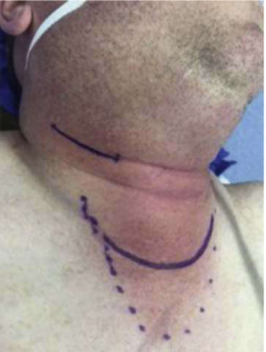
| Brawny, woody induration of floor of mouth | Hallmark; NOT soft and fluctuant; no palpable pus |
| Bilateral submandibular swelling | Tense, board-like, tender, non-pitting |
| "Bull neck" | Tense brawny edema from submandibular region to hyoid |
| Tongue elevation and protrusion | Pushed upward and backward by floor-of-mouth edema |
| Drooling | Inability to swallow secretions |
| Dysphagia / odynophagia | Floor-of-mouth and tongue displacement |
| Trismus | Pterygoid muscle involvement |
| Muffled / "hot potato" voice | Pharyngeal and tongue edema |
| Putrid halitosis | Anaerobic infection |
| Fever, rigors, systemic sepsis | High temperature, tachycardia |
| Dyspnea, stridor | Late sign - impending airway obstruction |
| Progressive anxiety | Hypoxia from airway impingement |
| "Sniffing position" | Patient instinctively maximizes airway diameter |
| Cervical adenopathy absent | Spreads via fascial planes, NOT lymphatics |
Rosen's EM, p. 962; KJ Lee's Otolaryngology, p. 712
Investigations
Clinical diagnosis - do not delay treatment for investigation.
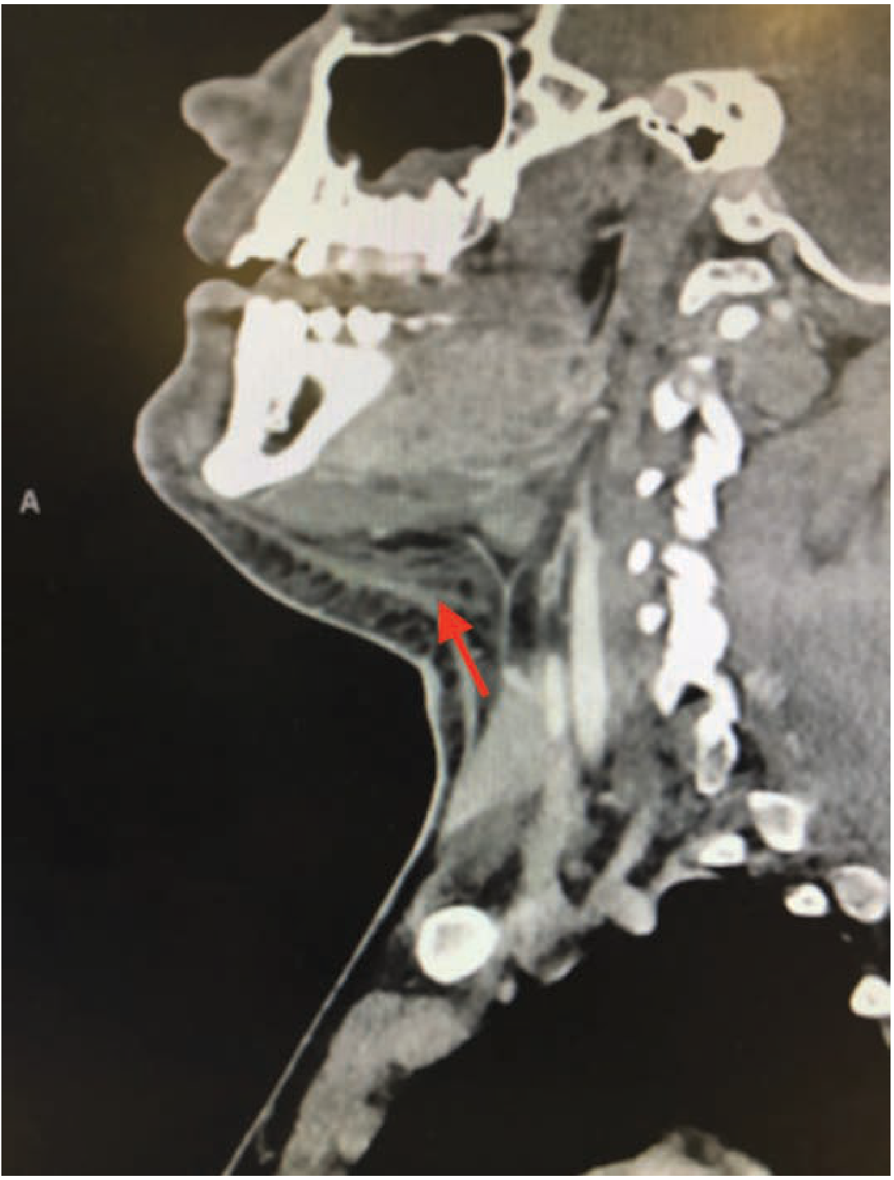
Contrast-enhanced CT neck (with or without chest):
- Extent of infection - which spaces involved, bilateral vs unilateral
- Presence of gas (necrotizing infection / gas-forming organisms) - poor prognostic sign
- Presence of a drainable collection vs. pure cellulitis/phlegmon
- Assess airway caliber and degree of tongue/supraglottic displacement
- Detect spread to parapharyngeal space, retropharyngeal space, or mediastinum
- Does NOT delay airway management if the patient is deteriorating
Blood tests:
- FBC (leukocytosis), CRP, ESR, blood cultures (before antibiotics), procalcitonin
- Electrolytes, renal function, LFTs, glucose (check for undiagnosed DM)
- Coagulation (if necrotizing infection suspected)
Microbiology: Culture and sensitivity from any drained material; send for aerobic + anaerobic culture
Management
Asphyxia from progressive edema is the leading cause of death. The management priority is therefore:
1. AIRWAY → 2. ANTIBIOTICS → 3. SURGERY (if needed) → 4. ICU
Step 1 - Airway Management (ABSOLUTE FIRST PRIORITY)
Never delay, never assume the airway will be maintained.
Signs mandating immediate airway intervention:
- Dyspnea / tachypnea
- Stridor (late and ominous)
- Inability to manage secretions / drooling
- Progressive agitation or altered mentation (hypoxia)
- Oxygen saturations falling
- Hoarse voice or voice change
Critical principles:
- Do NOT paralyze before securing the airway - a paralyzed patient with Ludwig's angina can be impossible to intubate or ventilate conventionally
- Conventional direct laryngoscopy is difficult or impossible due to trismus, tongue displacement, and edema
- Abscess rupture during laryngoscopy can flood the airway with pus - catastrophic
- Supine positioning worsens obstruction (tongue falls back further)
Airway options in order of preference:
| Option | Details |
|---|---|
| Awake fibreoptic nasotracheal intubation | Preferred method - nasal approach preferred due to trismus; requires adequate topicalization of nasal and pharyngeal mucosa; patient cooperative and breathing spontaneously throughout |
| Awake oral fibreoptic intubation | If nasal route not feasible |
| Video laryngoscopy (awake) | May be used as an adjunct |
| Awake tracheostomy under local anaesthesia | For severe cases with near-complete obstruction, or when fibreoptic fails; classic "if dated" approach - still valid |
| Emergency cricothyrotomy | Last resort if all intubation methods fail and patient losing airway |
The operating theatre should be prepared for emergency tracheostomy before any induction attempt, with surgical and anaesthetic teams present. Have pre-infiltrated the tracheostomy site with local anaesthetic + adrenaline.
Rosen's EM, p. 962; Miller's Anesthesia, p. 9800; KJ Lee's Otolaryngology, p. 712; Scott-Brown's ORL, p. 673
Step 2 - Intravenous Antibiotics (Immediate, Broad-Spectrum)
Start immediately - do not wait for culture results.
Must cover: gram-positive cocci, gram-negative rods, and crucially anaerobes.
| Scenario | Antibiotic Regimen |
|---|---|
| First-line (community-acquired) | Ampicillin-sulbactam 1.5-3.0 g IV every 6 hours |
| Penicillin allergy | Clindamycin 600-900 mg IV every 8 hours (excellent anaerobic cover) |
| Eikenella suspected (bites, tongue piercing) | Amoxicillin-clavulanate / moxifloxacin 400 mg QD |
| Severe/necrotizing | Penicillin G + metronidazole + clindamycin, or piperacillin-tazobactam 3g IV q6h |
| MRSA risk (IV drug users, colonized patients) | Add vancomycin 1g IV every 12 hours |
| Immunocompromised/nosocomial | Piperacillin-tazobactam or imipenem-cilastatin 500 mg IV every 6 h |
Review and narrow antibiotics once culture and sensitivity results are available.
KJ Lee's Otolaryngology, p. 710; Tintinalli's EM, p. 839
Step 3 - Adjunctive Corticosteroids
- Dexamethasone 0.6 mg/kg IV (max 10 mg) - reduces pharyngeal and floor-of-mouth edema, may reduce the need for surgical intervention and tracheostomy
- Evidence supports use in deep neck space infections, though Ludwig's-specific RCTs are lacking
Step 4 - Surgical Treatment
Usually NOT required if antibiotics are started early and the patient responds - Ludwig's angina is primarily a cellulitis, not an abscess.
Indications for surgery:
- Failure to respond to IV antibiotics within 24-48 hours
- Evidence of a drainable pus collection on CT
- Necrotizing infection (gas on CT, skin necrosis, rapid progression)
- Extraction of the causative tooth (source control - always do this)
- Progressive infection spreading to parapharyngeal or retropharyngeal spaces
Surgical technique:
Incision and drainage:
- Usually performed under general anaesthesia (preferred - more controlled; airway already secured)
- May be done under local in extremis
- Curved submental incision through skin and platysma below the mandible to access both submandibular triangles
- Multiple transcutaneous stab incisions may be needed bilaterally to adequately drain all spaces
- The mylohyoid muscle is incised to decompress the floor of mouth and drain the sublingual space
- Abscess wall is curetted; all loculations are broken down with digital dissection - break down any boundaries between retropharyngeal, parapharyngeal, and submandibular communicating spaces
- Soft, non-suction corrugated drains placed; wound lightly sutured around drains
- Drains removed gradually over days, monitored by number of dressing changes per 24 hours
- Deal with the causative tooth (extraction) at the same general anaesthetic
Intraoral drainage - limited role; can access sublingual space; insufficient for submylohyoid / bilateral involvement.
Scott-Brown's ORL, p. 673; Bailey & Love's Surgery, p. 808
Step 5 - ICU Admission and Monitoring
- All patients with Ludwig's angina are admitted to the ICU without exception
- Post-intubation monitoring: continuous pulse oximetry, arterial line, regular airway assessment
- Serial CT if clinical deterioration or concern of spreading infection
- Nutritional support (NG tube or parenteral - patient cannot swallow)
- Strict glucose control in diabetics
- IV fluid resuscitation and electrolyte management
- Daily wound care and drain management
Complications
| Complication | Mechanism |
|---|---|
| Asphyxiation / death | Progressive floor-of-mouth and supraglottic edema; #1 cause of mortality |
| Aspiration pneumonia / lung abscess | Pus aspiration during spontaneous rupture or intubation |
| Mediastinitis | Spread via styloglossus into parapharyngeal → retropharyngeal → danger space → mediastinum |
| Necrotizing fasciitis of the neck | Gas-forming organisms; rapidly fatal; requires radical debridement |
| Lemierre syndrome | Septic thrombophlebitis of the internal jugular vein; high spiking fevers + septic emboli |
| Cavernous sinus thrombosis | Retrograde spread; proptosis, EOM palsies, pupillary changes |
| Osteomyelitis of the mandible | Contiguous spread from dental source |
| Airway edema post-extubation | Delayed extubation for several days is standard |
Differential Diagnosis
| Condition | Distinguishing feature |
|---|---|
| Parapharyngeal / retropharyngeal abscess | Posterior pharyngeal wall displacement; tonsillar displacement |
| Peritonsillar abscess | Unilateral, fluctuant, uvular deviation |
| Submandibular sialadenitis | Glandular swelling, no floor-of-mouth induration |
| Sublingual haematoma | Trauma history, no fever |
| Angioedema | Rapid onset, urticarial, no fever, responds to epinephrine |
| Oral malignancy with infection | Underlying mucosal lesion visible |
Summary Algorithm
Suspected Ludwig's Angina
          |
     Assess airway
          |
   ┌──────┴──────┐
Stable           Compromised / stridor / desaturating
   |                     |
CT scan          AWAKE FIBREOPTIC INTUBATION
(confirm extent)  (nasal approach preferred due to trismus)
   |             Prepare emergency tracheostomy
   |                     |
   └──────────┬──────────┘
              |
    Broad-spectrum IV antibiotics
    (Ampicillin-sulbactam ± vancomycin if MRSA risk)
    + Dexamethasone 0.6 mg/kg IV
              |
    Reassess at 24-48h
              |
    Responding?         Failure / collection / gas / necrotizing
         |                           |
    Continue IV Abx          Surgical I&D
    ICU monitoring           Curved submental incision
    Source control           Mylohyoid incision
    (tooth extraction)       Multiple stab incisions bilaterally
                             Corrugated drains
                             Tooth extraction same GA
                             ICU post-op
Key Teaching Points
- Bilateral involvement of all submandibular spaces - the hallmark; not a simple dental abscess
- Spread via fascial planes - NOT lymphatics; hence no palpable fluctuation and no cervical lymphadenopathy
- Airway first, always - asphyxia is the #1 killer
- Never paralyze before securing the airway - cannot ventilate or intubate once muscle tone is gone
- Nasal fibreoptic intubation preferred - trismus prevents adequate mouth opening for oral approach
- Most cases respond to antibiotics alone - surgery only for failure, collection, necrotizing infection
- Extract the causative tooth - essential source control
- Diabetics and immunocompromised are at highest risk and progress fastest
Sources:
- Rosen's Emergency Medicine: Concepts and Clinical Practice, pp. 961-962
- Bailey & Love's Short Practice of Surgery, 28th ed., pp. 808-809
- Scott-Brown's Otorhinolaryngology Head & Neck Surgery, pp. 480, 671-673
- Scott-Brown's ORL Vol 2, p. 480
- Miller's Anesthesia, 10th ed., p. 9800
- Tintinalli's Emergency Medicine, p. 839
- KJ Lee's Essential Otolaryngology, pp. 712-713
- Bridwell R et al. (2021). Diagnosis and management of Ludwig's angina: an evidence-based review. Am J Emerg Med. PMID 33383265
This is a shared conversation. Sign in to Orris to start your own chat.