
Most common benign tumour of liver
hepatic hemangioma liver benign tumor ultrasound MRI
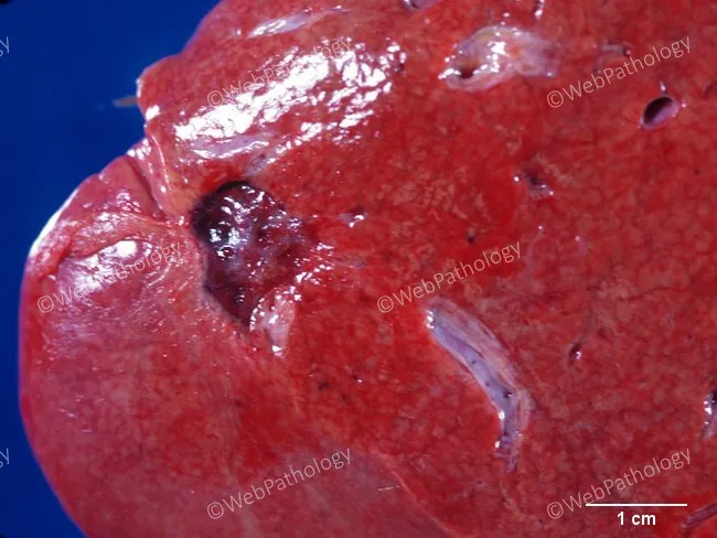
Gross pathology photography of an autopsy liver from an adult female. The image highlights a solitary hepatic lesion, most consistent with a cavernous hemangioma (benign vascular tumor). The lesion is red-brown, soft, and well circumscribed within the hepatic parenchyma. The cut surface is slightly depressed with a glistening, sponge-like appearance comprised of dilated vascular channels, as would be expected for cavernous hemangioma; no overt necrosis or calcification is evident. The scale bar at lower right indicates about 1 cm, suggesting a lesion approximately 1–1.5 cm in diameter. The surrounding liver shows otherwise preserved architecture without cirrhosis or steatosis in this view. There is no sign of invasion into adjacent structures. This incidental finding is usually asymptomatic and discovered during autopsy or imaging for unrelated reasons. Clinically, cavernous hepatic hemangiomas are the most common benign liver tumors and are typically identified by characteristic imaging features (hyperechoic on ultrasound, hypervascular on CT/MRI) but require histological confirmation if necessary. Differential considerations include focal nodular hyperplasia, hepatic adenoma, metastasis, angiosarcoma, and vascular malformations, though these are less likely given gross features. Documented here for archival pathology and educational search. Correlation with life history and imaging findings improves diagnostic confidence and educational value and practice today.
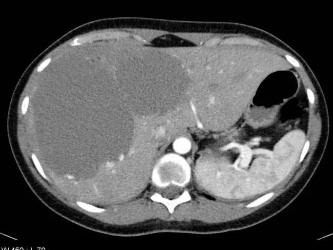
This axial abdominal CT demonstrates a solitary hepatic hemangioma in the liver. The image shown is pre-contrast (non-contrast) CT, where the lesion appears well defined and hypodense relative to the surrounding hyperdense liver parenchyma. Hemangiomas are common benign vascular liver tumors that must be distinguished from hepatic adenoma, hepatocellular carcinoma, and metastases. When contrast-enhanced CT is performed, typical behavior includes early peripheral nodular enhancement in the arterial phase with progressive centripetal fill-in during the portal venous and delayed phases; this enhancement pattern is highly characteristic. On MRI, hepatic hemangiomas usually show very high signal intensity on T2-weighted sequences and retain characteristic enhancement after gadolinium administration. Ultrasound commonly demonstrates a homogeneously hyperechoic lesion with sharp margins, reflecting internal vascular channels and slow flow. Clinically, imaging alone can often secure a confident diagnosis, avoiding biopsy due to bleeding risk from these vascular tumors. If atypical features are present or clinical suspicion remains for malignancy, additional imaging with contrast-enhanced CT, MRI with liver-specific contrast, or PET/CT can be pursued. The case from Radiopaedia (Case rID: 7014) highlights the canonical imaging appearances and reinforces the benign nature of typical liver hemangiomas, supporting radiologist confidence and appropriate patient management. This image exemplifies classic non-contrast imaging.
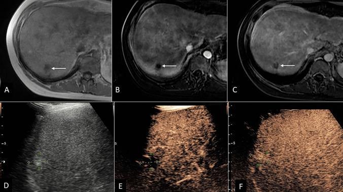
A multi-panel clinical imaging study comparing MRI and contrast-enhanced ultrasound (CEUS) for the evaluation of a hepatic hemangioma. Panels A-C display axial MRI views of the liver: (A) pre-contrast image showing a small, well-defined hypo-intense lesion in the right hepatic dome (white arrow); (B) arterial phase image exhibiting motion artifact with questionable peripheral enhancement; and (C) late phase image showing persistent hypoenhancement, leading to an initial LI-RADS 3 (LR-3) indeterminate categorization. Panels D-F present ultrasound imaging of the same 1.6 cm nodule: (D) grayscale ultrasound showing a classic hyperechoic appearance; (E) early CEUS showing peripheral interrupted arterial phase hyperenhancement (APHE); and (F) CEUS at 1 minute demonstrating persistent hyperenhancement filling the lesion. This enhancement pattern is characteristic of a benign cavernous hemangioma, allowing for definitive recategorization from MRI LR-3 to CEUS LR-1. The series illustrates the diagnostic utility of CEUS in clarifying indeterminate liver lesions found on cross-sectional CT or MRI.
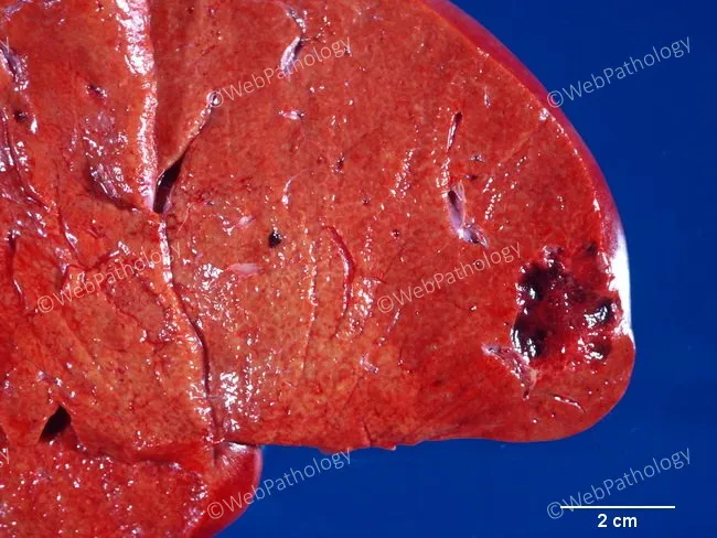
Gross pathology photograph of hepatic tissue showing a solitary, well-demarcated vascular lesion at the periphery of the liver. The lesion presents as a reddish-brown, sponge-like area approximately 2 cm in greatest diameter, as indicated by the 2 cm scale bar included in the image. Surrounding liver parenchyma appears largely preserved, with intact lobular architecture and no overt necrosis. The gross features are compatible with a cavernous hepatic hemangioma, a common benign vascular tumor of the liver composed of dilated, blood-filled vascular spaces lined by endothelium with fibrous septa. The lesion's sharply defined margins and vascular character help distinguish it from other hepatic neoplasms. Clinically, hepatic hemangiomas are often asymptomatic and discovered incidentally, especially in middle-aged women; they may enlarge during pregnancy or with estrogen therapy. Radiologic correlation (ultrasound, CT, MRI) typically reveals a well-defined, enhancing lesion with peripheral nodular progression. Differential diagnoses include focal nodular hyperplasia, hepatocellular adenoma, and hepatocellular carcinoma in cases with atypical imaging features. This image is relevant for education in gross pathology, hepatic vascular tumors, and correlates with imaging and clinical features noted in standard reviews of liver lesions. Clinical management decisions hinge on lesion size, symptoms, and radiologic behavior. This image therefore serves as a teaching example for correlating gross appearance with imaging and pathology.
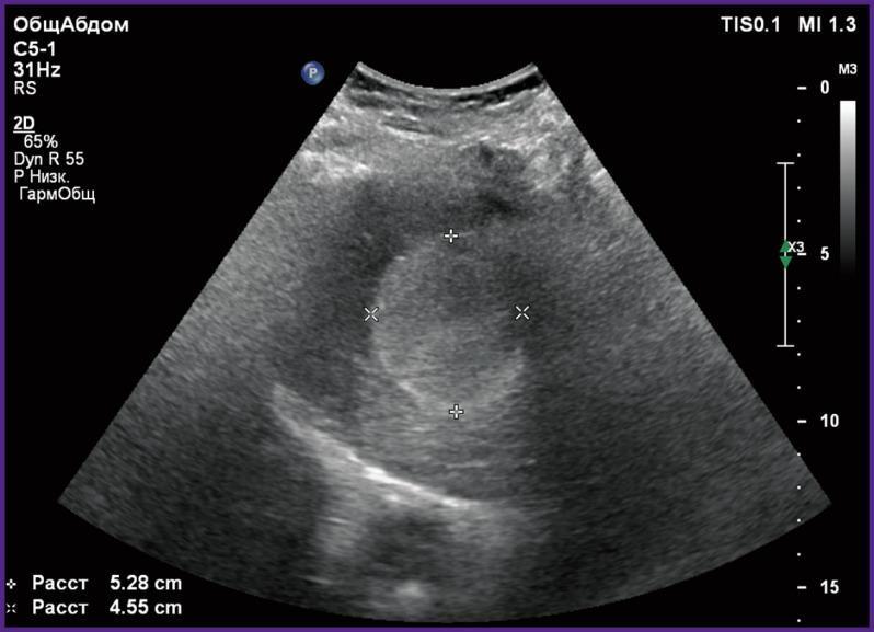
This diagnostic image is a 2D grayscale abdominal echosonogram (ultrasound) demonstrating a hepatic hemangioma in a 42-year-old patient. The lesion is located within the liver parenchyma and presents as a well-defined, predominantly hyperechoic mass with internal heterogeneous texture, including minor hypoechoic areas. The mass exhibits slightly irregular borders and appears to exert a mild mass effect on the surrounding hepatic tissue. Caliper measurements are visible on the image, marking the lesion's dimensions as 5.28 cm and 4.55 cm. Technical annotations include TIS 0.1, MI 1.3, and transducer settings for a C5-1 curved array probe. A depth scale on the right indicates the imaging field extends to approximately 15 cm. This image illustrates the typical sonographic appearance of a large cavernous hemangioma, a common benign vascular liver tumor, and is used in radiology education to distinguish focal liver lesions based on echogenicity and morphology.
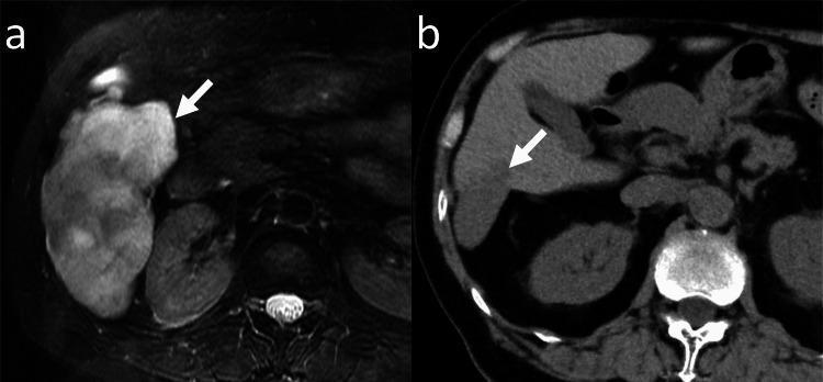
This diagnostic comparison demonstrates the longitudinal progression and shrinkage of a giant hepatic hemangioma in the right lobe of the liver. Image (a) is an axial fat-saturated T2-weighted magnetic resonance image (MRI) representing the baseline study. It reveals a large, hyperintense, lobulated mass (arrow) measuring 106 mm in diameter with a heterogeneous internal signal characteristic of cavernous hemangiomas. Image (b) shows the same patient 188 months later using axial unenhanced computed tomography (CT). The follow-up scan indicates significant involution of the lesion (arrow), now measuring 46 mm in diameter. On CT, the hemangioma appears as a well-defined, hypoattenuating region compared to the surrounding liver parenchyma. This case illustrates the natural history and potential for spontaneous regression in benign hepatic vascular tumors. Relevant landmarks include the vertebrae and renal silhouettes, providing anatomical context for the lesion's position within the right hepatic lobe. The content is suitable for radiology education regarding liver tumor monitoring and differential diagnosis.
Hepatic Hemangioma - Most Common Benign Liver Tumor
Key Facts
| Feature | Detail |
|---|---|
| Type | Benign vascular (mesenchymal) tumor |
| Prevalence | 2-20% general population |
| Sex predominance | Women (5:1 female:male ratio) |
| Multifocal | 10-30% of cases |
| Malignant potential | None |
| Symptoms | Vast majority are asymptomatic |
Pathogenesis
- These are congenital vascular malformations, not true neoplasms
- They enlarge by vascular ectasia (dilation of existing channels), NOT by angiogenesis
- Surrounded by a pseudocapsule; growth is expansile but non-invasive
- Growth may be associated with high estrogen states (puberty, OCP use, pregnancy, HRT), though causality is not firmly established
- Interestingly, hemangiomas can involute in the setting of chronic liver disease / cirrhosis
Gross & Histological Appearance
- Red-brown, soft, sponge-like lesion
- Well-circumscribed with sharp margins
- Cut surface shows dilated, blood-filled vascular channels lined by flat endothelium with fibrous septa
- No necrosis, no capsular invasion
Clinical Presentation
- Most are incidental findings on imaging done for unrelated reasons
- When symptomatic: right upper quadrant pain/discomfort, fullness, early satiety, nausea - attributed to distension of Glisson's capsule
- Rarely: giant hemangiomas (>10 cm) may present with a palpable mass or consumptive coagulopathy (Kasabach-Merritt syndrome)
Imaging Characteristics
| Modality | Findings |
|---|---|
| Ultrasound | Homogeneously hyperechoic, well-defined, sharp margins |
| CT (contrast) | Peripheral nodular enhancement in arterial phase → progressive centripetal fill-in on portal venous/delayed phases |
| MRI | Very high T2 signal ("light bulb" sign); peripheral nodular enhancement with gadolinium |
| CEUS | Peripheral interrupted arterial phase hyperenhancement → centripetal fill-in |
Management
- Vast majority: observation only - no treatment needed
- A large study of 388 patients followed conservatively showed 87% complete stability with no ruptures
- Indications for surgery: diagnostic uncertainty (suspicion of malignancy), severe symptoms due to size, or rare complications
- Surgical options: enucleation (preferred, exploits the pseudocapsule) or formal hepatic resection
Classification of Benign Liver Tumors (for context)
| Category | Tumors |
|---|---|
| Vascular (most common) | Cavernous hemangioma |
| Epithelial - hepatocellular | Focal nodular hyperplasia (FNH), Hepatic adenoma |
| Epithelial - biliary | Bile duct adenoma, Biliary cystadenoma |
| Cystic | Simple hepatic cyst |
| Other mesenchymal | Lipoma, Angiomyolipoma |
Images