
Types of syncope
types of syncope classification diagram
Types of Syncope
Classification (BOX 2.1 - Bradley and Daroff's Neurology in Clinical Practice)
1. Neurally-Mediated (Reflex) Syncope
a. Vasovagal (Vasodepressor) Syncope
- Prodrome of lightheadedness, nausea, pallor, diaphoresis, visual blurring, tinnitus
- Gradual onset allowing the patient time to fall safely
- Weak, slow pulse during the episode (transient bradycardia + vasodilation)
- Rapid recovery once supine; no postictal confusion
- Minor myoclonic jerks may occur but no tonic-clonic activity
b. Situational Syncope
- Cough syncope - forceful coughing raises intrathoracic pressure, reduces venous return
- Micturition syncope - sudden release of bladder pressure + vagal response; common in men at night
- Defecation syncope - Valsalva effect during straining
- Swallowing syncope - reflex bradycardia triggered by esophageal stimulation
- Diving/Valsalva syncope - breath-holding with increased intrathoracic pressure
c. Carotid Sinus Hypersensitivity (CSH)
2. Orthostatic (Postural) Syncope
a. Primary Dysautonomia
- Pure autonomic failure, Parkinson disease, multiple system atrophy (Shy-Drager syndrome)
- Impaired baroreceptor reflex arc
b. Secondary Dysautonomia
- Diabetes mellitus (autonomic neuropathy), amyloidosis, spinal cord injury
- A drop in systolic BP >20 mmHg (or diastolic >10 mmHg) within 3 minutes of standing is diagnostic
c. Drug-Induced Orthostatic Hypotension
- Antihypertensives (alpha-blockers, diuretics), nitrates, antidepressants, antipsychotics, alcohol
d. Volume Depletion
- Dehydration, hemorrhage, prolonged bedrest, Addison's disease
e. Postprandial Hypotension
- Blood pools in splanchnic circulation after eating; especially in the elderly
3. Cardiac (Cardiovascular) Syncope
a. Arrhythmias (most common cardiac cause)
- Bradyarrhythmias: Sick sinus syndrome (sinoatrial disorder), complete AV block (Stokes-Adams attacks), prolonged sinus pause, medication-induced
- Tachyarrhythmias: Ventricular tachycardia/fibrillation, supraventricular tachycardia (SVT), atrial fibrillation with rapid ventricular rate
- Reflex arrhythmias: Transient sinus pause from neural reflexes
- Long QT syndrome (hereditary or drug-induced) - especially consider in females with family history of sudden cardiac death
b. Structural/Obstructive Cardiac Causes
- Outflow obstruction: Aortic stenosis (syncope occurs with exertion due to inability to increase cardiac output + vasodepressor reflex), hypertrophic obstructive cardiomyopathy (HOCM), pulmonary stenosis, pulmonary hypertension
- Inflow obstruction: Left atrial myxoma/thrombus, mitral stenosis, cardiac tamponade, tension pneumothorax
- Cardiomyopathies: Dilated cardiomyopathy, amyloid cardiomyopathy
4. Cerebrovascular Syncope
- Vertebrobasilar insufficiency - drop attacks, diplopia, dysarthria, ataxia
- Subclavian steal syndrome - exercise of the arm on the side of subclavian stenosis diverts blood away from vertebral artery
- Takayasu arteritis (pulseless disease) - vasculitis causing stenosis of great vessels
- Carotid disease - unilateral carotid stenosis rarely causes syncope alone
- Cerebral vasospasm - e.g., in migraine (basilar-type migraine, more common in young females)
5. Metabolic Syncope
- Hypoglycemia - impaired glucose delivery to neurons; gradual onset, prolonged
- Hypoxia/Anoxia - respiratory failure, severe anemia (reduced oxygen-carrying capacity)
- Hypoventilation - hypocapnia from hyperventilation causes cerebral vasoconstriction (common in anxiety-related episodes)
6. Multifactorial Syncope
- Age-related reductions in baroreceptor responsiveness, cardiac compliance, and vestibulosympathetic reflexes
- Polypharmacy
- Multimorbidity (diabetes, Parkinson disease, heart disease)
- In elderly patients, syncope may present as an unexplained fall without a recognized prodrome
Key Distinguishing Features
| Feature | Vasovagal | Cardiac | Orthostatic |
|---|---|---|---|
| Onset | Gradual | Abrupt | On standing |
| Prodrome | Yes (nausea, pallor) | Often none | Brief presyncope |
| Position | Usually standing | Any position | Standing |
| Precipitants | Pain, heat, emotion | Exertion, palpitations | Posture change |
| Recovery | Prompt, no confusion | Prompt | Prompt if supine |
Syncope vs. Seizure (Key Differential)
- Syncope: pallor, diaphoresis, gradual or positional onset, minor irregular twitching only, no postictal confusion, no tongue biting, no urinary incontinence (rarely)
- Seizure: cyanosis, rhythmic jerking, tongue biting, urinary incontinence common, postictal confusion lasting minutes to hours
- Bradley and Daroff's Neurology in Clinical Practice, Box 2.1, Table 2.1
- Braunwald's Heart Disease - A Textbook of Cardiovascular Medicine
- Goldman-Cecil Medicine
- ROSEN's Emergency Medicine - Concepts and Clinical Practice
Innervation of facial nerve on the face, ipsilateral and bilateral lesion
facial nerve branches temporal zygomatic buccal marginal mandibular cervical
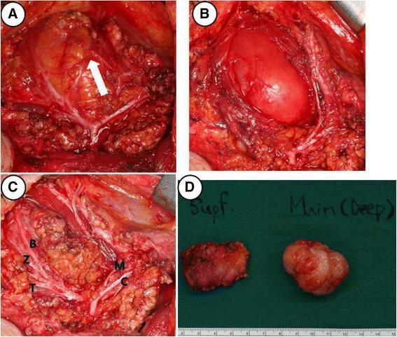
This composite of four clinical photographs illustrates intraoperative findings and gross pathology from a right parotidectomy for a deep-lobe tumor. Panel A shows the surgical field with a white arrow indicating a communicating nerve branch between the marginal mandibular and buccal branches of the facial nerve. Panel B demonstrates the tumor's location within the deep lobe of the parotid gland, adjacent to the masseter muscle. Panel C provides a comprehensive view of the preserved facial nerve branches following dissection, labeled as temporal (T), zygomatic (Z), buccal (B), marginal mandibular (M), and cervical (C). Panel D presents the gross surgical specimens against a green background with a metric scale; it differentiates the irregular superficial lobe parotid tissue ('Supf.') from the main, well-demarcated, multi-lobulated tumor mass ('Main (Deep)'). These images serve as educational material for head and neck surgery, specifically illustrating parotid gland anatomy, facial nerve preservation techniques, and surgical management of parotid neoplasms.
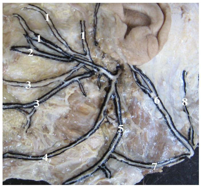
An anatomical dissection photograph illustrating the terminal branching patterns of the human facial nerve (CN VII) and its cervical connections. The image displays a 'Type I' branching variant characterized by a fan-like distribution over the facial musculature. Key structures labeled include the temporal branches (1) extending superiorly, zygomatic branches (2) coursing toward the midface, and buccal branches (3) distributed across the cheek area. Inferiorly, the marginal mandibular branch (4) follows the lower border of the mandible, while the cervical branch (5) descends toward the neck. The dissection also highlights the greater auricular nerve (6) and the lesser occipital nerve (8). Of clinical and surgical significance is the depicted connection (7) between the cervical branch of the facial nerve and the transverse cervical nerve of the cervical plexus. This anatomical resource serves to educate on neuroanatomical variations relevant to parotid surgery, facial reconstruction, and local anesthesia.
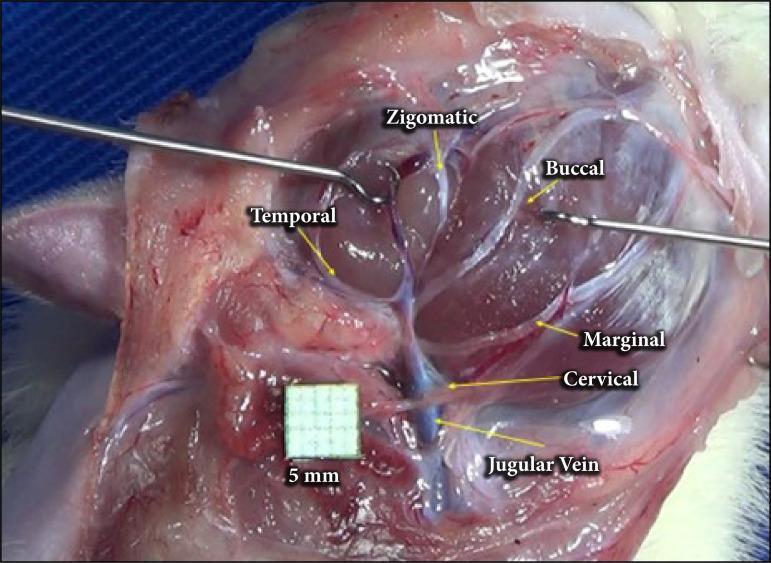
A clinical intraoperative photograph showing the anatomical arrangement of the facial nerve branches in a surgical dissection, following parotidectomy. The image provides a lateral view of the cervicofacial region where the parotid gland has been removed to expose the extratemporal facial nerve plexus. Labeled branches include the temporal branch extending superiorly, the zygomatic and buccal branches traveling toward the mid-face, and the marginal mandibular and cervical branches descending inferiorly. The cervical branch is depicted in close proximity to the jugular vein, which is visible in the lower surgical field. Surgical instruments (retractors) are positioned to maintain exposure of the deep tissues. A 5mm grid scale is included for anatomical measurement. This visual serves as an educational guide for surgical anatomy, identifying key landmarks for nerve preservation during parotid gland surgery or facial nerve decompression.
Facial Nerve (CN VII) - Innervation of the Face, and Lesion Patterns
1. Overview: Peripheral Distribution on the Face
| Branch | Region Supplied |
|---|---|
| Temporal | Frontalis (forehead wrinkling), orbicularis oculi (upper), corrugator supercilii |
| Zygomatic | Orbicularis oculi (lower), lower eyelid closure |
| Buccal | Buccinator, orbicularis oris, muscles of the upper lip and cheek |
| Marginal mandibular | Muscles of the lower lip and chin (depressor labii, mentalis) |
| Cervical | Platysma |
2. Central (Cortical/Supranuclear) Innervation - The Key Concept
Upper facial nucleus (forehead/eye division)
- Receives bilateral corticobulbar input - from BOTH ipsilateral and contralateral motor cortex
- Controls: frontalis (forehead wrinkling), orbicularis oculi (eye closure), corrugator
Lower facial nucleus (mouth/cheek division)
- Receives predominantly contralateral corticobulbar input
- Controls: buccinator, orbicularis oris, depressor muscles, platysma
3. Upper Motor Neuron (UMN) Lesion - Central Facial Palsy
- Forehead is SPARED - because the upper facial nucleus receives bilateral cortical input; the intact contralateral (now ipsilateral to the lesion) hemisphere still drives the upper face
- Lower face (mouth/cheek) is WEAK contralateral to the lesion - because this nucleus receives predominantly crossed input, which is now lost
- Voluntary movement is more impaired than emotional/spontaneous movement (e.g., patient cannot voluntarily retract the corner of the mouth, but may still smile asymmetrically during genuine emotion - because emotional facial pathways run separately through the thalamus/limbic system)
- No taste loss, no hyperacusis, no loss of lacrimation
4. Lower Motor Neuron (LMN) Lesion - Peripheral Facial Palsy
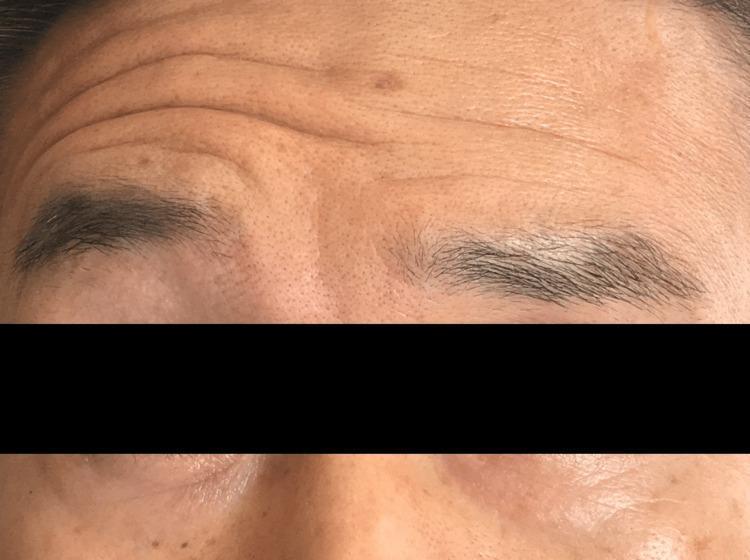
- Forehead IS affected - because both upper and lower facial nuclei are damaged or their axons are cut; the bilateral cortical input is irrelevant once the final common pathway is lost
- All ipsilateral muscles weak: frontalis (no forehead wrinkling), orbicularis oculi (cannot close eye - leading to corneal exposure/lagophthalmos), buccinator, orbicularis oris, platysma
- Eye closure failure is a key sign and risk - exposure keratitis can develop
- Depending on the level of the lesion, additional features may appear:
| Level of LMN Lesion | Extra Features |
|---|---|
| Pontine nucleus (e.g., Millard-Gubler syndrome) | Ipsilateral VI palsy (lateral gaze palsy) + contralateral hemiplegia (long tract signs) |
| Cerebellopontine angle (e.g., vestibular schwannoma) | Ipsilateral VIII palsy (hearing loss, tinnitus, vertigo) |
| Internal auditory canal | VIII involvement |
| Above stapedius branch (within petrous bone) | Hyperacusis (stapedius paralysis), taste loss (chorda tympani), dry eye (GSPN) |
| Above chorda tympani | Taste loss (anterior 2/3 tongue) + dry mouth |
| Below chorda tympani / at stylomastoid foramen | Pure motor facial palsy (Bell's palsy typical site) |
| Parotid gland (tumor, surgery) | Selective branch palsies |
5. Bilateral Lesion (Pseudobulbar Palsy / Bilateral LMN)
Bilateral UMN Lesion - Pseudobulbar Palsy
- Requires damage to corticobulbar fibers from both hemispheres (e.g., bilateral capsular strokes, MND, Steele-Richardson syndrome)
- After the first capsular stroke, residual 50% supranuclear innervation from the intact side maintains some function
- After a second contralateral stroke, both lower cranial nerve nuclei lose all supranuclear input
- Result: bilateral lower face weakness + dysarthria + dysphagia + emotional lability (pathological laughing/crying)
- The forehead may remain relatively spared (bilateral cortical input), but emotional expression is lost
Bilateral LMN Lesion
- Both peripheral facial nerves affected (e.g., Lyme disease, Guillain-Barré syndrome, sarcoidosis, bilateral Bell's palsy)
- Complete bilateral facial diplegia - expressionless "mask-like" face
- Both foreheads smooth, bilateral lagophthalmos, bilateral loss of taste
Summary Table
| Feature | UMN Lesion (Central) | LMN Lesion (Peripheral) |
|---|---|---|
| Side of weakness | Contralateral | Ipsilateral |
| Forehead | Spared | Affected |
| Eye closure | Intact | Weak/lost (lagophthalmos) |
| Taste | Preserved | Lost (if above chorda tympani) |
| Hyperacusis | No | Yes (if above stapedius branch) |
| Lacrimation | Preserved | Reduced (if above GSPN) |
| Voluntary vs. emotional | Voluntary worse | Both equally affected |
| Associated signs | Hemiplegia, UMN signs | CN VI palsy, hearing loss (by level) |
- Localization in Clinical Neurology, 8th Ed. - Anatomy of the Motor System
- Scott-Brown's Otorhinolaryngology Head & Neck Surgery Vol. 2 - Clinical Disorders of CN VII, Pseudobulbar Palsy
- Cummings Otolaryngology Head and Neck Surgery - Facial Nerve chapter
- Tintinalli's Emergency Medicine - CN VII lesion patterns
- Gray's Anatomy for Students - Facial nerve terminal branches