
Reading File
The image shows learning objectives (GM7.1 - GM7.6) covering joint pain pathophysiology, autoimmune disease, and articular/periarticular diseases. Let me search the medical library for comprehensive coverage of all these topics simultaneously.
Searching sources...
Finding Sources
Finding Sources
Finding Sources
Finding Sources
Finding Sources
Reading File
Reading File
Finding Sources
Reading File
Finding Sources
Reading File
Reading File
Finding Sources
Reading File
Finding Sources
Reading File
Loading Image
Finding Sources
I now have comprehensive textbook content to address all six learning objectives. Let me compile a thorough answer.
GM7 Learning Objectives: Joint Pain & Autoimmune Disease
GM7.1 - Pathophysiology and Genetic Basis of Autoimmune Disease
What is Autoimmune Disease?
Autoimmune disease results from a breakdown of self-tolerance - the normal state where the immune system distinguishes "self" antigens from "foreign" ones and does not attack the body's own tissues. When this fails, the immune system mounts sustained responses against self-antigens, causing tissue damage.
Key Mechanisms of Pathophysiology
1. Loss of Self-Tolerance
The immune system normally maintains tolerance through:
- Central tolerance (thymus/bone marrow): Self-reactive T and B cells are deleted (clonal deletion) or rendered anergic before leaving primary lymphoid organs
- Peripheral tolerance: Regulatory T cells (Tregs), anergy, and activation-induced cell death suppress any self-reactive cells that escape
When these mechanisms fail, autoreactive T and B cells become activated and target self-tissues.
2. Molecular Mimicry
Pathogens (viruses, bacteria) can share structural similarity to self-antigens. Immune responses triggered by infection cross-react with host tissues. Classic example: group A Streptococcus M protein resembles cardiac myosin (rheumatic fever).
3. Bystander Activation
During infection, local inflammation releases self-antigens. These are processed by activated antigen-presenting cells (APCs) and presented to previously ignorant (not tolerant) self-reactive T cells, breaking tolerance non-specifically.
4. Cytokine and Effector Mechanisms
- Autoreactive CD4+ Th1/Th17 cells produce IFN-γ and IL-17, driving inflammation and macrophage activation
- Autoantibodies (from autoreactive B cells) cause tissue damage through complement activation, opsonization (e.g., anti-GBM in Goodpasture's), or receptor blockade/stimulation (e.g., anti-TSH receptor in Graves' disease)
- Immune complexes deposit in vessel walls and tissues, fixing complement (SLE)
Genetic Basis
The genetic contribution is well established. From Firestein & Kelley's Textbook of Rheumatology:
"Polymorphisms in class I and II human leukocyte antigen (HLA) genes are known genetic risk factors for several autoimmune diseases... because the gene products influence T cell repertoire development, tolerance, and immune responses to foreign agents, certain polymorphisms are selected on the basis of environmental triggers."
Key genetic associations:
| Gene/Allele | Associated Disease |
|---|---|
| HLA-DR4 (DRB1*04) | Rheumatoid Arthritis |
| HLA-DRB10301, DQA10501 | Inflammatory myositis |
| HLA-B27 | Ankylosing spondylitis, spondyloarthritis |
| HLA-DR2, DR3 | SLE |
| HLA-DR3, DR4 | Type 1 Diabetes |
Other non-HLA genes implicated include:
- PTPN22 (regulates T cell signaling) - associated with RA, T1DM
- CTLA4 (T cell co-inhibition) - T1DM, Graves' disease
- STAT4, IRF5 - SLE
The concordance in identical twins (30-50%) vs fraternal twins (~5%) confirms polygenic inheritance modified by environment - no single gene is sufficient.
GM7.2 - Classification of Joint Pain Causes Based on Pathophysiology
Joint pain (arthralgia or arthritis) can be classified by the underlying pathophysiological mechanism. From Rheumatology, 2-Volume Set:
| Type | Pain Pattern | Causes |
|---|---|---|
| Bone pain | Present at rest and at night | Tumor, Paget disease, fracture |
| Mechanical joint pain | Pain related to joint use only | Unstable joint, osteoarthritic joint, hypermobility |
| Inflammatory joint pain | Present at rest and with use; worse at either end of the day | RA, gout, SLE, infection, spondyloarthritis |
| Soft tissue / Periarticular pain | Related to specific activity; point tenderness | Tendinitis, bursitis, enthesopathy |
| Neuropathic pain | Burning, stinging, paroxysmal, dermatomal distribution | Nerve entrapment, radiculopathy |
| Crystal-induced | Sudden severe onset (often nocturnal) | Gout (urate), pseudogout (CPPD) |
| Infectious | Acute, febrile, single joint | Septic arthritis, reactive arthritis |
Broader Pathophysiological Categories:
- Inflammatory - synovial inflammation driven by immune cells and cytokines (RA, SLE, gout, infection)
- Degenerative/Mechanical - cartilage breakdown and structural failure without primary inflammation (OA)
- Metabolic - crystal deposition (gout = uric acid; pseudogout = CPPD; hydroxyapatite)
- Infectious - direct microbial invasion (septic arthritis) or post-infectious immune reaction (reactive arthritis)
- Neoplastic - primary bone tumors, metastases, leukemic infiltration
- Endocrine/Systemic - hypothyroidism, acromegaly, hemochromatosis
GM7.3 - Systematic Clinical Approach to Joint Pain
The approach follows a logical stepwise framework, per The Washington Manual of Medical Therapeutics:
Step 1: Arthritis vs. Periarthritis?
- Arthritis: pain in all directions of motion; occurs with both active AND passive movement
- Periarthritis: pain at a single point or direction; primarily with active movement; passive movement preserved
Step 2: Inflammatory vs. Non-inflammatory?
| Feature | Inflammatory | Non-inflammatory |
|---|---|---|
| Morning stiffness | >1 hour, improves with activity | <30 min ("gelling") |
| Joint appearance | Swollen, red, warm | May be bony, cool |
| Systemic features | Fever, fatigue, weight loss, rash | Usually absent |
| ESR/CRP | Elevated | Normal or mildly raised |
| Synovial fluid WBC | >2,000/mm³ (often >10,000) | <2,000/mm³ |
Step 3: Number and Pattern of Joints?
- Monoarthritis (1 joint): Always consider septic arthritis first; also crystal-induced, trauma, hemarthrosis
- Oligoarthritis (<5 joints): Spondyloarthritis, crystal-induced, Still's disease, early polyarticular
- Polyarthritis (≥5 joints): RA, psoriatic arthritis, SLE, crystal-induced, viral
Step 4: Acute vs. Subacute vs. Chronic?
(see GM7.4 below)
Step 5: Investigations
- Bloods: FBC, ESR, CRP, uric acid, ANA, RF, anti-CCP, complement (C3/C4)
- Synovial fluid analysis (arthrocentesis in all acute monoarthritis):
| Measure | Normal | Non-inflammatory | Inflammatory | Septic |
|---|---|---|---|---|
| Appearance | Clear | Clear/yellow | Clear to opaque | Opaque |
| WBC/mm³ | <200 | 0-2,000 | >2,000 | >20,000 |
| PMN% | <25% | <25% | ≥50% | ≥75% |
- Imaging: X-ray (baseline), ultrasound (synovitis, effusion), MRI (soft tissue, early erosions)
GM7.4 - Acute, Subacute, and Chronic Causes of Joint Pain
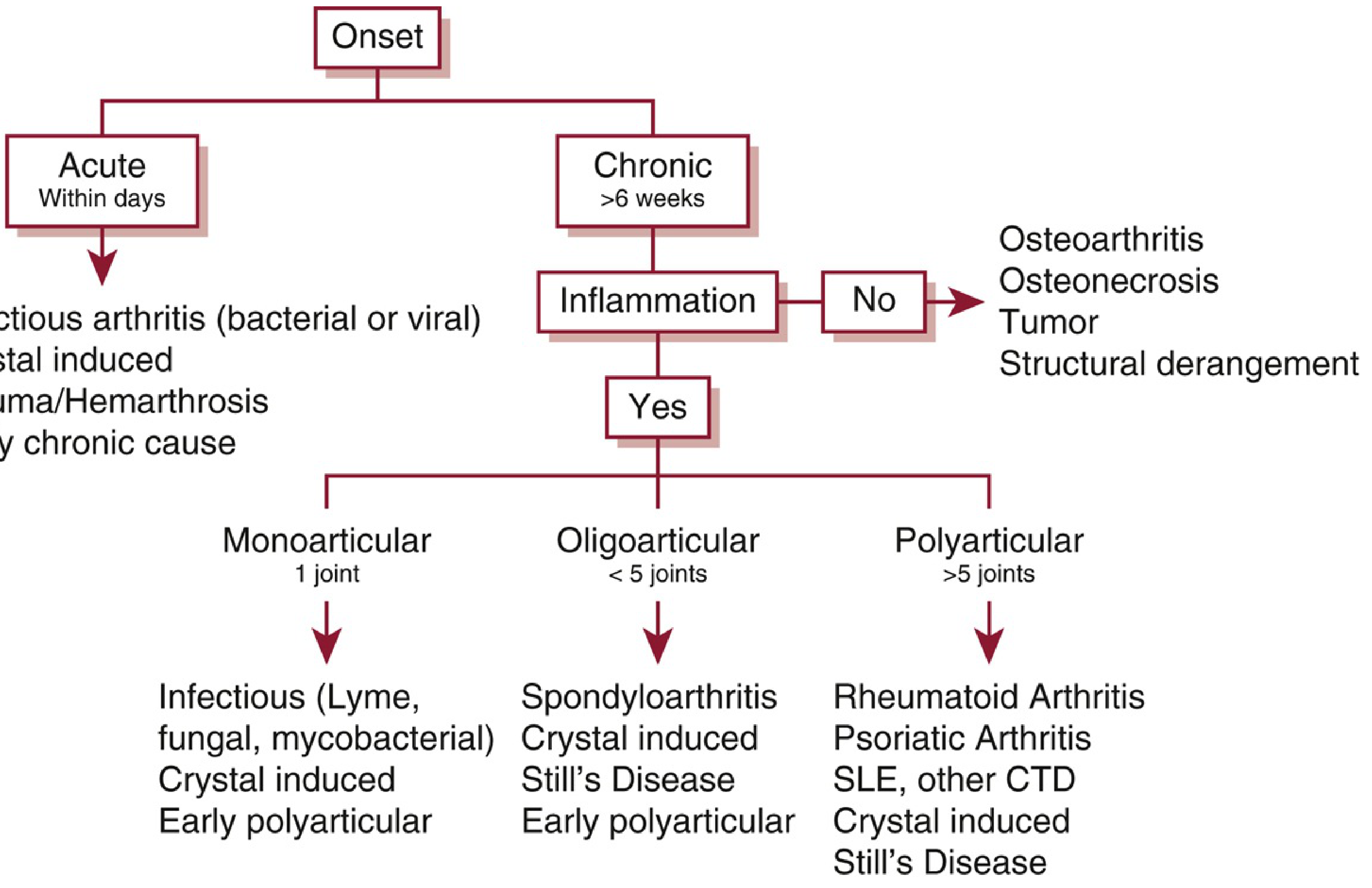
(Figure: Approach to joint pain - Washington Manual of Medical Therapeutics)
Acute (within days/hours)
- Septic arthritis (bacterial/viral)
- Crystal-induced: Gout (MSU crystals - negative birefringent needles), Pseudogout (CPPD - positive birefringent rhomboids)
- Trauma / Hemarthrosis (e.g., ACL tear)
- Reactive arthritis (Reiter syndrome - 1-4 weeks post-infection)
- Early presentation of any chronic cause
Subacute (weeks)
- Reactive arthritis
- Viral arthritis (hepatitis B/C, parvovirus B19, HIV)
- Lyme disease (Borrelia)
- Psoriatic arthritis (early)
- Palindromic rheumatism
Chronic (>6 weeks)
Inflammatory:
- Rheumatoid Arthritis (symmetric polyarthritis, MCP/PIP/wrists)
- Spondyloarthritis (HLA-B27 related: ankylosing spondylitis, psoriatic arthritis, IBD-associated)
- SLE and other CTDs (mixed CTD, Sjögren's, systemic sclerosis)
- Still's disease (adult-onset systemic JIA)
- Crystal arthropathy (chronic tophaceous gout)
Non-inflammatory (structural/mechanical):
- Osteoarthritis (DIP > PIP > hips > knees; Heberden/Bouchard nodes)
- Osteonecrosis (avascular necrosis)
- Bone tumors
- Structural derangements (torn meniscus, ligament laxity)
GM7.5 - Discriminating Arthralgia vs Arthritis; Articular vs Periarticular; Mechanical vs Inflammatory
Arthralgia vs. Arthritis
| Feature | Arthralgia | Arthritis |
|---|---|---|
| Definition | Joint pain WITHOUT objective findings | Joint pain WITH objective signs (swelling, warmth, erythema, restricted ROM) |
| Physical exam | Normal joint exam | Synovitis, effusion, tenderness on palpation |
| Significance | May be prodrome; or systemic disease without synovitis (e.g., SLE early) | Indicates active joint pathology |
Articular vs. Periarticular
| Feature | Articular | Periarticular |
|---|---|---|
| Pain with movement | All directions (active + passive) | Specific direction/plane; primarily active movement |
| Tenderness | Diffuse over joint line | Point tenderness at specific structure |
| Swelling | Intra-articular (effusion / synovial proliferation) | Periarticular soft tissue (bursa, tendon sheath) |
| Examples | RA, OA, gout | Bursitis (olecranon, prepatellar, trochanteric), tendinitis, enthesopathy, carpal tunnel |
Mechanical vs. Inflammatory
From Rheumatology, 2-Volume Set and Firestein & Kelley:
| Feature | Mechanical | Inflammatory |
|---|---|---|
| Pain pattern | With activity; relieved by rest | At rest AND with activity; worse at rest (morning) |
| Morning stiffness | Brief (<30 min, "gelling") | Prolonged (>1 hour) |
| Response to rest | Improves | Does not help, may worsen |
| Response to NSAIDs | Partial | Good |
| Response to steroids | Poor | Excellent (e.g., PMR responding to prednisolone) |
| Systemic features | Absent | Fever, fatigue, weight loss, elevated inflammatory markers |
| Joint appearance | Cool, bony enlargement | Warm, red, swollen |
| ESR/CRP | Normal | Elevated |
| Example | Osteoarthritis | Rheumatoid arthritis, gout, septic arthritis |
"Joint pain present at rest but worse with movement suggests an inflammatory process, whereas pain that occurs primarily with activity and is relieved by rest usually indicates a mechanical cause." - Firestein & Kelley's Textbook of Rheumatology
GM7.6 - Common Signs and Symptoms of Articular and Periarticular Diseases
Articular Disease - Signs and Symptoms
Symptoms:
- Pain: Character, site, radiation, diurnal pattern (morning = inflammatory; activity-related = mechanical)
- Stiffness: Duration of morning stiffness is a key inflammatory marker. Prolonged (>1 hr) = RA, PMR. Brief ("gelling") = OA
- Swelling: Patient-reported; may be effusion or synovial thickening
- Locking: Sudden inability to move (torn meniscus, loose body)
- Weakness/instability: Joint giving way; also consider myositis or neuropathy
Signs on Examination:
- Swelling: Effusion (fluctuant, ballottable), synovial proliferation (boggy/doughy), or bony enlargement
- Warmth and erythema: Active inflammation (gout = dramatic; RA = moderate)
- Tenderness: Joint line tenderness
- Deformity: Ulnar deviation, swan-neck deformity, boutonnière deformity (RA); Heberden/Bouchard nodes (OA)
- Restricted range of motion: Active and passive
- Crepitus: Felt or heard with movement (OA)
Periarticular Disease - Signs and Symptoms
Common Periarticular Conditions:
| Condition | Location | Key Signs |
|---|---|---|
| Olecranon bursitis | Posterior elbow | Fluctuant swelling over olecranon, non-tender ROM |
| Prepatellar bursitis ("housemaid's knee") | Anterior knee | Swelling anterior to patella |
| Trochanteric bursitis | Lateral hip | Point tenderness over greater trochanter; refers to lateral thigh |
| Subacromial bursitis | Shoulder | Painful arc 60-120° abduction |
| Lateral epicondylitis ("tennis elbow") | Lateral elbow | Pain on resisted wrist extension |
| Medial epicondylitis ("golfer's elbow") | Medial elbow | Pain on resisted wrist flexion |
| Achilles tendinitis | Posterior heel | Tenderness 2-6 cm above insertion |
| Plantar fasciitis | Heel | Worst on first steps in morning |
| Carpal tunnel syndrome | Wrist/hand | Tingling in radial 3.5 fingers; positive Tinel's/Phalen's |
Key distinguishing feature: In periarticular disease, passive ROM is preserved (or near-normal), while active ROM elicits pain. The pain can be reproduced by palpation of the specific structure. Referred pain from periarticular structures can mimic articular disease - for example, trochanteric bursitis may be mistaken for hip joint disease.
Summary Table: Key Discriminators
| Arthralgia | Arthritis | Periarthritis | Mechanical | Inflammatory | |
|---|---|---|---|---|---|
| Objective signs | ✗ | ✓ | ✓ (local) | ✓ | ✓ |
| Passive ROM | Normal | Reduced | Normal | Reduced (end) | Reduced |
| Morning stiffness | - | Variable | No | <30 min | >60 min |
| Warmth/redness | ✗ | ± | ✗ | ✗ | ✓ |
| ESR/CRP | Normal | Variable | Normal | Normal | Elevated |
| Rest improves pain | - | Mechanical yes; inflam no | Yes | ✓ | ✗ |
Sources: The Washington Manual of Medical Therapeutics, p. 942-943 | Rheumatology 2-Volume Set (Elsevier, 2022), p. 263 | Firestein & Kelley's Textbook of Rheumatology | Quick Compendium of Clinical Pathology 5th ed.
This is a shared conversation. Sign in to Orris to start your own chat.