
Pid for 20 marks ibs gyne
pelvic inflammatory disease PID salpingitis ultrasound diagram
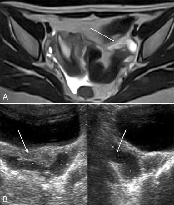
A dual-modality clinical diagnostic image illustrating Pelvic Inflammatory Disease (PID) through axial T2-weighted MRI (Panel A) and transabdominal ultrasound (Panel B). Panel A displays an axial T2W MRI of the pelvis, where a white arrow identifies a hyperintense, complex fluid collection in the left adnexa. This finding represents a damaged or distended fallopian tube, characteristic of salpingitis or hydrosalpinx, with associated free fluid noted in the Pouch of Douglas (POD). Panel B presents a transabdominal USG showing an anechoic to hypoechoic region in the posterior pelvis. White arrows point to internal echoes within the pelvic fluid collection, suggesting an inflammatory or exudative component. The educational focus is on identifying radiological signs of upper genital tract infection, specifically adnexal involvement and peritoneal fluid accumulation. This material is suitable for intermediate medical education in gynecology and radiology, demonstrating the multi-modal appearance of acute pelvic inflammatory pathology.
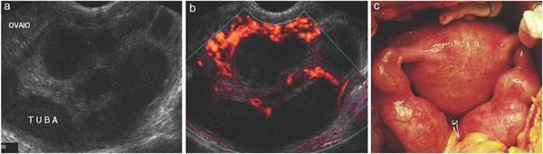
This composite figure illustrates a case of Pelvic Inflammatory Disease (PID) through ultrasound and intraoperative clinical photography. Panel (a) is a grayscale transvaginal ultrasound showing an adnexal mass. The ovary ('OVAIO') contains multiple small, anechoic follicles, while the adjacent fallopian tube ('TUBA') appears dilated and thickened with complex internal echoes, suggestive of salpingitis or a tubo-ovarian complex. Panel (b) utilizes Color Doppler ultrasound, demonstrating marked peripheral and internal hypervascularity (hyperemia) within the tubal and ovarian structures, a key diagnostic indicator of acute inflammation. Panel (c) provides clinical correlation via an intraoperative photograph during surgery. It shows a markedly erythematous and edematous uterus flanked by bilaterally enlarged, swollen, and congested fallopian tubes. The visual evidence of inflammatory exudate and pelvic congestion confirms the diagnosis of severe pelvic inflammatory disease. This material is intended for medical education regarding the diagnostic triad of grayscale findings, Doppler vascular assessment, and direct surgical visualization of acute pelvic infections.
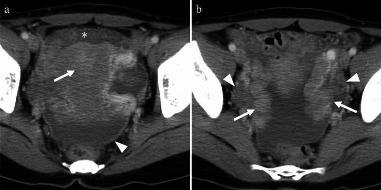
This dual-panel diagnostic image presents axial contrast-enhanced CT scans of the female pelvis, illustrating typical findings of Pelvic Inflammatory Disease (PID). Image A focuses on the uterus, demonstrating abnormal endometrial enhancement (arrow) consistent with endometritis. Associated inflammatory changes include fat stranding in the anterior pelvic planes (asterisk), peritoneal thickening, and the presence of complex free fluid in the dependent pelvic areas (arrowhead). Image B highlights the adnexal structures, showing bilateral thickening of the fallopian tubes exceeding 5 mm (arrows), which is a characteristic feature of salpingitis. The bilateral ovaries exhibit a polycystic morphology with multiple small follicles, accompanied by mild stromal haziness and hyperemia (arrowheads), findings indicative of oophoritis. The composite imaging illustrates the spectrum of PID manifestations, ranging from uterine and peritoneal involvement to tubal and ovarian inflammation, relevant for radiologists and clinicians specializing in gynecology and infectious diseases.
Pelvic Inflammatory Disease (PID) - 20 Marks
Definition
- Harrison's Principles of Internal Medicine 22E, 2025
Etiology
| Organism | Role |
|---|---|
| Neisseria gonorrhoeae | Primary cause, especially in high-gonorrhoea settings |
| Chlamydia trachomatis | Common; leads to immunopathologic tubal damage |
| Mycoplasma genitalium | Significantly associated with endometritis and salpingitis |
| Anaerobes (Prevotella spp., peptostreptococci) | Found in 25-33% of cases (BV-associated) |
| Facultative organisms | E. coli, H. influenzae, group B streptococci |
Epidemiology and Risk Factors
- Endocervical infection (gonorrhoea/chlamydia) or bacterial vaginosis (BV)
- History of prior PID (strongest predictor of recurrence)
- Recent IUD insertion (within 1 month)
- Multiple sexual partners; history of STIs
- Frequent vaginal douching
- Recent invasive uterine procedures (D&C, termination of pregnancy, hysterosalpingography)
- Onset during or just after menstruation
- Young age / adolescence
- Oral contraceptive pill use (decreases symptomatic PID)
- Tubal sterilisation (prevents intraluminal spread)
Pathogenesis
Clinical Features
Symptoms
- Lower abdominal/pelvic pain - bilateral, dull/aching, of <3 weeks duration
- Abnormal vaginal discharge - yellow or malodorous (mucopurulent cervicitis, MPC)
- Abnormal uterine bleeding (precedes or coincides with pain in ~40%)
- Dysuria (urethral involvement, ~20%)
- Nausea, vomiting, fever if peritonitis develops
- Right upper quadrant (RUQ) pain in 3-10% = Fitz-Hugh-Curtis syndrome (perihepatitis)
Signs
- Cervical motion tenderness (CMT) - pathognomonic clinical sign
- Uterine tenderness (endometritis)
- Adnexal tenderness - usually bilateral
- Adnexal swelling/mass in ~50%
- Fever >38°C in only ~1/3 of patients
- Speculum: MPC (yellow discharge, easily-induced endocervical bleeding)
Diagnosis
CDC Minimum Criteria (any ONE of the following in a sexually active woman with pelvic pain):
- Cervical motion tenderness, OR
- Uterine tenderness, OR
- Adnexal tenderness
"Treatment appropriate for PID must not be withheld from patients with an equivocal diagnosis - it is better to err on the side of overdiagnosis and overtreatment." - Harrison's 22E
Additional criteria (increase specificity):
- Temperature >38°C
- Elevated ESR (>15 mm/h) or CRP
- Elevated WBC count
- Palpable adnexal mass
- Laboratory confirmation of N. gonorrhoeae or C. trachomatis (NAAT)
- Evidence of MPC on speculum exam
- PMNs outnumbering epithelial cells on vaginal wet prep
Investigations
- NAAT of endocervical/vaginal swab for N. gonorrhoeae and C. trachomatis
- Wet preparation: PMNs in vaginal fluid
- Gram stain: PMNs and gram-negative diplococci
- ESR, CRP, CBC (leucocytosis in up to 60%)
- Urine beta-hCG (to exclude ectopic pregnancy)
- Pelvic USS: identifies TOA, free fluid, tubal thickening - first-line imaging
- MRI: more sensitive - shows increased tubal diameter, intratubal fluid, tubal wall thickening
- CT pelvis (contrast-enhanced): endometrial enhancement, bilateral tubal thickening >5mm, pelvic fat stranding
- HIV screening for all women with PID
Differential Diagnosis
- Ectopic pregnancy (must exclude first with beta-hCG)
- Appendicitis (favoured by anorexia/vomiting early, onset after day 14, unilateral RIF pain)
- Endometriosis
- Ovarian cyst/torsion
- Corpus luteum haematoma
Imaging in PID
Treatment
Indications for Hospitalisation (Parenteral Therapy)
- Diagnosis uncertain - cannot exclude surgical emergency (appendicitis, ectopic)
- Pregnancy
- Suspected pelvic abscess (TOA)
- Severe illness / nausea and vomiting precluding oral therapy
- HIV infection
- Patient unable to tolerate or comply with outpatient regimen
- Failure to respond to outpatient therapy within 72 hours
CDC Recommended Regimens (from Harrison's 22E / CDC MMWR 2021):
- Ceftriaxone 500 mg IM single dose PLUS
- Doxycycline 100 mg PO twice daily x 14 days PLUS
- Metronidazole 500 mg PO twice daily x 14 days
| Regimen | Drugs |
|---|---|
| Regimen A | Cefotetan 2g IV q12h (or Cefoxitin 2g IV q6h) + Doxycycline 100 mg IV/PO q12h |
| Regimen B | Clindamycin 900 mg IV q8h + Gentamicin (loading dose 2 mg/kg IV/IM, then 1.5 mg/kg q8h) |
- Parenteral therapy continued for at least 48 hours after clinical improvement
- Then transition to oral therapy to complete 14 days total
- In cases with TOA: prefer clindamycin (not doxycycline) to complete oral therapy (better anaerobic coverage)
Follow-up
- Outpatients: re-evaluate at 72 hours - if not improving, hospitalise
- Sex partners must be empirically treated for gonorrhoea and chlamydia
- NAAT for persistent/recurrent infection after treatment if symptoms recur
Fitz-Hugh-Curtis Syndrome (Perihepatitis)
- Occurs in 3-10% of PID cases
- Pleuritic RUQ pain and tenderness
- "Violin-string" adhesions between liver capsule and parietal peritoneum on laparoscopy
- Liver function tests and RUQ USS are nearly always normal
- Now most cases attributed to chlamydial (not gonococcal) salpingitis
- Diagnosed clinically: RUQ pain + MPC + adnexal tenderness in a young woman with normal gallbladder USS
Surgery
- Rupture or threatened rupture of a tubo-ovarian abscess (TOA)
- Drainage of an abscess not responding to antibiotics
- Options: posterior colpotomy (for pelvic abscess drainage) and peritoneal lavage for generalised peritonitis
Complications and Sequelae
Tubo-Ovarian Abscess (TOA)
- Most serious acute complication
- Develops in ~15% of hospitalised PID patients
- Requires parenteral antibiotics; surgical drainage if no response
Long-term Sequelae (from the Swedish salpingitis study):
| Episodes of PID | Risk of infertility (tubal occlusion) |
|---|---|
| 1 episode | 11% |
| 2 episodes | 23% |
| 3+ episodes | 54% |
- Ectopic pregnancy: 7-fold increased risk after PID (University of Washington data)
- Chronic pelvic pain: persistent in 20-30%
- Recurrent salpingitis
- Hysterectomy rate: 8-fold increased after PID
Prevention
- Annual NAAT screening for C. trachomatis in all sexually active women ≤25 years (CDC / US Preventive Services Task Force recommendation)
- Screening reduces PID incidence by 56% (RCT evidence)
- Safe sex practices; prompt treatment of STIs
- Careful aseptic technique during intrauterine procedures
Summary Table
| Feature | Details |
|---|---|
| Definition | Ascending infection: cervix → endometrium → tubes → peritoneum |
| Causative organisms | N. gonorrhoeae, C. trachomatis, M. genitalium, anaerobes (BV-related) |
| Key clinical sign | Cervical motion tenderness (CMT) |
| Minimum diagnostic criteria | CMT OR uterine tenderness OR adnexal tenderness |
| Gold standard diagnosis | Laparoscopy |
| Outpatient Rx | Ceftriaxone + Doxycycline + Metronidazole x 14 days |
| Parenteral Rx A | Cefotetan/Cefoxitin + Doxycycline |
| Parenteral Rx B | Clindamycin + Gentamicin |
| Perihepatitis | Fitz-Hugh-Curtis syndrome (violin-string adhesions) |
| Key complication | TOA, infertility, ectopic pregnancy |
| Sequelae after 3 episodes | 54% infertility |
Anemia in pregnancy 20 marks
anemia in pregnancy iron deficiency peripheral smear classification
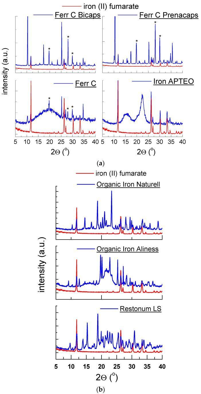
This figure presents X-ray powder diffraction (XRPD) patterns for various commercial iron dietary supplements, used in the management of iron deficiency anemia. The data are organized into two panels, (a) and (b), plotting diffraction intensity (arbitrary units) against the diffraction angle (2θ°) from 5° to 40°. Each graph compares a specific supplement (blue curve) against a reference standard of pure iron (II) fumarate (red curve). Panel (a) features Ferr C Bicaps, Ferr C Prenacaps, Ferr C, and Iron APTEO. Panel (b) includes Organic Iron Naturell, Organic Iron Aliness, and Restonum LS. Sharp, high-intensity peaks in products like Ferr C Bicaps indicate a high degree of crystallinity, primarily corresponding to iron (II) fumarate and vitamin C (indicated by asterisks). Conversely, the broad, diffuse humps observed in the Iron APTEO and Ferr C patterns signify a predominantly amorphous or poorly crystalline state. These diffraction fingerprints are critical for pharmaceutical quality control, verifying the presence of the active mineral form and excipients, and assessing the structural stability of the oral iron supplement.
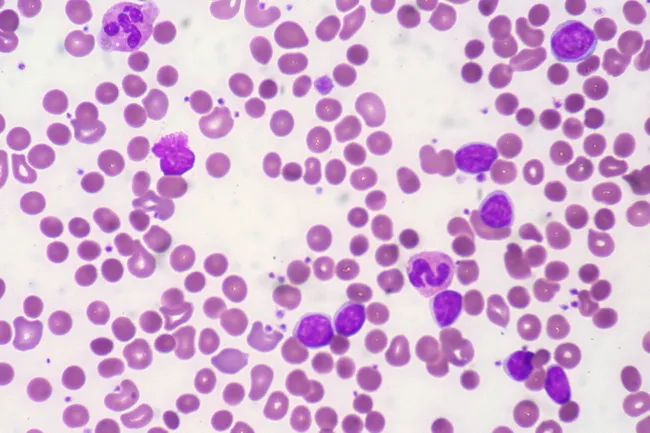
Peripheral blood smear from a patient with known chronic lymphocytic leukemia (CLL) showing autoimmune hemolytic anemia (AIHA). Brightfield microscopy of Wright-Giemsa stained smear at 1000x (oil immersion) reveals two dominant cell populations: numerous small mature lymphocytes typical of CLL and red blood cells showing spherocytic deformation. A smudge cell is evident just left of center, a classical clue for CLL. Red cells display reduced central pallor consistent with spherocytes, indicating intravascular or extravascular hemolysis driven by autoantibodies. The background shows mild polychromasia and rare nucleated RBCs, consistent with a robust reticulocyte response; the corrected reticulocyte count in this case is markedly elevated (>7%). Platelets are not significantly abnormal. These features collectively support autoimmune destruction of erythrocytes in the setting of CLL. Diagnostic significance lies in recognizing AIHA as a common autoimmune complication in CLL, with a clinical picture of sudden Hb decline and compensatory erythropoiesis. Differential considerations include drug-induced hemolysis, other causes of hemolytic anemia, or marrow infiltration by CLL. Clinical correlation includes therapy implications (steroids, rituximab) and close monitoring of hemolysis markers. This image is a valuable teaching resource for hematology morphology, AIHA in CLL, and differential diagnosis in anemia.
Anaemia in Pregnancy - 20 Marks
Definition
- <11 g/dL in the 1st and 3rd trimesters
- <10.5 g/dL in the 2nd trimester
- <10 g/dL is considered moderate; <7 g/dL is severe; <5 g/dL is very severe/critical
Physiological Basis - Normal Haematologic Changes in Pregnancy
| Parameter | Change |
|---|---|
| Total blood volume | Increases 40-45% (maximum at 34 weeks) |
| Plasma volume | Increases 47% |
| RBC mass | Increases only 17% (maximum at term) |
| Haemoglobin/Haematocrit | Falls (dilutional effect) |
| MCV, MCHC | No change (dilutional anaemia does NOT alter these) |
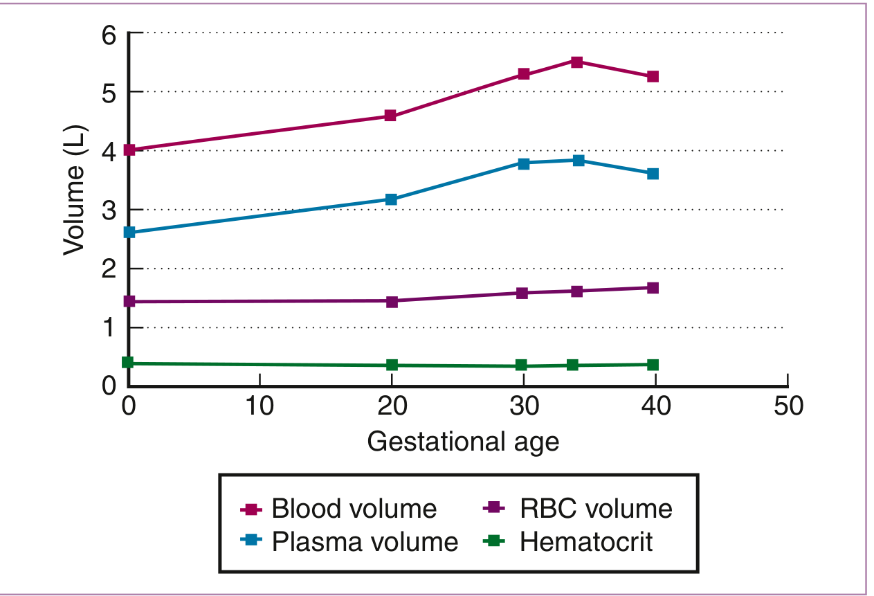
Key point: In physiological dilutional anaemia, MCV and MCHC remain normal. In true iron deficiency anaemia (IDA), both indices progressively decrease. This serial evaluation differentiates the two.
Classification
By Aetiology (most practical for exam):
- Iron deficiency anaemia (IDA) - most common
- Megaloblastic anaemia (folate or B12 deficiency)
- Combined iron + folate deficiency
- Antepartum haemorrhage
- Hookworm infestation (chronic blood loss)
- Malaria (most common haemolytic cause in India)
- Sickle cell disease
- Thalassaemia
- Autoimmune haemolytic anaemia
- G6PD deficiency
- Aplastic anaemia
- Leukaemia
By Severity (WHO/India NFHS classification):
| Grade | Hb (g/dL) | Action |
|---|---|---|
| Mild | 10-10.9 | Iron + folic acid supplementation |
| Moderate | 7-9.9 | Intensive iron therapy |
| Severe | <7 | Hospitalisation, IV iron/blood transfusion |
| Very severe/critical | <4 | Emergency blood transfusion |
Iron Requirements in Pregnancy
| Required for | Average (mg) |
|---|---|
| External iron loss | 170 |
| Expansion of RBC mass | 450 |
| Fetal iron | 270 |
| Placenta and cord iron | 90 |
| Blood loss at delivery | 150 |
| Total | 1130 |
- The estimated daily requirement is 22-23 mg/day but median dietary intake in pregnancy is only ~15 mg/day
- Without supplementation, IDA will develop in most women
- Iron supplementation with 60 mg elemental iron/day reduces anaemia incidence by up to 73%
Iron Deficiency Anaemia (IDA) - Stages
| Stage | Finding |
|---|---|
| Stage 1 - Pre-latent IDA | ↓ serum ferritin (reduced marrow stores), normal iron studies, normal Hb |
| Stage 2 - Latent IDA | ↓ serum iron, ↑ TIBC, ↓ % saturation, Hb still normal |
| Stage 3 - Frank IDA | ↓ Hb, microcytic hypochromic anaemia, ↓ MCV |
Megaloblastic Anaemia in Pregnancy
- Second most common nutritional anaemia in pregnancy
- Usually due to folate deficiency; rarely due to B12 deficiency (fetus preferentially takes B12 from mother)
- Folate deficiency defined as serum level <2.5-3 ng/mL; RBC folate <140-150 ng/mL
- Risk increased by: multiple pregnancy, haemolytic anaemia, poor diet, anticonvulsants, OCP use, alcohol, sulfa drugs (all reduce conjugase activity in intestine)
- Peripheral smear: macro-ovalocytes, hypersegmented neutrophils (>5 lobes or any 6-lobed cell), megaloblastic change in marrow
- Treatment: Folic acid 1-5 mg/day orally; prevention: 0.4-0.5 mg folic acid daily from pre-conception (4 mg/day if prior NTD history)
Risk Factors for Anaemia in Pregnancy
- Poor dietary iron and folate intake (low socioeconomic status)
- Repeated pregnancies at close intervals
- Menstrual losses (poor pre-pregnancy iron stores)
- Hookworm infestation (blood loss from gut)
- Malaria (haemolysis)
- Adolescent pregnancies
- Multiple gestation (increased demand)
- Prior history of anaemia
Clinical Features
Symptoms:
- Fatigue, weakness, pallor, exertional dyspnoea, palpitations
- Headache, dizziness, tinnitus
- Pica (craving non-food items like clay, chalk - classic in IDA)
- Glossitis, angular stomatitis, koilonychia (IDA specific)
- Glossitis, peripheral neuropathy (B12 deficiency specific)
Signs:
- Pallor (conjunctival, mucosal, palmar)
- Tachycardia, wide pulse pressure
- Systolic flow murmur
- Oedema (in severe cases - "nutritional oedema")
- Splenomegaly (haemolytic causes)
Investigations
- Hb at first antenatal visit, then at 28 and 36 weeks
| Test | Normal in Pregnancy |
|---|---|
| Hb | >11 g/dL (1st/3rd trimester); >10.5 g/dL (2nd) |
| MCV | 81-99 µm³ |
| MCHC | 32-35 g/dL |
| Serum ferritin | Most sensitive marker of iron stores |
| Serum iron | Decreased in IDA |
| TIBC | Increased in IDA |
| % transferrin saturation | Decreased in IDA |
| Serum folate | <2.5-3 ng/mL = deficient |
| RBC folate | <140-150 ng/mL = deficient |
| Reticulocyte count | 0.5-1.5% (elevated after treatment = response indicator) |
| Peripheral smear | Hypochromic microcytic (IDA), macro-ovalocytes (folate/B12) |
| Hb electrophoresis / HPLC | For thalassaemia, sickle cell |
| Direct Coombs test | For autoimmune haemolytic anaemia |
| Stool for ova/parasites | Hookworm |
| Malarial parasite (MP) smear | Malaria |
Effects on Mother and Fetus
Maternal effects:
- Increased risk of maternal mortality (RR 3.5 with Hb <5 g/dL)
- Postpartum haemorrhage (poor myometrial contraction + decreased blood reserve)
- Cardiac failure (high-output cardiac failure in severe anaemia)
- Increased risk of infection (iron deficiency impairs cellular immune function)
- Reduced work capacity and productivity
- Increased risk of preeclampsia
Fetal and neonatal effects:
- Preterm birth (>50% with Hb <5 g/dL in third trimester)
- Low birth weight / SGA neonates
- Stillbirth (with severe anaemia)
- Reduced fetal iron stores (cord blood ferritin correlates with maternal ferritin)
- Infant anaemia: 5.7-fold increased risk of anaemia at 1 year if mother was anaemic at delivery
- Neural tube defects (folate deficiency - hence periconceptional folic acid is mandatory)
Note: Paradoxically, very HIGH maternal Hb (>14.5 g/dL) is also associated with adverse outcomes (stillbirth, LBW, SGA) - thought to reflect inadequate plasma volume expansion and reduced intervillous blood flow.
Treatment
Oral Iron Therapy
- First-line treatment for mild to moderate IDA
- Ferrous sulphate 200 mg (65 mg elemental iron) TDS = 195 mg elemental iron/day
- Or ferrous fumarate / ferrous gluconate equivalent
- Take on empty stomach with Vitamin C (enhances absorption); avoid with tea, milk, antacids
- Expected response: Hb rises ~1 g/dL per week
- Reticulocytosis peaks at 7-10 days after starting therapy
- Continue for 3 months after Hb normalises to replenish stores
- IFA tablet: 100 mg elemental iron + 0.5 mg folic acid daily
- If Hb 10-12 g/dL: supplementation
- If Hb <10 g/dL: refer to PHC
Parenteral (IV) Iron Therapy
- Indications: oral iron intolerance, malabsorption, severe anaemia near term (<34 weeks), non-compliance, failure of oral therapy
- Iron sucrose is most commonly used (safest profile in pregnancy)
- Dose formula: Weight (kg) × (Target Hb - Actual Hb) × 0.24 + 500 mg
- Target Hb = 11-12 g/dL; max daily infusion 200 mg (split over alternate days)
- IV iron superiority over oral: meta-analysis of 11 RCTs (>600 patients per group) showed significantly higher Hb and more patients achieving target Hb at 4 weeks
- Six parenteral forms approved in USA: iron sucrose, iron isomaltoside, ferric gluconate, low-MW iron dextran, ferumoxytol, ferric carboxymaltose
- Test dose required only for low-MW iron dextran (anaphylaxis risk)
Blood Transfusion
- Indications: Hb <7 g/dL with severe symptoms, Hb <5 g/dL regardless of symptoms, Hb <8 g/dL near term (>36 weeks), cardiac failure due to anaemia
- Packed RBCs preferred over whole blood
- 1 unit packed cells raises Hb by ~1 g/dL
Erythropoietin
- Used in cases where oral iron has failed and Hb <8.5 g/dL
- Normalises Hb in ~2 weeks in 73% of cases when added to oral iron
- Risk of thromboembolism to be considered
Prevention
Antenatal:
- Routine IFA supplementation from first antenatal visit (100 mg elemental iron + 0.5 mg folic acid daily)
- Dietary counselling: increase iron-rich foods (green leafy vegetables, jaggery, meat), Vitamin C
- Deworming with albendazole 400 mg single dose (after first trimester)
- Malaria chemoprophylaxis in endemic areas
Preconceptional:
- Folic acid 0.4-0.5 mg/day from at least 1 month before conception (reduces NTD risk by ~70%)
- 4 mg/day if previous NTD-affected pregnancy
- Treat pre-existing anaemia before conception
Population level (India):
- National Nutritional Anaemia Prophylaxis Programme (NNAAP) - IFA tablets to pregnant women, lactating mothers, children
- Iron fortification of salt (National Institute of Nutrition, Hyderabad) - ferric orthophosphate or ferrous sulphate added to salt
- Screening at first ANC visit and at 28 and 36 weeks
Summary Table
| Feature | IDA | Megaloblastic (Folate) | Haemolytic |
|---|---|---|---|
| Smear | Microcytic hypochromic | Macro-ovalocytes, hypersegmented PMNs | Fragmented RBCs, spherocytes |
| MCV | Low | High | Variable |
| Ferritin | Low | Normal | Normal/High |
| TIBC | High | Normal | Normal |
| LDH | Normal | Elevated | Elevated |
| Bilirubin | Normal | Elevated (indirect) | Elevated (indirect) |
| Reticulocytes | Low | Low | High |
| Treatment | Oral/IV iron | Folic acid 1-5 mg/day | Treat cause |