
Mechanisms of labour
Reading File
The mechanisms of labour (or "cardinal movements") describe the sequence of positional changes a fetus makes as it navigates the birth canal during delivery. These include engagement, descent, flexion, internal rotation, extension, external rotation (restitution), and expulsion.
Searching sources...
Finding Sources
Finding Sources
Finding Sources
Finding Sources
Finding Sources
Finding Sources
Finding Sources
Finding Sources
Finding Sources
Reading File
Reading File
Reading File
Now let me fetch the diagram image for the cardinal movements:
Loading Image
Here is a thorough breakdown of the Mechanisms of Labour (Cardinal Movements), drawn from authoritative sources.
Mechanisms of Labour (Cardinal Movements)
The mechanism of labour describes the sequence of positional adaptations made by the fetus as it navigates the birth canal. It is determined by three interacting factors:
- Pelvic dimensions and configuration
- Fetal size
- Strength of uterine contractions
The core principle is that the fetus follows the path of least resistance - adapting the smallest achievable diameter of the presenting part to the most favourable contours of the birth canal at each level.
The following describes the classic sequence for a vertex (cephalic) presentation in the occiput anterior (OA) position, which accounts for ~97% of term deliveries.
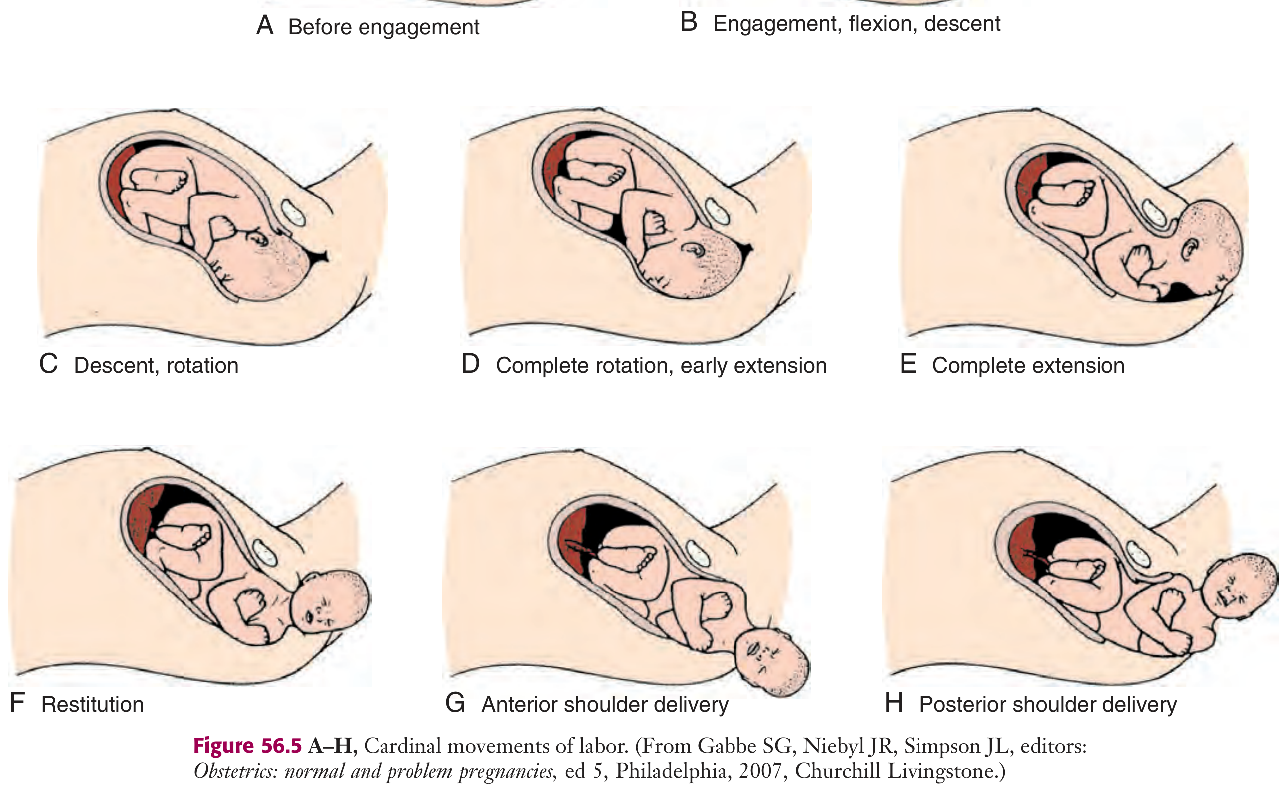
Fig. 56.5 - Cardinal movements of labour (A-H). From Roberts and Hedges' Clinical Procedures in Emergency Medicine.
The 7 Cardinal Movements
1. Engagement
The biparietal diameter (BPD) - the greatest transverse diameter of the fetal head - passes through the pelvic inlet (pelvic brim).
- The fetus is considered engaged when the presenting part is at station 0 (level of the ischial spines)
- In primiparous patients, engagement typically occurs in the last 2 weeks of pregnancy
- In multiparous patients, it may only occur at the onset of labour
- The head usually engages in the transverse or oblique diameter of the pelvic inlet
2. Flexion
- The fetal chin tucks onto the chest, converting the presenting diameter from the occipitofrontal (~11 cm) to the smaller suboccipitobregmatic diameter (~9.5 cm)
- This is mostly a passive movement driven by the resistance of the pelvic floor and walls
- Flexion is necessary for both engagement and descent through the narrowest pelvic dimensions
3. Descent
- The downward passage of the presenting part through the pelvis
- Progressive but not necessarily continuous - it accelerates during the second stage
- Driven by:
- Uterine contractions
- Abdominal wall contractions (maternal pushing effort)
- Straightening and extension of the fetal body
4. Internal Rotation
- As the head descends to the level of the ischial spines, it rotates so the occiput moves from a transverse position anteriorly toward the symphysis pubis (occiput anterior - most common)
- Less commonly, the occiput rotates posteriorly toward the sacral hollow (occiput posterior - persistent OP)
- This rotation brings the long axis of the fetal head into alignment with the anteroposterior (AP) diameter of the pelvic outlet
- Internal rotation is necessary to traverse the ischial spines
5. Extension
- Once the head reaches the vaginal introitus, the occiput comes to lie beneath the inferior margin of the pubic symphysis
- The head pivots around the symphysis - it is born by extension (not further flexion)
- The sequence of delivery: occiput → bregma → forehead → nose → mouth → chin
- After delivery, the head drops downward so the chin lies over the maternal anal region
6. External Rotation (Restitution)
- After the head is delivered, it rotates back to the transverse position it occupied at engagement - aligning with the fetal torso
- This is entirely passive and reflects the natural unwinding of the neck
- It signals that the fetal shoulders are now rotating internally to align with the AP diameter of the pelvic outlet
7. Expulsion
- The shoulders descend through the pelvis in the same path traced by the head
- The anterior shoulder is delivered beneath the symphysis pubis first
- The posterior shoulder is then delivered across the perineum
- The rest of the fetal body follows by lateral flexion
Breech Presentation - Key Differences
In breech presentations, the mechanics differ significantly:
- The bitrochanteric diameter is the widest engaging diameter (instead of the BPD)
- The hips usually engage in an oblique diameter of the inlet
- The anterior hip descends faster; internal rotation brings the bitrochanteric diameter into the AP diameter of the outlet
- Lateral flexion delivers the posterior hip first as the anterior hip catches under the symphysis
- Delivery of the aftercoming head (last part to deliver) is the most hazardous step - it requires controlled flexion, descent, and rotation, managed with techniques such as the Mauriceau-Smellie-Veit manoeuvre
Summary Table
| Step | Movement | Mechanism | Passive/Active |
|---|---|---|---|
| 1 | Engagement | BPD crosses pelvic inlet; presenting part at station 0 | Active (contractions) |
| 2 | Flexion | Chin-on-chest; smallest head diameter presented | Passive |
| 3 | Descent | Downward progression through birth canal | Active |
| 4 | Internal rotation | Occiput rotates anteriorly to AP outlet | Passive (pelvic floor) |
| 5 | Extension | Head pivots under pubic symphysis; delivered by extension | Passive |
| 6 | External rotation | Head realigns with torso (restitution) | Passive |
| 7 | Expulsion | Anterior then posterior shoulder delivered | Active |
Sources: - Roberts and Hedges' Clinical Procedures in Emergency Medicine, p.1351 | - Tintinalli's Emergency Medicine: A Comprehensive Study
This is a shared conversation. Sign in to Orris to start your own chat.