
Explain epiphysis
epiphysis bone anatomy diagram growth plate diaphysis
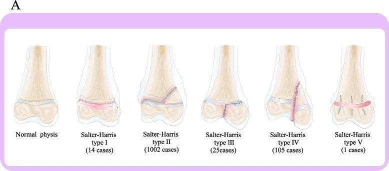
This medical illustration presents the Salter-Harris classification system for pediatric epiphyseal plate fractures, shown on a long bone model. Six diagrams compare the normal anatomy with fracture types I through V. The 'Normal physis' shows an intact growth plate between the metaphysis and epiphysis. Type I is a transverse fracture through the physis. Type II, the most frequent at 1002 cases, travels through the physis and exits through the metaphysis, creating a Thurston-Holland fragment. Type III travels through the physis and exits through the epiphysis into the joint space. Type IV is a vertical fracture crossing the metaphysis, physis, and epiphysis. Type V, the rarest with 1 case, is depicted as a crush or compression injury of the physis indicated by inward-pointing arrows. Each diagram is labeled with the corresponding fracture type and the number of cases from a clinical study, providing a visual guide for orthopedic classification and prevalence in pediatric populations.
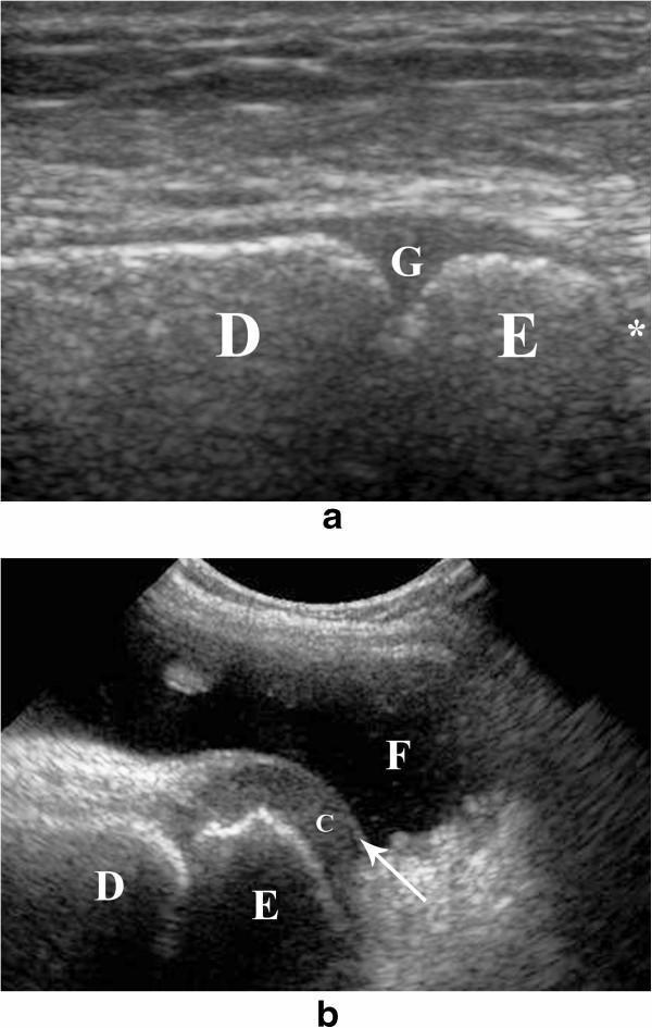
This dual-panel figure provides a musculoskeletal ultrasound comparison of the developing bone in pediatric patients. Image (a) is a longitudinal sonographic view of the distal tibia, illustrating the distal growth plate (G) as a characteristic hypo-echoic area located between the echogenic diaphysis (D) and epiphysis (E). The joint space (*) is visible distal to the epiphysis, demonstrating the importance of differentiating the physis from the intra-articular space. Image (b) presents a longitudinal view of the knee joint in a 5-year-old child. It shows the distal femoral epiphysis (E) and its surrounding peripheral non-ossified cartilage (c). A significant joint effusion (F), seen as an anechoic fluid collection, provides an acoustic window that enhances the visualization of the articular cartilage, which appears as a superficial echogenic line (indicated by an arrow). These images serve as educational references for identifying normal pediatric anatomy and pathological fluid during ultrasound-guided joint procedures.
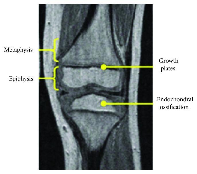
This diagnostic image is a T1-weighted or proton density-weighted MRI scan showing the longitudinal section of a pediatric knee joint, illustrating the key anatomical components of developing long bones. The image specifically highlights the distal femur and proximal tibia. Anatomical structures are identified with yellow labels and brackets: the Metaphysis is shown as the transitioning region of the diaphysis; the Epiphysis is indicated as the rounded end of the bone; and the Growth Plates (physeal cartilage) are visible as thin, dark horizontal bands of low signal intensity separating the metaphysis from the epiphysis. Additionally, an area of Endochondral Ossification is pointed out within the secondary ossification center of the proximal tibia. This image is used for educational purposes to demonstrate pediatric skeletal anatomy, specifically focusing on the maturation of the epiphyseal plate and the development of secondary ossification centers in children, which is critical for assessing bone age and skeletal trauma.
The Epiphysis
Definition and Location
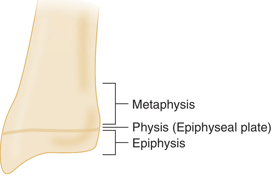
- Epiphysis - the bone end, between the physis and the joint
- Physis (epiphyseal plate) - the growth cartilage separating epiphysis from metaphysis
- Metaphysis - the flared transitional zone between physis and diaphysis
- Diaphysis - the tubular midshaft of the bone
- Apophysis - a bony outgrowth (usually with its own ossification center) serving as a tendon or ligament attachment point
Development (Endochondral Ossification)
- Cartilage anlage - mesenchymal precursor cells first form a cartilage mold of the future bone
- Primary center of ossification - around 8 weeks of gestation, the midshaft (diaphysis) begins ossifying beneath the periosteum, forming the cortex and medullary canal
- Secondary center of ossification - after birth (timing varies by bone), endochondral ossification begins at each bone end (the epiphysis) and proceeds in a centrifugal (outward) pattern
- Growth plate (physis) formation - a plate of cartilage becomes entrapped between the two expanding ossification centers. This is the physis, the engine of longitudinal bone growth
- Reserve zone - resting chondrocytes
- Zone of proliferation - active cell division
- Zone of hypertrophy - cells enlarge
- Zone of apoptosis and mineralization - matrix calcifies, capillaries invade
- Primary spongiosa - osteoblasts deposit new bone on calcified matrix
Epiphyseal Fusion
Blood Supply to the Epiphysis
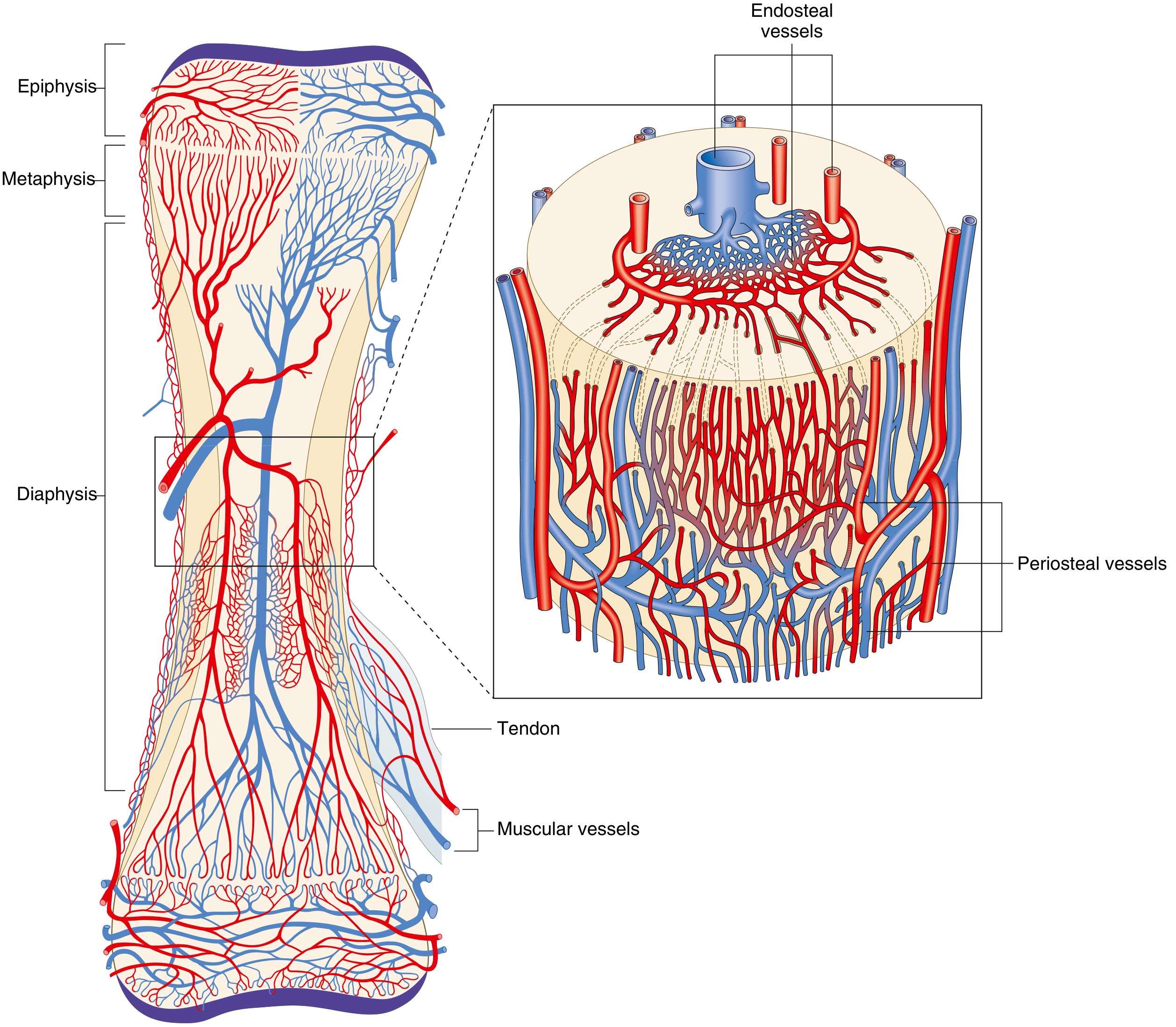
- Scaphoid (wrist)
- Talus (ankle)
- Femoral head
- Odontoid process (C2)
Clinical Significance
Diseases preferentially affecting the epiphysis:
| Condition | Notes |
|---|---|
| Epiphysitis | Inflammation of the epiphysis |
| Osteoclastoma (Giant Cell Tumor) | Characteristic epiphyseal location in adults after physeal closure |
| Slipped Capital Femoral Epiphysis (SCFE) | The femoral head epiphysis displaces on the femoral neck physis; common in overweight adolescents |
| Legg-Calvé-Perthes disease | Avascular necrosis of the femoral head epiphysis in children |
Salter-Harris Fractures (Physis and Epiphysis)
- Type I - fracture through the physis only
- Type II - through physis, exiting the metaphysis (most common; produces Thurston-Holland fragment)
- Type III - through physis, exiting through the epiphysis into the joint
- Type IV - vertical fracture crossing metaphysis, physis, and epiphysis
- Type V - crush/compression of the physis (rarest; worst prognosis for growth)
Summary
| Feature | Detail |
|---|---|
| Location | Rounded end of long bone, between physis and joint |
| Composition | Cancellous (spongy) bone, covered by articular cartilage |
| Development | Secondary center of ossification; centrifugal endochondral ossification |
| Grows via | Physis (epiphyseal growth plate) |
| Fuses | Late teens to mid-20s; signals end of longitudinal growth |
| Blood supply | Metaphyseal-epiphyseal periarticular plexus |
| Clinical relevance | SCFE, osteoclastoma, Salter-Harris fractures, avascular necrosis |