
Spinalcord tracts
Reading File
The spinal cord contains ascending (sensory) and descending (motor) tracts organized in white matter columns. Key ascending tracts include the dorsal columns (fine touch, vibration, proprioception) and spinothalamic tracts (pain, temperature, crude touch). Key descending tracts include the corticospinal tract (voluntary movement) and rubrospinal, reticulospinal, and vestibulospinal tracts.
Searching sources...
Finding Sources
Finding Sources
Reading File
Reading File
Reading File
Reading File
Loading Image
Loading Image
Reading File
Here is a complete overview of the spinal cord tracts, sourced from Gray's Anatomy for Students.
Spinal Cord Tracts
The white matter of the spinal cord is organized into three funiculi (columns) on each side - anterior, lateral, and posterior - each carrying specific ascending (sensory) or descending (motor) tracts.
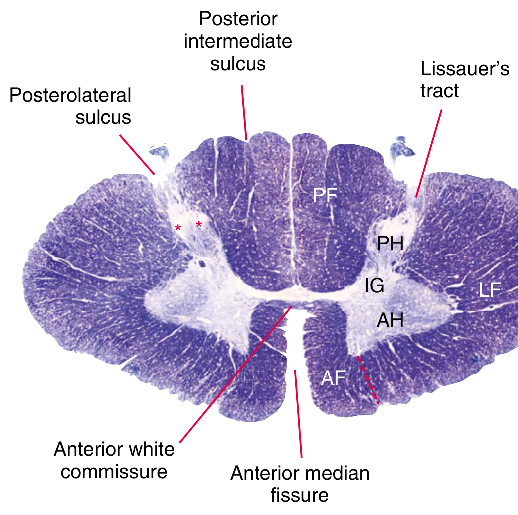
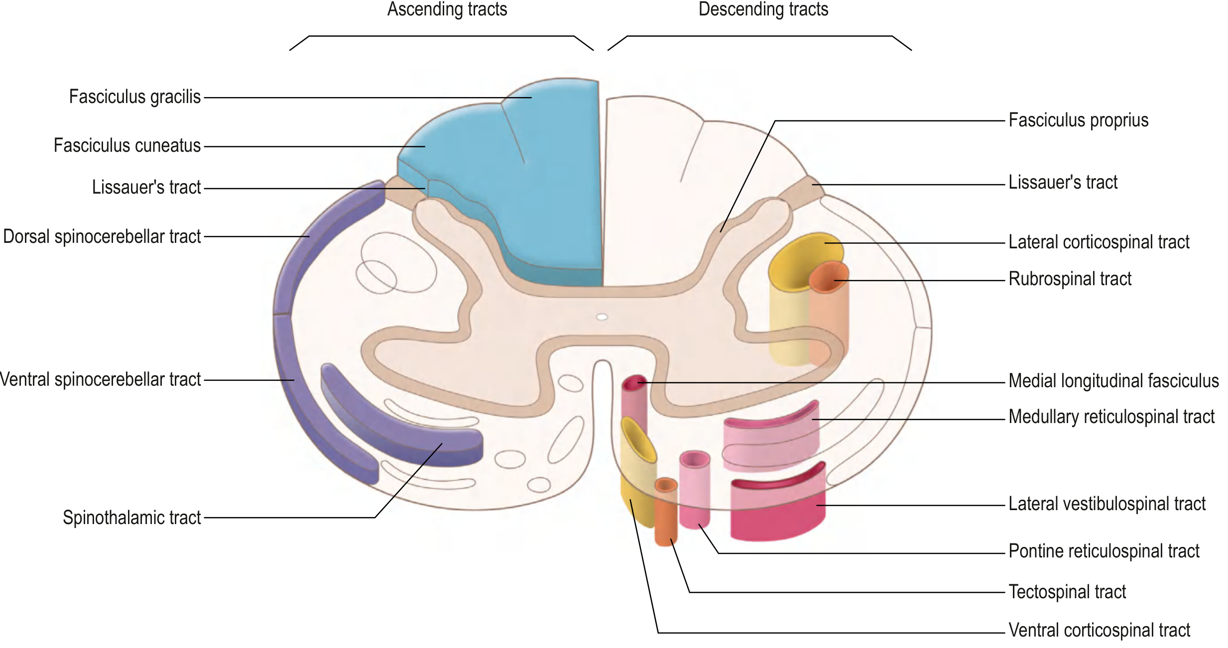
ASCENDING (Sensory) TRACTS
Sensory pathways use a 3-neuron chain: 1st-order (peripheral receptor → spinal cord/medulla), 2nd-order (spinal cord/medulla → thalamus), 3rd-order (thalamus → cortex).
1. Anterolateral Pathways (Anterior & Lateral Funiculus)
Carry pain, temperature, and crude touch.
Three tracts make up this system:
| Tract | Modality | Target |
|---|---|---|
| Spinothalamic | Pain, temperature, crude touch | VPL nucleus of thalamus → somatosensory cortex |
| Spinoreticular | Emotional/arousal aspects of pain | Reticular formation (brainstem) |
| Spinomesencephalic | Nociceptive modulation | Periaqueductal gray (midbrain) |
Course of the Spinothalamic Tract:
- 1st-order neuron: cell body in dorsal root ganglion → enters posterior horn
- Axons either synapse immediately in laminae I & V, or travel 1-2 segments in Lissauer's tract before synapsing
- 2nd-order neuron: crosses obliquely over 2-3 segments via the anterior white commissure to the contralateral anterolateral tract
- Ascends to VPL nucleus of thalamus
- 3rd-order neuron: projects via posterior limb of internal capsule to primary somatosensory cortex
Clinical key: Because crossing occurs over 2-3 segments, a hemisection of the cord (Brown-Sequard syndrome) causes contralateral pain/temperature loss 2-3 levels below the lesion.
2. Posterior Column-Medial Lemniscal Pathway (Posterior Funiculus)
Carries fine/discriminative touch, vibration, and conscious proprioception.
Two fasciculi:
- Fasciculus gracilis - lower limb and trunk (medial, present all levels)
- Fasciculus cuneatus - upper limb and neck (lateral, present from T6 upward)
Course:
- 1st-order neuron: cell body in dorsal root ganglion → axon ascends ipsilaterally in posterior column
- Synapses in nucleus gracilis / nucleus cuneatus in the caudal medulla
- 2nd-order neuron: crosses as internal arcuate fibers → forms medial lemniscus in contralateral medulla
- Ascends to VPL nucleus of thalamus
- 3rd-order neuron: projects via posterior limb of internal capsule → primary somatosensory cortex
Clinical key: Crosses in the medulla (not the cord). Ipsilateral proprioception/vibration loss occurs with cord lesions (e.g., subacute combined degeneration in B12 deficiency, tabes dorsalis).
3. Spinocerebellar Tracts (Lateral Funiculus)
Carry subconscious proprioception to the cerebellum.
| Tract | Origin | Decussation | Side |
|---|---|---|---|
| Dorsal spinocerebellar | Clarke's nucleus (C8-L3) | Does NOT cross | Ipsilateral |
| Ventral spinocerebellar | Lumbosacral cord | Crosses twice (net ipsilateral) | Ipsilateral (double-cross) |
DESCENDING (Motor) TRACTS
Descending tracts are divided into the lateral motor system (fine voluntary movement) and the medial motor system (posture, balance, axial movements).
LATERAL MOTOR SYSTEM
1. Lateral Corticospinal Tract (Lateral Funiculus)
The most clinically important tract - controls voluntary movement of the extremities.
Course:
- Upper motor neuron (UMN): cell body in primary motor cortex
- Axons converge in corona radiata → descend via posterior limb of internal capsule → crus cerebri of midbrain → anterior pons → pyramid of medulla
- ~85% decussate at caudal medulla (pyramidal decussation) → form lateral corticospinal tract
- Descend in lateral column → synapse on LMN cell bodies in lateral anterior horn
- Lower motor neuron exits via anterior root
2. Rubrospinal Tract (Lateral Funiculus)
- Origin: red nucleus (midbrain)
- Crosses at ventral tegmental decussation → descends in lateral column
- Only descends to cervical levels
- Facilitates flexor and inhibits extensor activity of the upper limb
MEDIAL MOTOR SYSTEM
Regulate axial/truncal muscles for posture, balance, and automatic movements. Project bilaterally.
| Tract | Origin | Extent | Function |
|---|---|---|---|
| Anterior corticospinal tract | Motor cortex (fibers that did NOT decussate at medulla) | To upper thoracic cord | Bilateral axial/proximal limb control |
| Tectospinal tract | Superior colliculus (dorsal midbrain) | Cervical cord only | Reflex postural responses to visual stimuli |
| Medial vestibulospinal tract | Medial vestibular nucleus | To thoracic cord, bilateral | Head/neck posture relative to vestibular input |
| Lateral vestibulospinal tract | Lateral vestibular nucleus (Deiters) | Entire cord, ipsilateral | Extensor muscle tone, balance |
| Reticulospinal tracts | Reticular formation (pons & medulla) | Entire cord | Modulate muscle tone, autonomic functions |
Summary Comparison Table
| Tract | Type | Location | Crosses? | Modality |
|---|---|---|---|---|
| Fasciculus gracilis | Ascending | Post. funiculus | In medulla | Fine touch, vibration, proprioception (LL) |
| Fasciculus cuneatus | Ascending | Post. funiculus | In medulla | Fine touch, vibration, proprioception (UL) |
| Spinothalamic | Ascending | Anterolateral | In cord (2-3 seg) | Pain, temperature, crude touch |
| Dorsal spinocerebellar | Ascending | Lateral funiculus | No | Subconscious proprioception (LL) |
| Ventral spinocerebellar | Ascending | Lateral funiculus | Double cross | Subconscious proprioception (LL) |
| Lateral corticospinal | Descending | Lateral funiculus | In medulla (~85%) | Voluntary movement (extremities) |
| Anterior corticospinal | Descending | Anterior funiculus | In cord (bilateral) | Axial/proximal voluntary movement |
| Rubrospinal | Descending | Lateral funiculus | In midbrain | UL flexion |
| Vestibulospinal (lateral) | Descending | Anterior funiculus | No | Extensor tone, balance |
| Reticulospinal | Descending | Anterior/lateral | Mixed | Tone, autonomic |
| Tectospinal | Descending | Anterior funiculus | In midbrain | Head/neck reflexes to visual stimuli |
Source: Gray's Anatomy for Students, Neuroanatomy section (eFigs. 9.39-9.47)
So what is, uh, you know, what, uh, tell me what is corticocerebral areas and what are, is like, say, is cerebro, uh, bulbar, uh, no, is corticobulbar also a pyramidal, pyramidal tract? No.
Yes! The corticobulbar tract IS part of the pyramidal system. Let me look this up properly from the textbooks.
Searching sources...
Finding Sources
Finding Sources
Finding Sources
Finding Sources
Reading File
Reading File
Finding Sources
Finding Sources
Reading File
I now have everything needed for a thorough answer. Let me put it all together.
Cortical Motor Areas & the Corticobulbar Tract
Part 1: Cortical Motor Areas
The motor cortex is in the posterior frontal lobe, anterior to the central sulcus. It has three main areas:
1. Primary Motor Cortex (M1) - Brodmann Area 4
- Located in the precentral gyrus
- Contains a somatotopic map (motor homunculus) - face/mouth laterally near the Sylvian fissure, then hand, arm, trunk, leg medially into the longitudinal fissure
- Over half of M1 is devoted to the hands and speech muscles
- Stimulation produces discrete, contralateral movements
- Contributes ~31% of corticospinal tract fibers
2. Premotor Area (PMA) - Brodmann Area 6 (lateral)
- Lies 1-3 cm anterior to M1
- Same rough topography as M1
- Controls coordinated multi-joint movements and postural adjustments that prepare for fine movements
- Functions based on sensory feedback (relies on input from outside the body)
- Contributes ~29% of corticospinal tract fibers
3. Supplementary Motor Area (SMA) - Brodmann Area 6 (medial)
- Lies in the longitudinal fissure on the medial surface, extending onto the superior frontal cortex
- Often produces bilateral movements (e.g., bilateral grasping)
- Involved in internally generated, self-initiated movements and motor planning/sequencing
- Damage → difficulty initiating voluntary movements (akinesia)
Key difference: Premotor cortex = externally cued movements. SMA = internally planned movements.
Other specialized regions include Broca's area (word formation, just anterior to M1 face area), voluntary eye movement fields, head rotation area, and the hand skills area.
Part 2: Is the Corticobulbar Tract a Pyramidal Tract? YES.
Yes, absolutely. The corticobulbar tract is part of the pyramidal system, alongside the corticospinal tract.
Here is how they relate:
| Feature | Corticospinal Tract | Corticobulbar Tract |
|---|---|---|
| Part of pyramidal system? | Yes | Yes |
| Origin | Motor cortex (M1, PMA, SMA, parietal) | Motor cortex (same origin) |
| Neuron shape | Pyramidal (layer V) | Pyramidal (layer V) |
| Internal capsule | Posterior limb | Genu |
| Target | Spinal cord anterior horn (LMN) | Cranial nerve motor nuclei (brainstem) |
| Controls | Limb and trunk muscles | Face, jaw, tongue, pharynx, larynx muscles |
| Which cranial nerves? | - | V (trigeminal), VII (facial), IX/X (nucleus ambiguus), XI, XII (hypoglossal) |
Why both are "pyramidal"
Both tracts originate from pyramidal-shaped neurons in layer V of the cortex, and both pass through the medullary pyramids (corticospinal) or travel alongside them (corticobulbar). The word "pyramidal tract" refers to this shared origin and pathway.
The corticobulbar tract diverges from the corticospinal at various brainstem levels to synapse on cranial nerve nuclei - either directly on motor neurons or via interneurons. Most cranial nerve nuclei receive bilateral corticobulbar input (from both hemispheres), which is why a unilateral stroke usually does NOT completely paralyze muscles like the jaw or larynx.
Exception - Lower face (CN VII) and tongue (CN XII): These receive predominantly contralateral corticobulbar innervation. This is why a cortical stroke causes:
- Contralateral lower face weakness (forehead is spared - bilateral input)
- Contralateral tongue deviation on protrusion
Summary
PYRAMIDAL SYSTEM
├── Corticospinal tract → spinal cord → limb/trunk muscles
└── Corticobulbar tract → brainstem cranial nerve nuclei → face/mouth muscles
Both originate from the same cortical areas (M1 = 31%, premotor = 29%, parietal/somatosensory = 40%), travel through the internal capsule, and are classified as upper motor neurons (UMNs). Damage to either produces UMN signs.
Sources: Ganong's Review of Medical Physiology; Guyton & Hall Textbook of Medical Physiology, Ch. 56
How does bilateral innervation explain forehead sparing in strokes?
The forehead muscles (frontalis) receive corticobulbar input from both hemispheres, so a stroke affecting one hemisphere still leaves the other hemisphere's input intact - the forehead is spared. The lower face only gets input from the contralateral hemisphere, so a stroke knocks it out completely.
Searching sources...
Finding Sources
Finding Sources
Reading File
Finding Sources
Loading Image
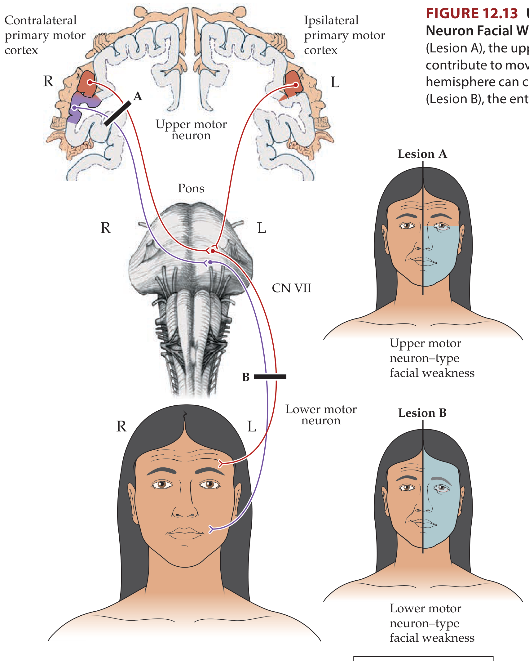
Here is the full explanation with the textbook diagram.
Forehead Sparing in UMN Facial Palsy - Explained by Bilateral Innervation
The Core Anatomy
The facial nucleus (CN VII) in the pons has two divisions:
| Division | Muscles supplied | Cortical input |
|---|---|---|
| Upper division | Frontalis (forehead), orbicularis oculi (eye) | Both contralateral AND ipsilateral motor cortex |
| Lower division | Orbicularis oris (lips), buccinator, platysma (lower face) | Only contralateral motor cortex |
The Mechanism, Step by Step
Normal state:
- Your forehead (frontalis muscle) receives corticobulbar signals from the left hemisphere AND the right hemisphere simultaneously - bilateral input
- Your lower face only receives signals from the opposite (contralateral) hemisphere
During a unilateral cortical stroke (e.g., right hemisphere stroke):
- The right motor cortex is knocked out
- Lower face: loses its only cortical input (right hemisphere) → paralysed on the left side
- Forehead: loses input from the right hemisphere, but the left hemisphere's input is still intact → forehead still works fine → spared
This is the clinical hallmark of an upper motor neuron (UMN) facial palsy.
Why Is the Upper Face Bilaterally Innervated?
The evolutionary/functional reason is that forehead and eye movements (like raising eyebrows, blinking) are often bilateral, symmetric, and emotionally driven - they need coordination from both hemispheres. The lower face, by contrast, is used for fine unilateral voluntary movements like speech, which require dominant-hemisphere control.
The Critical Clinical Distinction
| Feature | UMN Palsy (stroke, cortical lesion) | LMN Palsy (Bell's palsy, CN VII lesion) |
|---|---|---|
| Forehead | Spared (can wrinkle) | Paralysed (cannot wrinkle) |
| Eye closure | Mild weakness only | Cannot close eye (lagophthalmos) |
| Lower face | Weak - contralateral | Weak - ipsilateral |
| Whole face involved? | No - upper spared | Yes - entire half of face |
| Bell's phenomenon | Absent | Present (eyeball rolls up on attempted closure) |
| Other UMN signs | Often arm/leg weakness, aphasia | None |
One important nuance: The corticobulbar fibers do not synapse directly onto facial nucleus LMNs in most cases - they project onto pontine interneurons, which then relay to the LMNs. The bilateral arrangement applies to these interneuron connections for the upper face.
Memory Hook
"The forehead has two bosses (both hemispheres) - lose one boss, and the other keeps it working. The lower face has only one boss - lose that boss and it stops working entirely."
Source: Neuroanatomy through Clinical Cases, 3rd Edition (KCC 12.3, Fig. 12.13)
This is a shared conversation. Sign in to Orris to start your own chat.