
Explain me muscle contraction for MBBS 1 st year student
Reading File
Now let me search the medical library for muscle contraction content.
Finding Sources
Finding Sources
Guyton & Hall and Costanzo Physiology are the best for MBBS 1st year. Let me read those sections in parallel.
Finding Sources
Reading File
Reading File
Now let me read the core muscle contraction sections from Guyton & Hall:
Reading File
Reading File
Excellent content! Let me now read the neuromuscular junction and calcium/troponin regulation sections:
Reading File
Reading File
I now have excellent content. Let me fetch a couple of the key diagrams from Guyton to include in the response:
Loading Image
Loading Image
I now have all the information and images needed. Let me also fetch the cross-bridge cycling / sliding filament diagram:
Reading File
I now have everything I need to write a thorough, MBBS 1st year level explanation of muscle contraction. Let me compile the full answer now.
Muscle Contraction - Complete Guide for MBBS 1st Year
Source: Guyton and Hall Textbook of Medical Physiology (Chapter 6)
Overview
Skeletal muscle makes up about 40% of body mass. The fundamental principle of contraction is the sliding filament theory - thin actin filaments slide over thick myosin filaments, shortening the sarcomere and thus the whole muscle. No filaments actually shorten; they just slide past each other.
1. Structure of Skeletal Muscle (Anatomical Levels)
Here is the organizational hierarchy from gross to molecular level:
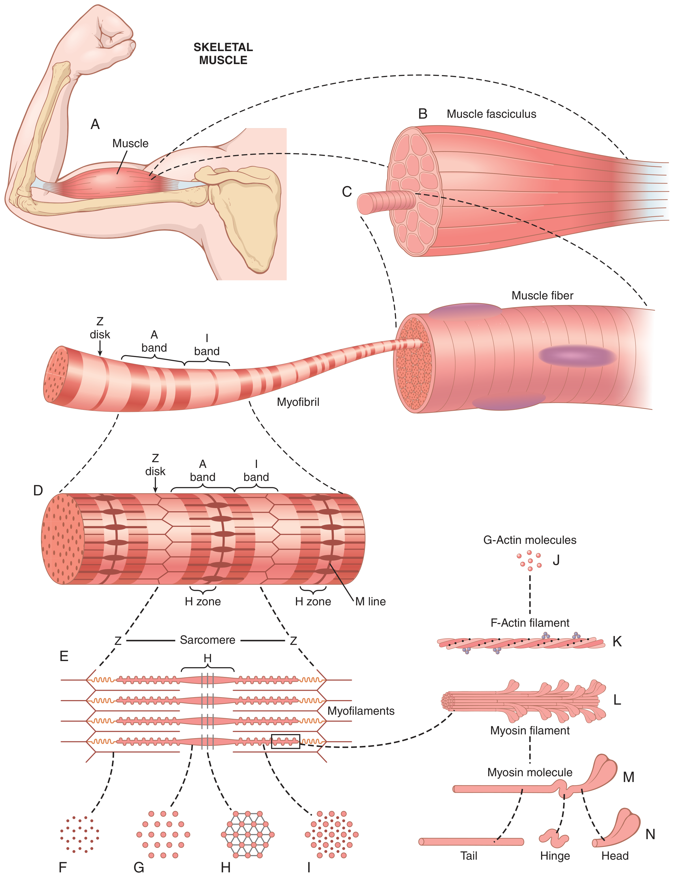
Figure 6.1 from Guyton & Hall - Organization of skeletal muscle from gross to molecular level
| Level | Description |
|---|---|
| Muscle | Entire muscle (e.g. biceps brachii) |
| Fasciculus | Bundle of muscle fibers |
| Muscle fiber (cell) | 10-80 µm diameter; runs full length of muscle |
| Myofibril | 1500 myosin + 3000 actin filaments per myofibril |
| Sarcomere | Functional unit between two Z disks (~2 µm long) |
Key Structural Components
Sarcolemma - the cell membrane of the muscle fiber. It fuses with tendons at each end, which then attach to bone.
Myofibrils - contain the contractile proteins. Each myofibril has alternating light and dark bands:
- A band (dark) - contains myosin + overlapping ends of actin; anisotropic to polarized light
- I band (light) - contains only actin; isotropic to polarized light
- H zone - center of A band; myosin only, no actin overlap
- Z disk - anchors actin filaments; one sarcomere = Z disk to Z disk
- M line - center of H zone; holds myosin filaments together
Titin - giant elastic protein (~3.9 million molecular weight) that tethers myosin to the Z disk and acts like a molecular spring, maintaining the sarcomere's structural integrity.
2. The Contractile Proteins
Myosin (Thick Filament)
- Each myosin filament is ~1.6 µm long and made of ~200 myosin molecules
- Each myosin molecule has a tail (bundled together to form filament body) and two heads (the cross-bridges)
- The heads project outward and are arranged in pairs, each pair rotated 120° from the previous - this ensures cross-bridges extend in all directions
- The head acts as an ATPase enzyme - it cleaves ATP to provide energy for contraction
- The cross-bridges have two hinges: one where the arm leaves the filament body, and one where the head meets the arm
Actin (Thin Filament)
- Made of 3 proteins: F-actin, tropomyosin, and troponin
- F-actin = two strands of G-actin molecules (MW ~42,000 each) wound in a double helix
- Each G-actin has one ADP molecule attached - these are the active binding sites for myosin cross-bridges
- ~1 active site every 2.7 nanometers along the filament
Tropomyosin
- Runs spirally along the groove of the F-actin double helix
- In the resting state, tropomyosin physically covers the active sites on actin, blocking myosin from binding
- This is the "OFF" switch for contraction
Troponin Complex
- Three subunits with distinct roles:
- Troponin I - binds to actin (anchors the complex)
- Troponin T - binds to tropomyosin
- Troponin C - binds to Ca²⁺ (this is the key switch!)
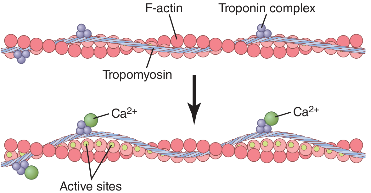
Figure 6.6 from Guyton & Hall - How Ca²⁺ activates the actin filament by moving tropomyosin to expose active sites
3. The Neuromuscular Junction (NMJ)
Before contraction happens, a nerve signal must reach the muscle:
- A motor neuron action potential arrives at the motor nerve terminal (pre-synaptic)
- ACh (Acetylcholine) is released from vesicles into the synaptic cleft
- ACh binds to nicotinic receptors on the motor end plate (post-synaptic sarcolemma)
- This opens Na⁺/K⁺ channels → depolarization of the sarcolemma → an end-plate potential
- The end-plate potential triggers a muscle action potential that spreads across the entire fiber surface
Key pharmacology to remember:
- ACh is broken down by acetylcholinesterase to stop the signal
- Curare blocks nicotinic receptors → muscle paralysis
- Neostigmine inhibits acetylcholinesterase → prolongs ACh action
4. Excitation-Contraction (E-C) Coupling
This is how the electrical signal (action potential) is converted into mechanical contraction:
- The action potential travels along the sarcolemma and dips inward via the T-tubules (transverse tubules) - these invaginate deep into the fiber at the level of each A-I band junction
- The T-tubule signal reaches the sarcoplasmic reticulum (SR) - the intracellular Ca²⁺ store
- The SR releases Ca²⁺ ions into the sarcoplasm (cytoplasm of muscle fiber)
- Ca²⁺ concentration rises from ~10⁻⁷ to ~10⁻⁵ mol/L (100-fold increase)
- Ca²⁺ binds to Troponin C on the thin filament
- This causes a conformational change → troponin complex tugs tropomyosin deeper into the groove between actin strands
- Active sites on actin are exposed → myosin cross-bridges can now bind
5. The Sliding Filament Theory - Cross-Bridge Cycle
This is the walk-along (ratchet) mechanism of contraction:
Step-by-Step Cross-Bridge Cycle:
| Step | Event |
|---|---|
| 1. Energized state | ATP binds to myosin head → head cleaves ATP to ADP + Pi → energy cocks the head at 90° (perpendicular, high-energy position) |
| 2. Attachment | Energized myosin head attaches to the exposed active site on actin |
| 3. Power stroke | ADP + Pi are released → head swivels from 90° to 45° → pulls actin filament ~10 nm toward the center of sarcomere |
| 4. Detachment | A new ATP molecule binds to the myosin head → head detaches from actin |
| 5. Re-cocking | The new ATP is cleaved → head is cocked back to 90° ready for the next cycle |
| 6. Repeat | Head binds the next active site and repeats the process |
Memory tip: "ATP Attaches, Power stroke, ATP detaches" - each power stroke moves the actin ~10 nm. With hundreds of cross-bridges cycling asynchronously, the muscle shortens smoothly.
Important: If ATP is absent (e.g. after death), myosin stays attached to actin permanently → rigor mortis
6. Relaxation
When the nerve stops firing:
- Ca²⁺ is actively pumped back into the SR by Ca²⁺-ATPase pumps (requires ATP)
- Sarcoplasmic Ca²⁺ falls
- Ca²⁺ detaches from Troponin C
- Tropomyosin moves back to cover actin active sites
- Cross-bridge cycling stops → muscle relaxes
7. The Length-Tension Relationship
The force a muscle generates depends on sarcomere length at the time of contraction:
| Sarcomere length | Force generated | Reason |
|---|---|---|
| ~2.0-2.2 µm | Maximum | Optimal overlap of actin over all myosin cross-bridges |
| >2.2 µm (stretched) | Decreasing | Fewer cross-bridges overlap with actin |
| <2.0 µm (compressed) | Decreasing | Actin filaments from both sides overlap each other; Z disks may hit myosin ends |
This is clinically relevant in the Frank-Starling Law of the heart (optimal sarcomere length for maximum cardiac output).
8. Types of Muscle Fibers
| Feature | Type I (Slow, Red) | Type II (Fast, White) |
|---|---|---|
| Size | Smaller | Larger |
| Speed of contraction | Slow | Fast |
| Fatigue resistance | High | Low |
| Energy source | Oxidative | Glycolytic |
| Mitochondria | Many | Few |
| Myoglobin | High (red color) | Low (white) |
| SR Ca²⁺ release | Slower | Faster, more extensive SR |
| Example | Soleus (posture) | Gastrocnemius (jumping) |
9. Motor Unit
- A motor unit = one motor neuron + all the muscle fibers it innervates
- A single axon branches to innervate several muscle fibers
- All fibers of a motor unit contract together ("all or none" for the unit)
- Fine movement muscles (e.g. extraocular muscles) = small motor units (3-6 fibers per neuron)
- Power muscles (e.g. quadriceps) = large motor units (hundreds of fibers per neuron)
10. Quick Summary - The Sequence
Nerve AP → ACh released at NMJ → End-plate potential
    → Muscle AP → spreads down T-tubules
    → Ca²⁺ released from SR
    → Ca²⁺ binds Troponin C
    → Tropomyosin moves → actin active sites exposed
    → Myosin cross-bridges attach to actin
    → Power stroke (ATP energy)
    → Actin slides → sarcomere shortens → CONTRACTION
    → Nerve stops → Ca²⁺ pumped back to SR
    → Tropomyosin covers active sites → RELAXATION
High-Yield Points for Exams
- Sliding filament theory - filaments slide, they do NOT shorten
- Troponin C binds Ca²⁺ (not troponin I or T)
- ATP is needed for BOTH contraction AND relaxation (SR Ca²⁺ pump)
- Rigor mortis - no ATP, myosin cannot detach from actin
- T-tubules link surface action potential to deep SR Ca²⁺ release
- Optimal sarcomere length = 2.0-2.2 µm for maximum tension
- A band does NOT change length during contraction (contains myosin); I band and H zone shorten
Based on: Guyton and Hall Textbook of Medical Physiology, 14th Ed. (Chapters 6-8)
This is a shared conversation. Sign in to Orris to start your own chat.