
Vocal cord palsy
Reading File
Searching sources...
Finding Sources
Finding Sources
Finding Sources
Good finds. Now let me read the comprehensive sections on vocal cord palsy from the ENT textbooks and neurology text in parallel.
Reading File
Reading File
Finding Sources
Now let me read the main dedicated section on vocal cord palsy from the ENT textbook and the treatment/management section.
Reading File
Reading File
Finding Sources
Let me find the main vocal cord palsy chapter in Cummings Otolaryngology and the block with comprehensive etiology and management.
Finding Sources
Reading File
Let me find the dedicated vocal cord palsy chapter in the block10 file and also search in Cummings.
Finding Sources
Reading File
Excellent - I found the key dedicated chapters. Let me now read the most comprehensive sections in parallel.
Reading File
Reading File
Good content coming in. Let me read more from both sources - treatment in KJ Lee and the bilateral section in Scott-Brown's.
Reading File
Reading File
Now let me read the bilateral section and the treatment section from Scott-Brown's, and also get the KJ Lee treatment section.
Reading File
Reading File
I now have comprehensive information to provide a thorough answer. Let me compile everything into a well-structured response.
Vocal Cord Palsy (Vocal Fold Paralysis)
Vocal cord palsy (VCP) refers to the loss of voluntary movement of one or both vocal folds due to disruption of their nerve supply. The condition ranges from a minor voice problem to a life-threatening airway emergency depending on whether it is unilateral or bilateral.
Anatomy and Nerve Supply
The vocal folds receive motor supply via the vagus nerve (CN X) through two branches:
- Recurrent laryngeal nerve (RLN): Supplies all intrinsic laryngeal muscles except cricothyroid. The left RLN has a longer course, descending into the thorax and looping under the aortic arch before ascending in the tracheo-oesophageal groove - making it more vulnerable to injury.
- Superior laryngeal nerve (internal and external branches): The external branch supplies the cricothyroid; the internal branch is sensory to the supraglottis. High vagal injury affecting both nerves is more functionally devastating than isolated RLN injury.
Causes
Unilateral VCP
Studies show the three main causes are:
| Cause | Approximate frequency |
|---|---|
| Iatrogenic/Surgical trauma | 22-75% |
| Neoplasm | 7-52% |
| Idiopathic | 10-41% |
| Intubation injury | 5-11% |
(Scott-Brown's Otorhinolaryngology Head & Neck Surgery)
Surgical causes:
- Thyroidectomy (most common in ENT practice); risk factors include Graves' disease, post-operative haematoma, revision surgery
- Oesophagectomy, tracheal resection, thymectomy, mediastinal procedures
- Cervical spine surgery
- Carotid endarterectomy
Neoplastic causes:
- Lung carcinoma (esp. left hilar/apical), thyroid cancer, oesophageal carcinoma, lymphoma, mediastinal tumours
Cardiovascular:
- Aortic aneurysm (left RLN)
- Patent ductus arteriosus surgery
- Cardiac valve surgery
- Enlarged left atrium - Ortner syndrome (cardiovocal syndrome)
Neurological:
- Cerebrovascular accident (usually with other deficits)
- Wallenberg syndrome (lateral medullary infarct): vocal fold palsy + dysphagia + vertigo + ataxia + Horner syndrome + hemifacial sensory deficit
- Arnold-Chiari malformation
- Multiple sclerosis, Parkinson's disease, motor neurone disease, myasthenia gravis, Guillain-Barré syndrome
Other:
- Inflammatory: sarcoidosis, Lyme disease, syphilis, collagen vascular disease
- Organophosphate exposure, neurotoxic chemotherapy (vincristine, cisplatin)
- Congenital VCP: presents with hoarse cry + feeding/aspiration difficulties in infants
Clinical Features
Unilateral VCP
- Dysphonia: breathy, weak voice; vocal fatigue, diplophonia (double voice), decreased projection
- Aspiration: occasionally, especially liquids; worse with high vagal lesions affecting laryngeal sensation
- Wet voice quality: pooled secretions indicate laryngeal penetration - a warning sign
- Exertional breathlessness: from air escape during phonation and loss of the Valsalva mechanism
- Occasionally asymptomatic (found incidentally)
Key point: Dysphagia risk is greater with "high" vagal injuries (affecting both SLN and RLN), adding hemi-laryngeal anaesthesia, pharyngeal constrictor atony, and cricopharyngeal hyperfunction. (Scott-Brown's Otorhinolaryngology Head & Neck Surgery)
Bilateral VCP
Symptoms depend on the position of the paralysed cords:
- Medialized cords: Normal or near-normal voice, but stridor and dyspnoea (the key danger)
- Lateralized cords: Weak voice, possible dysphagia, but better airway
- Acute bilateral VCP (e.g., post-thyroidectomy) can cause life-threatening airway obstruction
- Chronic/progressive cases (e.g., neurological disease) may be better tolerated as patients compensate
- Inspiratory stridor is characteristic: negative pressure on inspiration pulls denervated folds together
Pathophysiology
After nerve injury, the vocal fold may initially sit in a lateral position and medialize over ~6 months due to muscle atrophy and fibrosis. The final "paramedian" resting position reflects residual tone from any partial reinnervation. Importantly, reinnervation does not restore voluntary motion - it restores muscle bulk/tone only. Synkinesis (misdirected nerve regeneration) is common: the fold fails to move because of uncoordinated simultaneous activation of abductors and adductors. (K.J. Lee's Essential Otolaryngology)
Diagnosis
-
Flexible nasolaryngoscopy/laryngoscopy: Gold standard; directly visualizes cord mobility. Palpate the vocal process to assess for mechanical fixation (cricoarytenoid joint fixation, posterior glottic scar - must be distinguished from true neurogenic palsy).
-
Laryngeal EMG (LEMG): Evaluates nerve integrity; absence of activity or synkinetic patterns predict poor recovery. Guides prognosis before committing to permanent surgery.
-
Imaging:
- CT neck and thorax (from skull base to thoracic inlet/aortic arch) to identify compressive/infiltrative lesions
- MRI brain if central/neurological cause suspected
- Chest X-ray for lung malignancy
-
Flow-volume loop: Bilateral VCP shows variable extra-thoracic obstruction pattern (passive adduction on inspiration, abduction on expiration).
-
Address underlying cause: full history including surgical history, chest symptoms, neurological review, smoking history.
Management
Unilateral VCP
Goal: improve glottic closure for voice and airway protection.
1. Conservative (observe and wait):
- Many cases show spontaneous recovery or natural compensation within 6-12 months
- Voice therapy as first-line: strengthening techniques, pushes/pulls to improve adduction
- Temporary injection augmentation while awaiting recovery
2. Injection Laryngoplasty (medialization by injection):
Can be done awake, under sedation, or GA; transcutaneous or transoral routes.
- Temporary materials (while awaiting recovery): Carboxymethylcellulose (2-3 months), hyaluronic acid (2-4 months), micronized cadaveric dermis (2-6 months)
- Longer-lasting: Calcium hydroxyapatite (Radiesse Voice; ~18 months) - requires deep injection
- Permanent: Autologous fat (unpredictable absorption; 30-50% absorbed early)
- Teflon: historical only - no longer used due to granuloma formation
3. Medialization Laryngoplasty (Type I Thyroplasty):
- Permanent medialization via external approach; Gore-Tex or silastic implant placed through a window in the thyroid cartilage
- Done under local anaesthesia + light sedation to allow real-time voice monitoring
- Implant placed in the inferior half of the thyroid cartilage (vocal folds are deep to the inferior half)
- Complications: airway oedema, implant extrusion, poor voice if implant too high/anterior
- Not adequate alone if there is a large posterior glottic gap (lateralized arytenoid)
4. Arytenoid Adduction:
- Mimics the lateral cricoarytenoid muscle action
- Indicated when there is a large posterior glottic gap with lateralized vocal process
- Muscular process of arytenoid exposed, suture passed to rotate it medially
- Often combined with Type I thyroplasty (Zeitels technique)
5. Reinnervation (Ansa cervicalis to RLN anastomosis):
- Restores muscle bulk and tone rather than functional motion
- Improves voice long-term; takes months to show effect
Bilateral VCP
Goal: secure the airway while preserving as much voice quality as possible.
- Tracheotomy: Gold standard; speech possible with Passy-Muir valve or digital occlusion
- Endoscopic arytenoidectomy (medial or total): Enlarges the glottis; preserves membranous cord for phonation
- Transverse cordotomy (posterior cordotomy): Incision across the vocal fold at the vocal process to widen the airway; can be extended as needed
- Suture lateralization: Lateralizes one cord if no joint fixation present
- Arytenoid abduction (external approach)
- Laryngeal pacing: Experimental implantable stimulator to restore abductor function
Acute post-thyroidectomy bilateral VCP: Immediate re-intubation or emergency tracheotomy.
Vocal Fold Paresis (Incomplete Palsy)
Partial denervation causing weakness (not complete paralysis). Presents with dysphonia, vocal fatigue, diplophonia, and reduced vocal range. Laryngoscopy shows restricted motion, reduced cord bulk ("bowing"), and asymmetric movement. Diagnosed with LEMG. Treatment parallels unilateral VCP (injection augmentation, thyroplasty). (K.J. Lee's Essential Otolaryngology)
Prognosis
- Spontaneous recovery most likely within 6-12 months of onset
- LEMG findings guide prognosis: absent or synkinetic activity = poor recovery
- Idiopathic cases have a better prognosis than neoplastic causes
- Congenital unilateral VCP often improves as the child develops compensatory mechanisms
Sources: Scott-Brown's Otorhinolaryngology Head & Neck Surgery (Vol 1 & 2, Chapters 67/78); K.J. Lee's Essential Otolaryngology (9th ed.); Cummings Otolaryngology Head and Neck Surgery.
Features of rln and sln palsy
Searching sources...
Finding Sources
Finding Sources
Reading File
Reading File
Finding Sources
Finding Sources
Reading File
Reading File
Finding Sources
Reading File
Finding Sources
Finding Sources
Reading File
Now let me fetch the key laryngoscopic images and get the Seimon's law/vagal lesion content.
Loading Image
Finding Sources
Finding Sources
Reading File
I now have all the information needed for a comprehensive, well-structured comparison. Let me compile the answer.
Features of RLN and SLN Palsy
Anatomy First - What Each Nerve Does
| Nerve | Motor | Sensory |
|---|---|---|
| RLN | All intrinsic laryngeal muscles except cricothyroid (PCA, LCA, IA, TA) | Subglottis and trachea |
| SLN - external branch (EBSLN) | Cricothyroid muscle only | None |
| SLN - internal branch (IBSLN) | None | Supraglottis and glottis down to vocal cord level |
The PCA (posterior cricoarytenoid) is the only abductor of the vocal fold. All other RLN-innervated muscles are adductors/tensors.
RLN Palsy
Unilateral RLN Palsy
Voice:
- Hoarseness - breathy, weak, flaccid dysphonia
- Reduced loudness, short phrases due to excessive air escape
- Diplophonia (two simultaneous pitch levels from unequal vibration of the two cords)
- Mild inspiratory stridor during contextual speech
- "Paralytic falsetto": if SLN is intact, the patient may compensate by raising pitch via cricothyroid - generates a higher-pitched, strained voice quality
Laryngoscopy:
- Paralysed cord lies near the midline (paramedian position) at rest
- Normal cord crosses the midline to meet the paralysed cord during phonation - glottic gap remains
- Spindle-shaped gap if the membranous cord is predominant; V-shaped (posterior) gap if the vocal process is lateralized
- Increasing tension on the paralysed cord when the patient raises pitch suggests the SLN/cricothyroid is intact (Figure 78.1 below)
- Prolapsed arytenoid: the arytenoid on the paralysed side may fall forward into the laryngeal introitus - sign of profound denervation and muscular support loss
- Supraglottic hyperfunction: ventricular fold adduction as compensation for glottic insufficiency
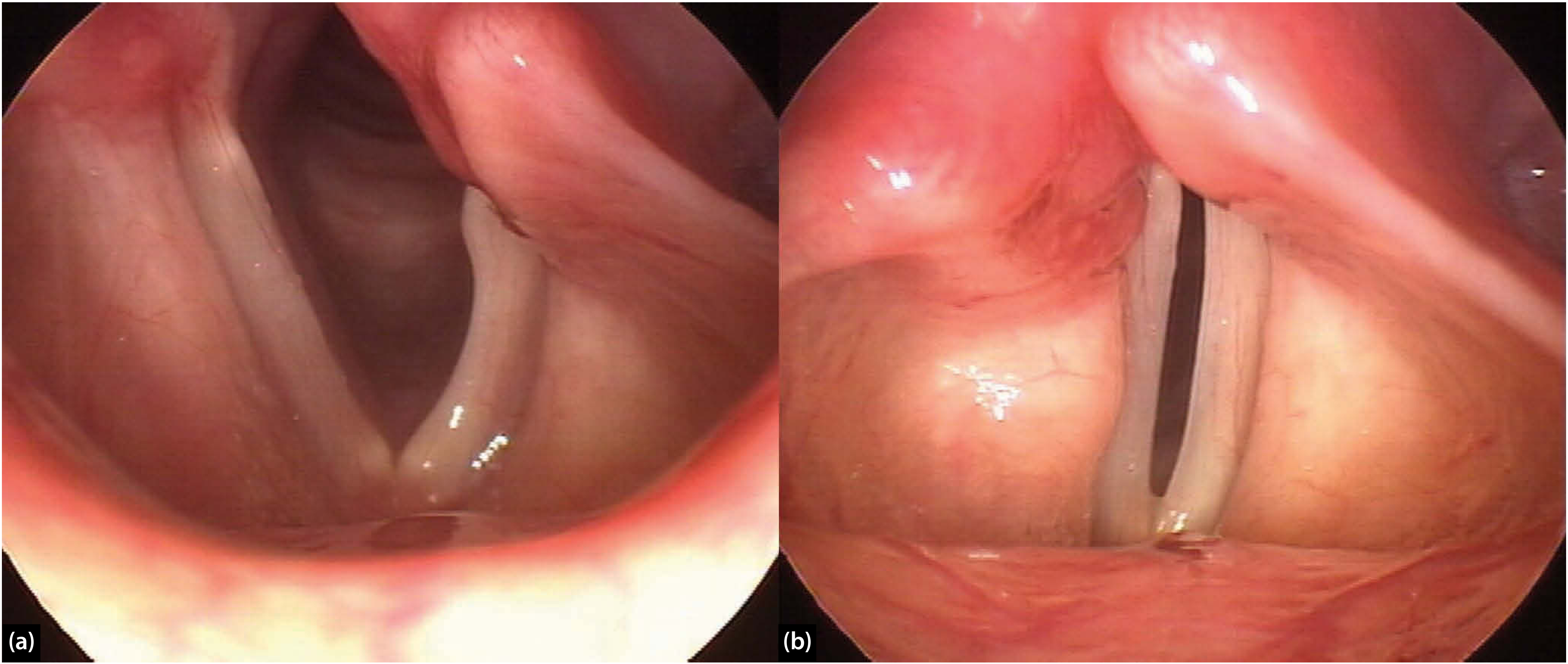
Figure 78.1: Left vocal fold paralysis (post anterior cervical approach). (a) Quiet respiration - cord in paramedian position. (b) Phonation - increased tension in the paralysed cord suggests cricothyroid (SLN) is intact. (Scott-Brown's Otorhinolaryngology)
Swallowing:
- Occasional aspiration, mainly liquids
- Dysphagia worse if it is a "high vagal" lesion (SLN also involved) due to added ipsilateral laryngeal anaesthesia, pharyngeal constrictor weakness, and cricopharyngeal hyperfunction
Key law - Semon's law:
With progressive/peripheral RLN injury, adductor muscles are affected first, then abductors last. So in early/partial palsy the cord may still adduct but cannot abduct (abductor paralysis). In complete palsy, all muscles are paralysed and the cord lies paramedian.
Position of cord:
- Paramedian in most cases (partial tone from intact cricothyroid + muscle atrophy over time)
- More lateral (cadaveric position) with high vagal lesions (complete tone loss)
Bilateral RLN Palsy
Voice: Weak but often surprisingly clear (both cords near midline, still approximate)
Airway - the dominant clinical problem:
- Inspiratory stridor - negative inspiratory pressure sucks denervated cords medially
- Progressive dyspnoea and exercise intolerance
- In acute bilateral palsy (e.g., post-thyroidectomy extubation): life-threatening respiratory distress - emergency
- In chronic (e.g., progressive neurological disease): patients may compensate as paralysis develops slowly
Swallowing: Usually not affected
Flow-volume loop: Variable extra-thoracic obstruction (dynamic - folds adduct on inspiration, abduct passively on expiration)
Abductor paralysis specifically:
- Bilateral abductor paralysis = cords approximated in midline
- Severe airway limitation, often requiring tracheostomy
- Can occur in spinocerebellar ataxia type I (associated with nocturnal stridor and mild dysphagia)
SLN Palsy
The SLN has two branches with distinct features when injured:
External Branch SLN (EBSLN) Palsy - Motor
The EBSLN supplies the cricothyroid muscle, which:
- Lengthens and tenses the vocal folds
- Is responsible for increasing pitch and projecting the voice
- Maintains longitudinal tension of the cord
Unilateral EBSLN palsy:
- Very subtle clinically - often missed
- Slight hoarseness, reduced upper vocal range
- Loss of high notes; reduced voice projection
- Loss of ability to increase pitch voluntarily
- On laryngoscopy: oblique glottis - the anterior commissure rotates toward the paralysed side because the opposite cricothyroid pulls it across; asymmetric cord tension
- Cricothyroid muscle atrophy on the affected side
Bilateral EBSLN palsy:
- Hoarseness, easy voice fatigue
- Significant loss of pitch control and voice projection
- Cannot reach high notes
- Airway is not jeopardized (no abductor/adductor function affected)
- Particularly disabling for singers and professional voice users
(Morgan & Mikhail's Clinical Anesthesiology; Localization in Clinical Neurology)
Internal Branch SLN (IBSLN) Palsy - Sensory
The IBSLN provides sensation to the supraglottis and glottis down to the cord level.
Features:
- Loss of laryngeal sensation above the vocal cords
- Impaired laryngeal protective reflexes
- Silent aspiration - fluid/food enters the airway without triggering cough/laryngospasm
- Penetration and aspiration of secretions without awareness
- Increased aspiration pneumonia risk
Combined SLN + RLN Palsy (High Vagal Lesion)
This is the most functionally severe single-nerve injury pattern:
| Feature | Effect |
|---|---|
| RLN loss | Immobile vocal cord, hoarseness |
| EBSLN loss | Loss of cricothyroid tension, further weakens voice |
| IBSLN loss | Loss of supraglottic sensation - silent aspiration |
| Pharyngeal motor (vagal) | Pharyngeal constrictor weakness, cricopharyngeal hyperfunction |
Results in: severe dysphonia + dysphagia + high aspiration risk - the combination is more dangerous than isolated RLN palsy alone.
Occurs in: jugular foramen syndrome, skull base tumours/surgery, high cervical trauma, lateral medullary (Wallenberg) infarct.
Summary Comparison Table
| Feature | Unilateral RLN | Bilateral RLN | EBSLN (unilateral) | EBSLN (bilateral) | IBSLN |
|---|---|---|---|---|---|
| Voice | Hoarse, breathy, weak | Weak but clear | Mildly hoarse, reduced range | Hoarse, fatigable | Normal |
| Pitch control | Partially preserved (via intact SLN) | Poor | Lost | Lost | Normal |
| Airway | Safe | Compromised - stridor, dyspnea | Safe | Safe | Safe |
| Aspiration | Mild (liquids) | Not usually | Not usually | Not usually | Silent aspiration |
| Laryngoscopy | Paramedian cord, glottic gap | Both cords near midline | Oblique glottis, asymmetric tension | Symmetric cord bowing | Normal mobility |
| Main danger | Voice disability | Airway obstruction | Vocal disability (esp. singers) | Vocal disability | Silent aspiration/pneumonia |
Sources: Scott-Brown's Otorhinolaryngology Head & Neck Surgery (Chapters 58, 78); Localization in Clinical Neurology 8e; Morgan & Mikhail's Clinical Anesthesiology 7e; K.J. Lee's Essential Otolaryngology
Position of vocal cord
Searching sources...
Finding Sources
Finding Sources
Finding Sources
Finding Sources
Reading File
Reading File
Finding Sources
Reading File
Reading File
Finding Sources
Finding Sources
Finding Sources
Reading File
Finding Sources
I have all the key information. Let me now compile a comprehensive, well-structured answer on vocal cord positions.
Positions of the Vocal Cord
The vocal cord (vocal fold) can occupy several positions along the spectrum from full adduction (midline) to full abduction (maximum lateral). These positions are clinically significant because they directly determine voice quality, airway adequacy, and the underlying nerve lesion involved.
The Classic Positions (Medial to Lateral)
MIDLINE ←————————————————————→ FULL ABDUCTION
  |        |           |         |          |
Median  Paramedian  Intermediate  Lateral  Full abduction
(closed) (nearly    (cadaveric)           (deep inspiration)
          closed)
| Position | Also Called | Angle from midline | Clinical significance |
|---|---|---|---|
| Median | Adducted / closed | 0° | Normal phonation; both cords here during voicing |
| Paramedian | Near midline | ~3-4 mm from midline | RLN palsy; breathy but functional voice |
| Intermediate | Cadaveric position | Midway (~3.5 mm) | Vagal/complete flaccid palsy; severe glottic incompetence |
| Lateral | ~7 mm | Deep inspiration; PCA at full action | |
| Full abduction | Maximum | Normal deep breathing / sniffing |
Position in Different Nerve Lesions
1. RLN Palsy - Paramedian position
- RLN carries both abductor (PCA) and adductor fibres
- In RLN palsy: all intrinsic muscles paralysed except cricothyroid (SLN intact)
- The intact cricothyroid still acts on the cord, maintaining some tension and pulling it toward midline
- Result: cord rests just lateral to the midline (paramedian)
- Voice: hoarse but present; reasonable phonation because normal cord can cross to meet the paralysed one
- Semon's law: in progressive/incomplete RLN injury, abductor fibres (PCA) are affected before adductors - so early partial RLN palsy shows abductor paralysis with preserved adduction (cord can adduct but not abduct)
(K.J. Lee's Essential Otolaryngology; Cummings Otolaryngology)
2. Vagal (High) Lesion - Intermediate / Cadaveric position
- Complete vagal lesion = RLN + SLN both paralysed
- Loss of all intrinsic laryngeal muscle tone, including cricothyroid
- No residual tension pulling the cord medially
- Cord lies midway between adduction and abduction - the "cadaveric position" - so called because it mimics the position seen at post-mortem
- This position causes severe glottic incompetence with a wide gap
- Voice: very breathy or aphonic; cough weak/absent
- Associated features: ipsilateral palatal paralysis, nasal voice ("nasal twang"), dysphagia, absent ipsilateral gag reflex
(Localization in Clinical Neurology 8e; K.J. Lee's Essential Otolaryngology)
3. Normal Physiological Positions
| Activity | Position |
|---|---|
| Quiet breathing | Slightly abducted (paramedian) |
| Deep inspiration / sniffing | Full abduction (lateral) |
| Normal phonation / singing | Median (full adduction) |
| Whispering | Posterior glottis open, anterior closed (whisper triangle) |
| Valsalva / cough / straining | Tight adduction (median + supraglottic closure) |
| Sleep | Slight abduction |
Why Position Matters Clinically
| Scenario | Cord position | Voice | Airway |
|---|---|---|---|
| Unilateral RLN palsy | Paramedian | Hoarse, weak | Safe |
| Unilateral vagal palsy | Intermediate (cadaveric) | Very hoarse / aphonic | Usually safe (one side only) |
| Bilateral RLN palsy (both paramedian) | Both paramedian (near midline) | Weak but clearer | Compromised - stridor, dyspnoea |
| Bilateral RLN palsy (abducted position) | Both lateral | Hoarse, breathy | Air movement possible but poor cough/aspiration risk |
| Bilateral vagal palsy | Both cadaveric/lateral | Aphonic | Severely compromised |
Important Notes on Traditional Terminology (Modern Revision)
Cummings Otolaryngology (current edition) notes:
"The traditional terms for vocal fold position - intermediate, paramedian, and cadaveric - do not adequately characterize glottic configuration, and the distance of the vocal fold from the midline is not a reliable indicator of lesion site."
The cord's final resting position depends on:
- Completeness of nerve injury (complete vs. partial)
- Degree of re-innervation and synkinesis over time
- Muscle atrophy (partial medialization over months as muscle fibres are replaced by fibrofatty tissue)
- Presence/absence of intact SLN/cricothyroid (pulls cord toward midline)
So in clinical practice, EMG and laryngoscopy together are more reliable for lesion localisation than cord position alone.
Gutman Sign (SLN Palsy)
A useful clinical sign for SLN palsy:
- Normal: lateral pressure on thyroid cartilage → increased pitch; anterior pressure → decreased pitch
- SLN palsy: the reverse - anterior pressure increases pitch, lateral pressure decreases it
(K.J. Lee's Essential Otolaryngology)
Sources: K.J. Lee's Essential Otolaryngology; Scott-Brown's Otorhinolaryngology Head & Neck Surgery; Cummings Otolaryngology Head and Neck Surgery; Localization in Clinical Neurology 8e; Schwartz's Principles of Surgery 11e
Position of vocal cord
This was covered in detail in my previous response. Here's a concise summary:
Positions of the Vocal Cord
Spectrum (Medial → Lateral)
MIDLINE ←─────────────────────────────→ FULL ABDUCTION
   │           │              │              │
Median    Paramedian    Intermediate      Lateral
(phonation) (RLN palsy)  (vagal palsy /   (deep breath)
                          cadaveric)
Positions in Nerve Lesions
| Lesion | Cord Position | Reason |
|---|---|---|
| RLN palsy | Paramedian | All muscles lost except cricothyroid (SLN intact) - CT still pulls cord toward midline |
| Vagal (high) lesion | Intermediate / Cadaveric | RLN + SLN both lost - no residual tone at all; cord lies midway between adduction and abduction |
| SLN (EBSLN) palsy alone | Near normal / subtle oblique glottis | Only cricothyroid lost; other muscles intact |
Normal Physiological Positions
| Activity | Position |
|---|---|
| Quiet breathing | Slightly abducted |
| Deep inspiration / sniff | Full abduction |
| Phonation | Full adduction (median) |
| Whispering | Posterior gap open (whisper triangle), anterior closed |
| Cough / Valsalva | Tight adduction |
Key Points
- Paramedian = just off midline; voice hoarse but present; airway safe
- Cadaveric (intermediate) = midway; very breathy/aphonic; severe glottic gap
- Bilateral RLN palsy with both cords paramedian = airway compromised (stridor/dyspnoea) despite relatively preserved voice
- Semon's law: in progressive RLN injury, abductors fail before adductors (PCA fibres most vulnerable)
- Modern view (Cummings): cord position alone is not a reliable indicator of lesion site - EMG + laryngoscopy are needed for accurate localisation
This is a shared conversation. Sign in to Orris to start your own chat.