
Penile yeast infection treatment
"candidal balanitis" OR "balanitis"[MeSH Terms] AND candida
candidal balanitis treatment guideline topical clotrimazole fluconazole
candidal balanitis glans penis erythema
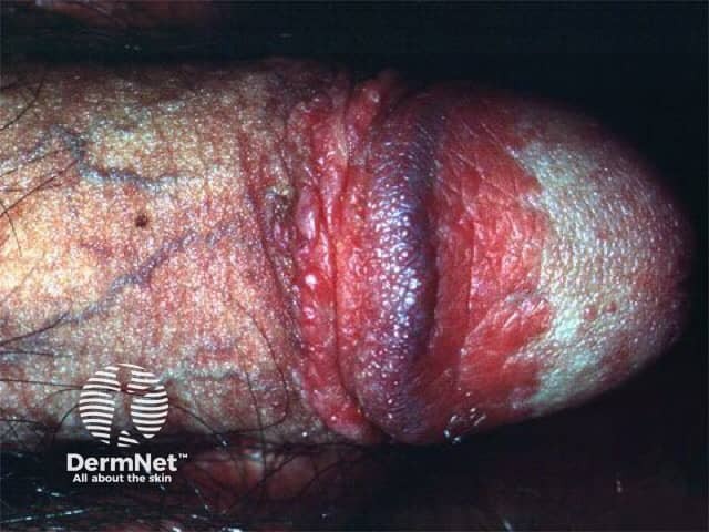
Clinical photography of penile skin demonstrates an external genital dermatosis. Imaging modality: Clinical photography using macro close-up illumination to capture surface texture. Anatomy: Penile skin encompassing the glans and distal shaft, with partial foreskin retraction exposing the coronal sulcus. Location/accessory terms: genital skin, balanitis-prone areas; body system integumentary. Visual features: diffuse erythema with raised red plaques, shiny, moist surface, mild edema, and areas of desquamation along the penile shaft and preputial skin. There is no discrete ulceration or purulent discharge evident in this image. The features are suggestive of balanitis-like dermatitis, irritation, or inflammatory dermatopathy rather than acute infection; however, candidal balanitis or bacterial balanitis remains in differential. Inflammatory patterns may include contact dermatitis, eczema, or psoriasis variants localized to genitalia. Diagnostic significance: Highlights a common genital dermatosis amenable to topical therapy; assists in distinguishing inflammatory dermatoses from infectious etiologies. Clinical use cases: educational reference for dermatology and urology trainees; differential diagnosis in patients with erythema, edema, and plaques on the penis; guides management such as hygiene measures, barrier creams, antifungal or anti-inflammatory agents; aids patient counseling about symptom monitoring and treatment response. Clinical correlation with history, symptoms, and potential secondary infection is essential for accurate diagnosis and treatment planning.
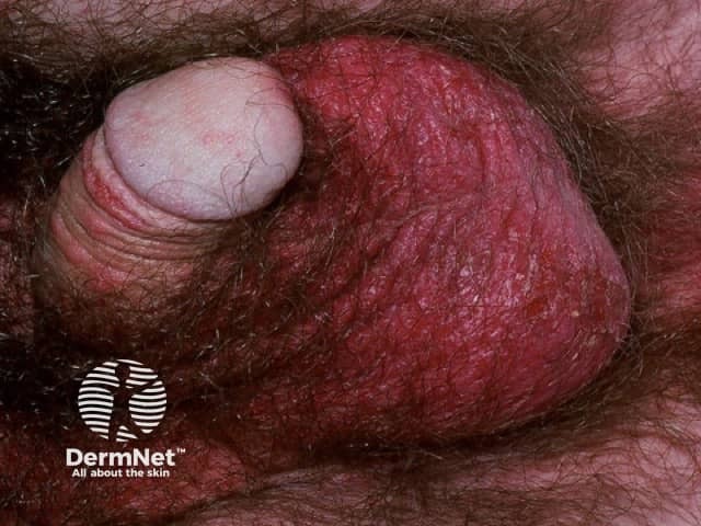
Imaging modality: Clinical photography; Technique: Close-up digital image with color-calibrated lighting; Region: genital skin, penis including shaft, glans, and foreskin; Observation: well-demarcated erythematous plaques with subtle desquamation on penile skin; Surface ranges from smooth to mildly flaky; Moist environment may reduce scale; Fissuring possible in severe cases; Differential morphology includes psoriatic plaque versus candidal balanitis; Thickness and vascularity reflect epidermal hyperplasia and dermal capillary dilation; Absence of pustules; Coloration is deep red in active plaques; Surrounding non-affected skin shows mild erythema; The image highlights genital psoriasis pattern involving glans and prepuce, aiding differentiation from infections and dermatitis; Clinically relevant for management guidance, including topical corticosteroids, vitamin D analogs, and calcineurin inhibitors, and for documenting extent for monitoring response; Diagnostic significance centers on distinguishing psoriasis from infections and dermatitis, guiding therapy; Relevance spans dermatology, urology, primary care, and medical education; Potential use cases include documentation of disease extent, treatment response monitoring, telemedicine triage, and educational illustration of genital plaque morphology.
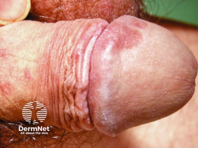
This is a high-resolution clinical photograph of penile skin, demonstrating balanopreputial involvement consistent with genital psoriasis. Modality: Clinical photography; technique: macro close-up, diffuse frontal lighting; in vivo imaging of the balanopreputial complex with glans and foreskin. Anatomical location: genital integument, balanopreputial region of the penis, including the glans penis and preputial skin. Visual features: well-demarcated erythematous plaques with fine silvery-scale, superficial scaling, and mild fissuring at the mucocutaneous junction; surrounding skin shows mild moisturized hyperemia but no overt erosion. The plaques appear plaque-like, hyperkeratotic, and can be annular or circumferential around the prepuce. Pathologic correlation: The photo supports a clinical diagnosis of genital psoriasis; histology (if biopsy is performed) would typically show epidermal hyperplasia (acanthosis), parakeratosis, thinning of the granular layer, and dermal inflammatory infiltrates; differential diagnoses include candidal balanitis, irritant dermatitis, eczema, lichen planus. Diagnostic significance: Genital psoriasis requires differentiation from infectious causes; recognizing characteristic scaling and erythema aids early dermatology referral and topical therapy. Potential clinical use cases: educational image for dermatology and urology training, telemedicine triage, image-based documentation of genital psoriasis response to topical corticosteroids or vitamin D analogs, and patient counseling. Clinical context: correlate with history of plaque psoriasis elsewhere on the body systemically.
First-line treatment
- Topical imidazole cream: Use clotrimazole 1% or miconazole 2%, applied twice daily for 7 to 14 days. The BASHH 2025 balanoposthitis guideline recommends both. The 2008 version said to apply until symptoms settle.
- Marked inflammation: Add a topical imidazole combined with 1% hydrocortisone. The BASHH 2008 guideline suggests this (evidence grade IV, C). Campbell-Walsh says short courses of low-potency topical steroids are occasionally used - Campbell Walsh Wein Urology, "Balanitis and Balanoposthitis".
Alternatives
- Severe symptoms: Oral fluconazole 150 mg as a single dose. The older BASHH guideline lists this, based on a trial comparing it with topical clotrimazole. Avoid it in pregnancy.
- Suspected resistance or imidazole allergy: Nystatin cream (100,000 units/g).
Hygiene and self-care
- Wash gently with water or a soap substitute, and dry the area well, especially under the foreskin.
- Avoid irritants such as perfumed products, antiseptic creams and excess soap.
- Avoid tight synthetic clothing.
Partners
- Treat a partner who has symptoms. BASHH says this can be considered, since candida is common in sexual partners.
- Dermatology (5th ed.) says that for recurrent balanitis, clearing candida from the partner's genital tract is generally recommended.
- Andrews' Diseases of the Skin notes that candidal balanitis may occur in the uncircumcised partner of a woman with candidal vulvovaginitis.
If it keeps coming back or doesn't improve
- Check for diabetes. Rosen's Emergency Medicine advises testing glucose in recurrent candidal balanitis. Look for other risk factors too, such as being uncircumcised, antibiotic use, or immunosuppression.
- Take a swab or culture. Candidal balanitis is often over-diagnosed. The Scottish Sandyford guideline says to diagnose it only when yeasts are seen on microscopy or culture.
- Consider other causes:
- irritant or allergic dermatitis
- psoriasis
- lichen sclerosus
- bacterial or anaerobic balanitis
- herpes
- Zoon balanitis (Campbell-Walsh, Box 59.1)
- Recent evidence: A 2026 systematic review (PMID 41026603) found that male genital lichen sclerosus is often misreported as candidal or other infective balanitis. It was published after most of the sources above. It doesn't change the antifungal regimen, but it supports getting a biopsy or specialist review if the problem is persistent.
When to see a doctor
- no improvement after 1 to 2 weeks
- frequent recurrences
- a foreskin that won't retract, or white or scarred patches
- ulcers, discharge, pain on urinating, or fever
- known diabetes or a weakened immune system