
Treatment of hair fall in detail And procedures and surgeries
hair fall treatment 2025 medical procedures surgeries comprehensive
alopecia treatment minoxidil finasteride hair loss
androgenetic alopecia hair loss treatment stages
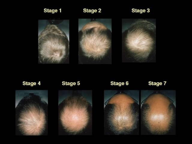
Clinical photography depicting a staged sequence of male pattern hair loss on the vertex and crown regions, illustrating Norwood-Hamilton stages 1 through 7. The modality is photography, captured in top-down views with uniform lighting to document progressive scalp thinning and hair shaft miniaturization in an adult male. The primary subject is androgenetic alopecia, with a gradual increase in scalp visibility, decrease in hair density, and widening of the distal hair shaft diameter as stages advance. Stage 1 shows minimal recession with intact vertex area; Stage 2-3 demonstrates mild to moderate thinning at the vertex; Stage 4-7 shows extensive balding with marked crown thinning and temple recession. The images emphasize non-scarring, non-inflammatory hair loss without visible scarring or perifollicular erythema. Clinically this pattern is highly characteristic of hereditary male pattern baldness, linked to androgen sensitivity and genetic predisposition. Diagnostic significance lies in providing a visual framework for staging, prognostication, and treatment planning. Differential considerations include telogen effluvium, diffuse alopecia, and traction alopecia, though these typically lack the symmetrical vertex-dominant pattern. The montage is valuable for dermatology education, patient counseling, and research on pharmacologic responses to finasteride, dutasteride, or topical minoxidil, as well as for quantitative hair density assessments and longitudinal monitoring.
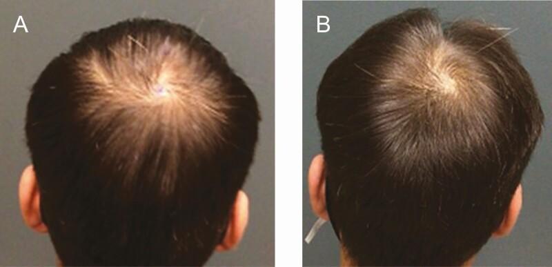
Clinical photograph comparison (before and after) depicting androgenetic alopecia treatment outcomes in a 27-year-old male. Image A (pre-treatment) shows the vertex of the scalp with characteristic patterned hair loss consistent with Norwood-Hamilton Stage III. There is visible thinning and miniaturization of hair follicles, leading to increased scalp exposure and reduced hair density in the central crown area. Image B (one-year post-treatment) demonstrates a significant increase in hair density and thickness following the administration of extracellular vesicle-based therapy (Exoflo). The central scalp region shows improved coverage with more robust, pigmented terminal hairs, resulting in a marked reduction in visible scalp surface. This comparison serves as a clinical illustration of therapeutic response in regenerative medicine for hair restoration, highlighting the transition from sparse hair distribution to denser scalp coverage.
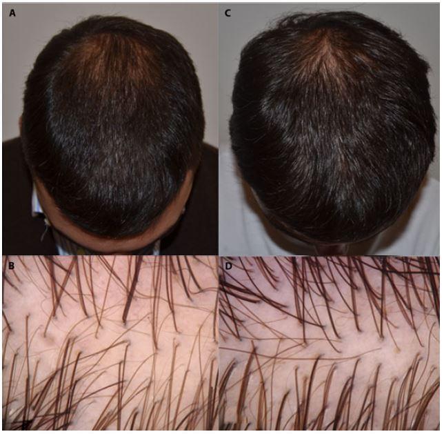
This composite image illustrates the progression of Androgenetic Alopecia (AGA) treatment in a 31-year-old male over a six-month interval. Panels A and C are clinical photographs of the scalp from a vertex view. Panel A (baseline) shows characteristic male-pattern hair loss with significant thinning at the vertex and recession in the frontal-temporal region. Panel C (6-month follow-up) demonstrates clinical improvement with increased hair density and improved scalp coverage in the same regions. Panels B and D provide high-magnification trichoscopic views of the scalp. Panel B (baseline) displays hair shaft diameter diversity (anisotropy) and several miniaturized hairs, which are hallmark diagnostic features of AGA. Panel D (post-treatment) reveals a notable increase in hair follicle density and a higher proportion of terminal hairs compared to miniaturized ones, indicating a positive therapeutic response. This comparison serves as a diagnostic and monitoring tool in dermatology, highlighting the role of trichoscopy in assessing hair shaft diameter and follicular unit density.
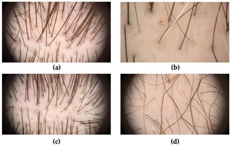
This composite of four clinical trichoscopy images (a-d) illustrates key diagnostic features of androgenetic alopecia (AGA) at various stages of progression. Image (a) demonstrates hair shaft heterogeneity (anisotrichosis), characterized by a significant variation in hair diameter, the loss of triple-hair follicular units, and the presence of yellow dots (follicular infundibula filled with sebum or keratin). Image (b) highlights the presence of short, thin, non-pigmented vellus hairs replacing terminal hairs. Image (c) shows early-stage AGA featuring peripilar signs, appearing as darkened, brownish-grey halos (perifollicular hyperpigmentation) around the hair shaft exit points. Image (d) illustrates late-stage AGA, marked by a severe reduction in hair density, the predominance of single-hair units over multiple-hair units, and numerous prominent yellow dots. These images are essential for the dermatological assessment of pattern hair loss, providing visual evidence of follicular miniaturization and characteristic scalp skin changes used to differentiate AGA from other forms of alopecia.
hair transplant FUE follicular unit extraction surgical procedure
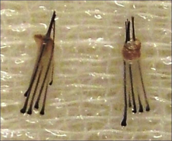
This clinical photograph shows two high-density follicular unit extraction (FUE) grafts used in hair restoration surgery. The macroscopic view demonstrates two distinct follicular units resting on a textured surgical surface. Each graft consists of multiple dark terminal hairs emerging from a common base of dermal and subcutaneous tissue. The graft on the left contains five terminal hairs, while the graft on the right contains six, illustrating the rare extraction of larger follicular units. The surrounding perifollicular tissue appears intact, semi-translucent, and slightly bulbous at the base, which is critical for graft viability and successful transplantation. The dark, rounded distal ends of the hair shafts are visible, along with uniform shaft thickness. This image serves as a teaching tool for dermatologists and hair transplant surgeons to identify graft integrity and composition following the use of micropunches (typically 0.8 to 1.0 mm) during the FUE procedure.
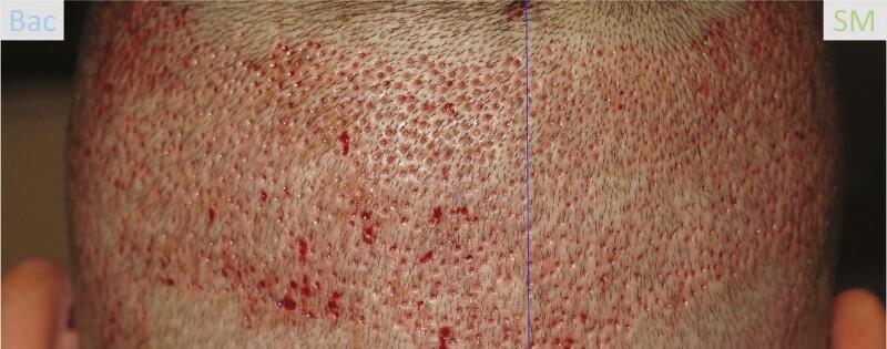
This clinical photograph displays a split-scalp comparison in a 41-year-old male patient two days after a Follicular Unit Extraction (FUE) hair transplant procedure. The image depicts the donor area of the scalp, characterized by a uniform distribution of numerous small, circular punctate wounds resulting from pneumatic hair follicle harvesting. A central blue line divides the scalp into two treatment zones: the left side labeled 'Bac' (treated with Bacitracin) and the right side labeled 'SM' (treated with Stratamed). The 'Bac' side shows more prominent areas of focal erythema and serosanguinous crusting at the extraction sites. In contrast, the 'SM' side exhibits a drier appearance with less visible discharge, suggesting accelerated initial wound healing and clot formation. This visual is used in dermatology and hair restoration education to evaluate post-procedural wound care protocols and compare the efficacy of topical treatments in reducing post-operative erythema and promoting re-epithelialization.
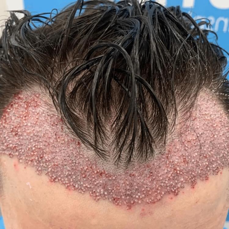
This clinical photograph displays a top-down view of the anterior scalp immediately following a hair transplant procedure. The image focuses on the recipient area, where a new hairline has been established using single follicular units. The transplanted follicles are arranged in a high-density, dotted pattern across the frontal and temporal regions. Visually, the recipient site exhibits characteristic immediate postoperative signs: widespread erythema (pinkish-red coloration) indicative of inflammation and small punctate scabs or micro-incisions at each graft insertion point, creating a rough texture. The patient's existing dark hair is damp and combed forward, partially covering the surgical site. This photograph serves as an educational example of follicular unit transplantation (FUT) or follicular unit extraction (FUE) outcomes, illustrating the immediate surgical manifestation, graft distribution density, and the anatomical reconstruction of the anterior hairline in the treatment of androgenetic alopecia.
Treatment of Hair Fall: A Comprehensive Guide
Understanding Hair Loss First
Types of Hair Loss
| Type | Pattern | Key Feature |
|---|---|---|
| Androgenetic alopecia (AGA) | Frontotemporal/vertex (male); diffuse crown (female) | DHT-driven follicular miniaturization |
| Alopecia areata | Patchy, well-circumscribed | Autoimmune, "exclamation mark" hairs |
| Telogen effluvium | Diffuse shedding | Triggered by stress, illness, postpartum |
| Traction alopecia | Marginal/hairline | Mechanical - tight hairstyles |
| Cicatricial (scarring) alopecia | Permanent follicle destruction | Inflammatory; not suitable for transplant |
| Trichotillomania | Irregular patches | Compulsive hair-pulling |
Medical / Pharmacological Treatments
1. Minoxidil (Topical & Oral)
-
Mechanism: Opens potassium channels, increases follicle size, prolongs anagen phase, stimulates blood flow to follicles
-
Formulations:
- Topical 2% solution (women), 5% solution or foam (men/women)
- Oral low-dose minoxidil: 0.25-2.5 mg/day - effective and usually well tolerated
-
Onset: Must be used for at least 6 months before assessing efficacy
-
Warning: Stopping minoxidil causes an acute telogen efflux 3-4 months later; hair returns to pre-treatment state
-
Cummings Otolaryngology, p. 445
2. Finasteride (Oral & Topical)
- Mechanism: Inhibits 5α-reductase, blocking conversion of testosterone to DHT
- Dose: 1 mg/day oral (FDA-approved for men)
- Topical finasteride spray: As effective as oral with less systemic DHT reduction
- Stopping finasteride causes gradual hair loss with return to pre-treatment status within 1 year
- Side effects: Post-finasteride syndrome - persistent sexual dysfunction and depression in some men (important counseling point)
3. Dutasteride
- Dose: 0.5 mg/day oral; approved in some countries (not yet FDA-approved for AGA)
- More effective than finasteride with a similar safety profile - inhibits both Type 1 and Type 2 5α-reductase
- A 2025 network meta-analysis (PMID 40586152) confirmed comparative efficacy among 5-alpha reductase inhibitors vs minoxidil
4. JAK Inhibitors (for Alopecia Areata)
-
Baricitinib (2-4 mg/day) - FDA-approved for severe alopecia areata
-
Ruxolitinib (10-20 mg twice daily)
-
Tofacitinib (5-10 mg twice daily)
-
Riticitinib and deuxolitinib under FDA review
-
Mechanism: Inhibit the JAK/STAT pathway that drives the autoimmune follicular attack
-
Highly effective but relapses occur if stopped
-
Goldman-Cecil Medicine, p. 4334
5. Corticosteroids (for Alopecia Areata)
- Intralesional triamcinolone: First-line for localized patches and eyebrow involvement
- High-potency topical steroids under occlusion for acute/severe disease
- High-dose pulse systemic corticosteroids: Effective in acute AA; not useful in chronic ophiasis or totalis/universalis
Norwood & Ludwig Classification (Staging for Treatment Planning)
- Norwood (men): Stages I-VII; I = minimal frontotemporal recession, VII = narrow horseshoe band of hair
- Ludwig (women): Grades I-III; I = minimal, III = severe generalized thinning with intact anterior hairline
Non-Surgical Procedures
1. Platelet-Rich Plasma (PRP) Injections
- Patient's own blood is centrifuged to concentrate platelets/growth factors
- Injected into the scalp at follicle level
- Growth factors stimulate follicular cells, extend anagen phase, improve density
- Typically 3-4 sessions monthly, then maintenance every 3-6 months
- Can combine with 5% minoxidil and finasteride for synergistic effect
- A 2026 systematic review (PMID 41603616) confirmed injectable therapies (including PRP) show benefit in AGA
2. Low-Level Laser Therapy (LLLT)
- FDA-cleared non-invasive option
- Specific wavelengths (typically 650-670 nm) stimulate follicular activity, increase blood flow
- Devices include helmets, caps, combs (e.g., 304-diode laser caps)
- Safe for both men and women with pattern hair loss
- 30 minutes/day, 3x/week; results in improved density within 60 days in some patients
3. Mesotherapy
- Micro-injections of vitamins, minerals, amino acids, and growth factors into the scalp
- Improves local circulation and delivers nutrients directly to follicles
4. Exosome Therapy (Emerging)
- Nanoscale extracellular vesicles applied/injected to promote follicular cell proliferation and differentiation
- Growing evidence base; still considered experimental
5. Scalp Micropigmentation (SMP)
- Specialized tattooing technique creating the appearance of hair follicles/stubble
- Not a hair growth treatment - cosmetic camouflage
- Ideal for complete baldness or to add density illusion
- Requires touch-ups every few years
Surgical Procedures
Overview
A. Follicular Unit Transplantation (FUT) - "Strip Method"
- A strip of hair-bearing scalp (approximately 1-2 cm wide, 15-20 cm long) is harvested from the occipital/parietal donor area under local anesthesia
- The wound is closed primarily, leaving a linear scar (camouflaged by surrounding hair)
- The strip is dissected under ×10 binocular magnification into individual follicular units (FUs) containing 1-4 hairs with their sebaceous glands and connective tissue
- Grafts are kept moist in saline-soaked Petri dishes
- Recipient site incisions are made, and grafts are placed
-
Duration: 4-6 hours under local anesthesia
-
1,000 grafts = 2,000-3,000 hairs typically
-
Higher graft yield per session than FUE
-
Allows harvesting of the highest quality grafts (microscopic dissection)
-
Linear scar is the main drawback; not ideal for patients preferring short hair
-
Cummings Otolaryngology, p. 451
B. Follicular Unit Extraction (FUE)
- Individual follicular units are extracted one by one using small circular punches (0.8-1.2 mm diameter)
- Punch first scores the epidermis to center on the FU, then advances to isolate it from perifollicular fat
- FU is gently grasped with microforceps and removed
- Heals with small, scattered circular scars (rather than a linear scar)
- Sharp punches: Higher transection risk (follicle not visible during cutting)
- Blunt/unsharp punches: Lower transection rates
- Hybrid trumpet punch: Latest advancement - further reduced follicular transection rates
- Robot-assisted (ARTAS): AI-guided robotic systems improving accuracy and consistency
- No linear scar - ideal for short hairstyles
- Less post-operative donor-site pain
- Can harvest hairs from other body areas (beard, chest) as supplementary donor sites
- Faster recovery
- Cummings Otolaryngology, p. 452
C. Robotic & AI-Assisted FUE (2025 Update)
- Newer robotic systems (e.g., ARTAS iX) use AI and image-guided algorithms to select and extract follicular units
- Reduces human error, consistent graft depth and angle
- Smaller incisions, faster healing, more natural hairline designs
- Adaptable to different hair textures (including African-American curl patterns)
D. No-Shave FUE / Precision Follicular Extraction
- Donor area does not need to be fully shaved
- Uses micro-FUE tools and Ultra-Fine Diamond Sapphire Blades
- Virtually unchanged appearance of donor area
- Typically combined with high-density PRP at no extra cost
- Ideal for patients who want discretion during recovery
E. Scalp Reduction (Limited Modern Use)
- Surgical removal of the bald scalp area, pulling hair-bearing areas together
- Rarely used today as a first-line option
- Still relevant in specific cases (e.g., post-burn cicatricial alopecia reconstruction)
F. Scalp Flaps (Limited Modern Use)
- Rotation or transposition of hair-bearing scalp flaps to cover bald areas
- Provides immediate dense coverage in one stage
- Reserved for specific reconstructive indications
- Higher complication risk (vascular compromise, scar, alopecia of flap margins)
G. Corrective Hair Transplant Surgery
- Add large numbers of follicular unit grafts (1-4 hairs) between existing larger plugs to soften the appearance
- Surgically remove the large grafts and replace with smaller FU grafts
- Combination approach
H. Eyebrow & Specialty Transplants
- Same FU principles applied to eyebrows, beard, eyelashes, pubic region
- Growing in popularity for alopecia areata affecting brows, post-trauma reconstruction, or cosmetic enhancement
- Requires precise angulation (brow hairs grow at very flat, acute angles)
Emerging & Investigational Treatments
| Treatment | Mechanism | Status |
|---|---|---|
| Stem cell therapy | Reactivate dormant follicles using follicular stem cells | Phase II trials ongoing |
| Stem cell banking | Non-invasively preserve hair follicle cells for future use | Available commercially (Acorn Biolabs) |
| Hair cloning | Multiply follicular cells ex vivo for unlimited donor supply | Research phase |
| Exosome therapy | Intercellular signaling vesicles to stimulate follicles | Emerging clinical evidence |
| Topical immunotherapy | DNCB, DPCP, SADBE for alopecia areata | Available but not FDA-approved |
| Melatonin topical | Antioxidant; some evidence for AGA | Under study |
Treatment by Hair Loss Type: Summary
| Condition | First-Line | Second-Line | Surgery? |
|---|---|---|---|
| AGA (male) | Minoxidil 5% topical + Finasteride 1 mg | Dutasteride, oral minoxidil, PRP | Yes - FUT/FUE for moderate-severe |
| AGA (female) | Minoxidil 2-5% topical | Spironolactone, low-dose oral minoxidil | Possible but limited by diffuse thinning |
| Alopecia areata | Intralesional steroids, topical steroids | JAK inhibitors (baricitinib), topical immunotherapy | Not usually until stable |
| Telogen effluvium | Treat underlying cause | Minoxidil supportive | No |
| Traction alopecia | Remove traction source | Minoxidil | If scarring present - FUE possible |
| Cicatricial alopecia | Disease-specific Rx (steroids, antibiotics) | Immunosuppressants | Only after disease arrest |
Key Treatment Principles
- Start early: Hair loss that causes 50% density reduction before becoming visible means follicular miniaturization is well advanced by the time symptoms appear
- Commit long-term: All treatments require 6+ months before efficacy assessment; stopping reverses gains
- Combine modalities: Minoxidil + finasteride + PRP has additive benefit over monotherapy
- Rule out reversible causes first: Drug-induced alopecia (from ACE inhibitors, anticoagulants, antiepileptics, beta-blockers, etc.), thyroid disease, anemia, and nutritional deficiencies should be excluded
- Surgery is not a cure: Transplanted hairs are permanent, but native hairs continue to miniaturize; ongoing medical treatment post-transplant (especially finasteride 1 mg/day) improves long-term surgical results