
Corticospinal tract
corticospinal tract anatomy diagram descending motor pathway
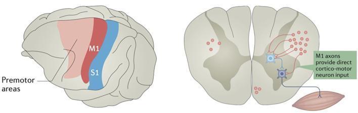
Educational neuroanatomical and physiological diagram illustrating the corticospinal pathway and cortical localization. The left side features a lateral view of the brain with color-coded functional regions: the primary motor cortex (M1) in red, the primary somatosensory cortex (S1) in blue, and anterior premotor areas in light red. The right side depicts a transverse section of the spinal cord (ventral/dorsal horn anatomy) showing descending motor control. M1 axons are visualized as red descending fibers that synapse in the ventral horn. The diagram highlights two distinct connectivity patterns: direct cortico-motor neuron input, where M1 axons synapse directly onto a lower motor neuron, and indirect input mediated by spinal interneurons (light blue). The lower motor neuron is shown exiting the spinal cord to innervate skeletal muscle. This visual demonstrates the neurobiology of voluntary motor control and the functional organization of the precentral and postcentral gyri.
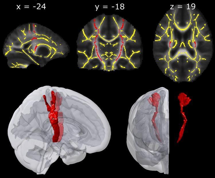
Educational neuroimaging figure illustrating the Regions of Interest (ROIs) for the corticospinal tract (CST) and Tract-Based Spatial Statistics (TBSS). The upper row presents 2D multiplanar slices in sagittal (x=-24), coronal (y=-18), and axial (z=19) views. These slices are group maps displaying mean Fractional Anisotropy (FA) in stereotactic MNI-152 space. Overlaid on the grayscale anatomy is a yellow-coded TBSS skeleton representing central white matter tracts and red-coded ROIs specifically delineating the bilateral corticospinal tracts. The lower panel provides a 3D volumetric rendering of the red CST ROIs. Two views show the ROIs in situ within a translucent gray brain template, demonstrating their descent from the primary motor and sensory hand areas through the internal capsule. A third isolated 3D model on the far right displays the distinct morphology of the tract. This visual is designed for advanced neuroanatomy or radiology education, focusing on white matter tractography and the spatial distribution of the descending motor pathway.
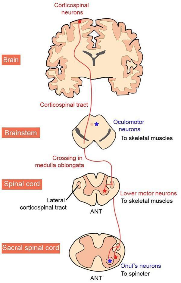
This medical anatomical diagram illustrates the organization of the human corticospinal tract and the selective vulnerability of motor neuron (MN) groups in Amyotrophic Lateral Sclerosis (ALS). The diagram follows the tract's descent through four anatomical levels: the Brain (cortex), Brainstem, Spinal Cord, and Sacral Spinal Cord. The pathway begins at upper motor neurons (corticospinal neurons) and descends through the brainstem, where it exhibits decussation at the medulla oblongata before continuing as the lateral corticospinal tract. The diagram highlights three specific targets: oculomotor neurons in the brainstem (innervating skeletal muscles), lower motor neurons in the spinal cord ventral horn, and Onuf's neurons in the sacral spinal cord (innervating sphincters). A color-coding scheme distinguishes neurodegeneration patterns: neurons labeled in red (corticospinal and typical lower motor neurons) represent groups vulnerable to degeneration in ALS, while those in blue (oculomotor and Onuf's neurons) represent groups characteristically resistant to the disease. The cross-sections of the spinal cord are labeled with 'ANT' to indicate anterior orientation.
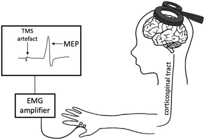
This medical schematic illustrates the physiological pathway of Transcranial Magnetic Stimulation (TMS). The diagram depicts a figure-of-eight TMS coil placed over the scalp, targeting the primary motor cortex (M1). It visualizes the induction of an electric field that depolarizes cortical neurons, initiating a descending volley along the corticospinal tract, which is labeled as a pathway extending from the brain to the upper limb. Surface electrodes placed on a target muscle in the hand capture the resulting motor activity, which is transmitted to an EMG amplifier. The output is displayed on a monitor as an electromyography (EMG) waveform. Key features of the waveform are annotated, including the initial 'TMS artefact' (representing the stimulator's electrical pulse) and the subsequent 'MEP' (Motor Evoked Potential), which demonstrates a biphasic deflection corresponding to the muscle's response. This illustration is intended for educational use in neurology and neurophysiology to explain non-invasive cortical excitability testing.
Corticospinal Tract (Pyramidal Tract)
Origin and Cortical Sources
| Source | Contribution |
|---|---|
| Primary motor cortex (Brodmann area 4, precentral gyrus) | ~30% (Guyton) / >50% (Neuroanatomy textbooks vary) |
| Premotor and supplementary motor areas (area 6) | ~30% |
| Somatosensory cortex (areas 3, 1, 2) + parietal areas (5, 7) | ~40% |
- Guyton and Hall Textbook of Medical Physiology, p. 688
- Neuroanatomy through Clinical Cases, 3e, p. 256
Course (Top to Bottom)
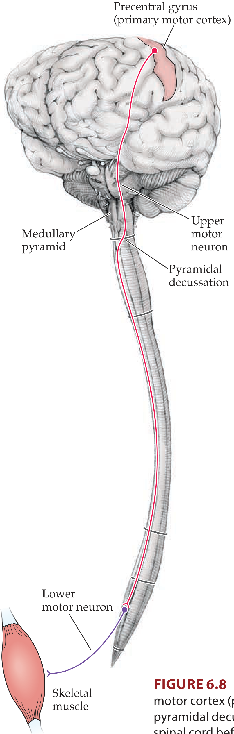
1. Corona Radiata
2. Internal Capsule
3. Cerebral Peduncles (Midbrain)
4. Pons
5. Medullary Pyramids
6. Pyramidal Decussation (Caudal Medulla)
- ~85-90% decussate → descend as the lateral corticospinal tract in the contralateral lateral funiculus
- ~10-15% do NOT decussate → descend ipsilaterally as the ventral (anterior) corticospinal tract
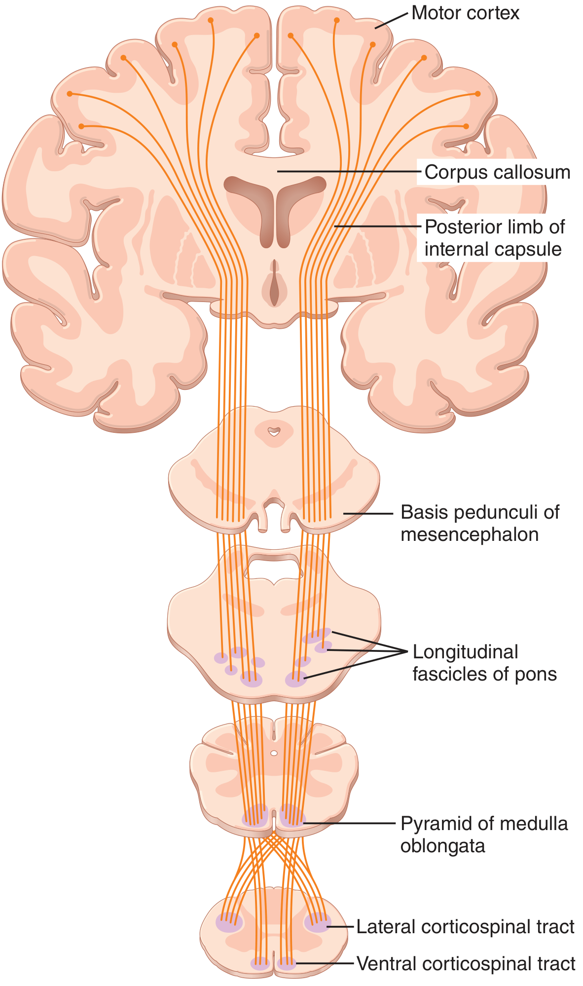
- Guyton and Hall, p. 689
- Localization in Clinical Neurology, 8e, p. 246-247
Two Divisions in the Spinal Cord
| Feature | Lateral CST | Ventral (Anterior) CST |
|---|---|---|
| Location | Lateral funiculus (contralateral) | Ventral funiculus (ipsilateral) |
| Proportion | ~85-90% | ~10-15% |
| Decussation | At pyramidal decussation (medulla) | At ventral white commissure of cord (cervical/upper thoracic) |
| Function | Fine voluntary movements of limbs | Bilateral axial/postural movements |
| Extent | All spinal levels (C1 to sacral) | Only cervical + upper thoracic |
| Termination | Laminae IV-VII and IX | Lamina III (cervical/upper thoracic) |
Termination in Spinal Cord
-
Axial muscle motor neurons: extreme ventromedial lamina IX
-
Limb-girdle muscles: intermediate position
-
Intrinsic extremity (distal limb) muscles: dorsolateral lamina IX
-
Localization in Clinical Neurology, 8e, p. 247
-
Guyton and Hall, p. 689
Function
- Primary pathway for skilled voluntary movements, especially fine fractionated movements of the hands and fingers
- Excites flexor movements and inhibits extensor tone
- Modulates spinal reflex arcs via interneuronal connections
- The pre-SMA and pre-PMd do not send axons directly to the cord - they reach it indirectly through subcortical projections
- Principles of Neural Science, 6e (Kandel), p. 868
Upper Motor Neuron (UMN) vs. Lower Motor Neuron (LMN) Lesions
| Feature | UMN Lesion (CST damage) | LMN Lesion |
|---|---|---|
| Weakness | Contralateral (after decussation) | Ipsilateral |
| Tone | Spastic (increased) | Flaccid (decreased) |
| Reflexes | Hyperreflexia | Hyporeflexia/areflexia |
| Babinski sign | Present (upgoing plantar) | Absent |
| Atrophy | Minimal/disuse only | Prominent |
| Fasciculations | Absent | Present |
Key Clinical Correlations by Level
| Level | Lesion | Result |
|---|---|---|
| Motor cortex | Focal infarct | Monoplegia (arm or leg, depending on somatotopy) |
| Internal capsule | Small lacunar infarct | Dense contralateral hemiplegia (face + arm + leg together) |
| Midbrain peduncle | Infarct + CN III palsy | Weber syndrome (contralateral hemiplegia + ipsilateral CN III palsy) |
| Pontine | Infarct + CN VI/VII | Contralateral hemiplegia + ipsilateral facial/abducens palsy |
| Medulla (above decussation) | Infarct | Contralateral hemiplegia |
| Medulla (at decussation) | Infarct | "Cruciate paralysis" - crossed arm/leg deficits |
| Cervical cord | Trauma/MS | Ipsilateral spastic paresis below lesion |
Summary Diagram (additional visual)
- Neuroanatomy through Clinical Cases, 3e (Blumenfeld) - Chapter 6
- Guyton and Hall Textbook of Medical Physiology - Chapter 56
- Principles of Neural Science, 6e (Kandel) - Chapter 34
- Localization in Clinical Neurology, 8e - Corticospinal Tract section