

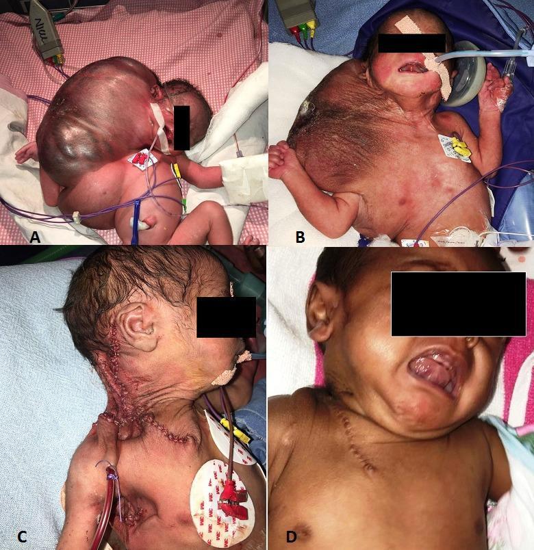
Act as top neonatologist and give diagnosis , investigations and treatment plan for the condition shown in image
frontonasal encephalocele neonate sincipital encephalocele nasal bridge swelling
Neonatal Frontonasal (Sincipital) Encephalocele
Clinical Findings in This Neonate
DIAGNOSIS
Primary Diagnosis: Sincipital (Frontoethmoid) Encephalocele - Nasofrontal/Glabellar Type
- Most prevalent in Southeast Asia (Thailand, Vietnam, Cambodia, Myanmar) - incidence 1:3,500-5,000 live births (vs. 1:35,000 in Western countries)
- Results from failed closure of the anterior neuropore or poor migration of neural crest cells
- Communicates with the subarachnoid space through a skull base defect
Differential Diagnoses to Exclude (Before Any Intervention):
| Condition | Key Features | Risk |
|---|---|---|
| Sincipital Encephalocele | Soft, compressible, pulsatile, Furstenberg sign positive | Intracranial connection in 100% |
| Nasal Glioma | Firm, non-compressible, not pulsatile | May have intranasal connection |
| Nasal Dermoid Cyst | Dome-shaped, midline, may have pit/hair | Intracranial connection in up to 25% |
| Deep Infantile Hemangioma | Soft, warm, grows in first weeks | No intracranial connection |
CRITICAL WARNING: Never biopsy, incise, or needle-aspirate this lesion before imaging. Encephaloceles have 100% intracranial connection - violation risks meningitis, CSF leak, and death. - Fitzpatrick's Dermatology, p. 1781
INVESTIGATIONS
Urgent (Bedside/Immediate):
- Furstenberg Test - Compress both internal jugular veins: positive if mass enlarges/pulsates (confirms intracranial communication). Also observed with Valsalva/crying.
- Transillumination - Helps assess if predominantly fluid-filled
- Vital signs + SpO2 - Assess for respiratory compromise (nasal obstruction in neonates who are obligate nose breathers)
- Full neurological examination - Tone, reflexes, fontanelle tension, head circumference
Imaging (Priority Order):
| Investigation | Purpose | Priority |
|---|---|---|
| MRI Brain + Face (with contrast) | Gold standard - defines herniated contents (brain tissue vs. meninges only), sac dimensions, neck of sac, intracranial anatomy (associated hydrocephalus, corpus callosum agenesis, Chiari) | FIRST CHOICE |
| CT Brain + Face (3D reconstruction) | Defines bony defect size and location, cranial base anatomy - essential for surgical planning | Required pre-op |
| Cranial Ultrasound | Bedside, no radiation, assesses for hydrocephalus, midline shift | Immediate adjunct |
| MR Angiography | If vascular structures may be within sac | Pre-op if needed |
Laboratory Investigations:
- CBC, CRP, blood culture - Baseline, rule out concurrent infection
- Serum electrolytes, glucose, calcium - Neonatal metabolic baseline
- Chromosomal microarray / Karyotype - Associated with trisomy 13, 18; Meckel-Gruber syndrome (autosomal recessive - encephalocele + polydactyly + polycystic kidneys)
- Coagulation profile (PT, APTT) - Pre-surgical
- Blood group and crossmatch - Pre-surgical
Specialist Consultations:
- Paediatric Neurosurgery (primary)
- Neonatology/NICU
- Paediatric Ophthalmology (visual pathway assessment, hypertelorism)
- Genetics
- Anaesthesia (paediatric)
TREATMENT PLAN
Immediate Stabilization (NICU):
- Protect the sac - Cover with warm, moist sterile gauze; do NOT compress
- Positioning - Lateral/prone to avoid direct pressure on mass
- Airway management - Assess for nasal obstruction; prepare for oral airway/intubation if needed (obligate nasal breathing in neonates)
- IV access + fluids - Maintain euglycemia and normal electrolytes
- Temperature regulation - Servo-controlled incubator
- Anti-seizure prophylaxis - Only if seizures observed; monitor with aEEG
- Prophylactic antibiotics - Consider if sac appears thin/leaking (risk of meningitis); e.g., IV cefotaxime
- Avoid LP until intracranial anatomy is defined
Definitive Treatment: Surgical Repair
| Lesion Type | Approach |
|---|---|
| External glabellar encephalocele (as in this case) | Open craniotomy + craniofacial repair - bifrontal approach |
| Intranasal encephalocele | Endoscopic transnasal repair |
| Small defects | Combined open + endoscopic |
- Bifrontal craniotomy
- Reduction of herniated brain/meningeal tissue (non-viable tissue resected; viable tissue returned to intracranial compartment)
- Closure of skull base defect in multilayer fashion (fat graft, fascia lata, pericranium, or alloplastic material)
- Craniofacial reconstruction of the glabella/nasal bridge
- Orbital osteotomies if significant hypertelorism is present (may be staged to 12-18 months)
- If present pre- or post-operatively: ventriculoperitoneal (VP) shunt or endoscopic third ventriculostomy (ETV)
Post-operative Care:
- ICU monitoring post-craniotomy
- Serial head circumference measurements
- Ophthalmology follow-up for visual outcomes
- Neurodevelopmental surveillance (speech, motor, cognitive milestones)
- MRI at 3-6 months post-op to assess repair integrity
PROGNOSIS
| Factor | Impact |
|---|---|
| Nasofrontal location | Better prognosis than occipital (less brain tissue involved) |
| Meningocele only (no brain herniation) | Excellent neurological outcome |
| Meningoencephalocele | Higher risk of developmental delay, epilepsy |
| Hydrocephalus | Worsens outcome if uncontrolled |
| Associated anomalies (Meckel-Gruber, trisomy 13/18) | Poor prognosis |
Summary: This neonate has a sincipital nasofrontal encephalocele - a herniation of CNS tissue through a bony defect at the glabella. The priorities are: protect the sac, obtain urgent MRI + CT, consult paediatric neurosurgery, and plan elective craniofacial repair at 3-6 months after full stabilisation and imaging workup.
What are your thoughts about the condition cystic hygroma? Can this be the diagnosis?
Can This Be Cystic Hygroma? - A Definitive Analysis
What is Cystic Hygroma?
Comparison: Cystic Hygroma vs. This Case
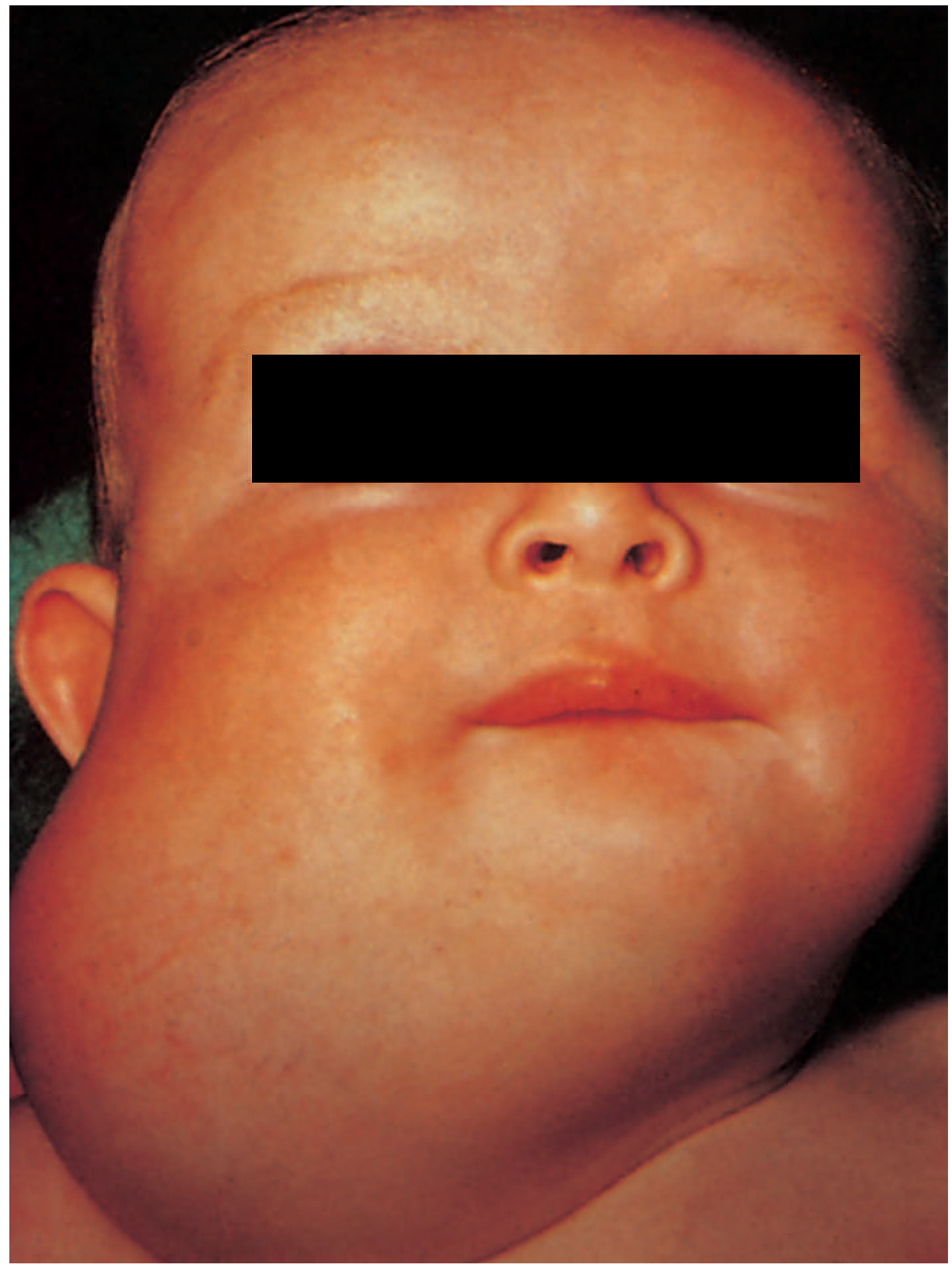
Point-by-Point Comparison
| Feature | Cystic Hygroma | This Neonate's Mass | Verdict |
|---|---|---|---|
| Location | Posterior neck (most common), also axilla, groin, mediastinum, floor of mouth | Midline glabella / frontonasal | ❌ Wrong site for hygroma |
| Shape | Diffuse, lobulated, poorly marginated - conforms to tissue planes | Well-defined, dome-shaped, discrete | ❌ Wrong morphology |
| Skin color | Normal overlying skin, may appear bluish if large | Normal skin, no bluish tinge | ✓ Compatible - but non-specific |
| Transillumination | Brilliantly transilluminant (filled with clear lymph - this is the hallmark sign) | Encephaloceles are NOT brilliantly transilluminant | ❌ Test would differentiate |
| Compressibility | Soft and partially compressible; increases with cough/cry | Encephaloceles are soft AND increase with crying (via Furstenberg mechanism - but via CSF, not lymph) | ⚠️ Overlapping but different mechanism |
| Anatomical embryology | Derived from lymphatic sac failure (mesoderm/endothelium) | Derived from failed neural tube closure (neuroectoderm) | ❌ Entirely different origin |
| Furstenberg test | Negative - does not expand with jugular compression | Positive in encephalocele | ❌ Test would definitively separate |
| Pulsatility | Non-pulsatile | Can be pulsatile (transmitted from CSF) | ❌ Favors encephalocele |
| Bony defect on imaging | No skull defect | Skull base defect present | ❌ Definitive on CT/MRI |
| Midline facial anomaly | Does NOT cause hypertelorism or nasal bridge widening | Causes hypertelorism by expanding through the foramen cecum | ❌ Structural explanation only fits encephalocele |
| Intracranial connection | None | 100% in encephalocele | ❌ Critical safety difference |
Why Location Alone Excludes Cystic Hygroma Here
"Swelling usually occurs in the neck and may involve the face, submandibular region, tongue and floor of the mouth... The cheek, axilla, groin and mediastinum are other less frequent sites."
- Bailey & Love's Short Practice of Surgery, 28th Ed., p. 827
The One Shared Feature That Could Cause Confusion
- Soft
- Present at birth
- Skin-covered
- May increase in size with crying
Bedside Test to Definitively Separate Them
| Test | Cystic Hygroma | Encephalocele |
|---|---|---|
| Transillumination | Brilliant (clear lymph fluid) | Dull or partially transilluminant (brain/meningeal tissue) |
| Furstenberg test (bilateral jugular compression) | Negative - no enlargement | Positive - mass enlarges |
| Pulsation | Absent | Present (CSF transmitted) |
Conclusion
A premature 29 weeks with weight 1.2 kg newborn developed murmur on 10th day. His hb is 8.8
Preterm Neonate (29 weeks, 1.2 kg) with New Murmur on Day 10 and Hb 8.8 g/dL
Clinical Reasoning
DIAGNOSIS
Primary: Patent Ductus Arteriosus (likely hemodynamically significant)
- Ductus arteriosus normally closes within 48 hours in 96% of healthy term neonates, but in extreme prematurity (29 weeks), ductal tissue is immature and unresponsive to normal closure stimuli (rising PaO2, falling prostaglandins)
- Classic murmur: 1-4/6 continuous "machinery" murmur, loudest at the left upper sternal border (LUSB), often with a wide pulse pressure and bounding peripheral pulses - Harriet Lane Handbook, Table 7.10
- Onset around day 7-10 of life is typical, coinciding with resolving RDS and increasing left-to-right shunt as pulmonary resistance drops
- Hb 8.8 g/dL at day 10 is low even accounting for physiologic nadir, and anemia can itself unmask or worsen a PDA murmur by increasing cardiac output and flow velocity across the duct
- Anemia of prematurity results from diminished erythropoietin response, shortened RBC lifespan, iatrogenic phlebotomy losses, and rapid growth in extremely preterm infants
Differential Diagnoses to Consider
| Condition | Distinguishing Feature |
|---|---|
| VSD | Holosystolic murmur at LLSB, not continuous |
| ASD | Fixed split S2, systolic ejection murmur at LUSB (no continuous component) |
| Peripheral pulmonary stenosis (innocent) | Common benign murmur in preterms, resolves by 3-6 months |
| Sepsis-related flow murmur | Consider if signs of systemic illness present |
INVESTIGATIONS
Immediate Bedside:
- Four-limb blood pressure - wide pulse pressure supports PDA; assess for hypotension (diastolic runoff)
- Pre/post-ductal SpO2 - assess for differential saturation
- Precordial palpation - hyperdynamic precordium, bounding pulses, active precordium with large shunt
Confirmatory Imaging:
- Echocardiography with Doppler (gold standard) - confirms ductal patency, measures ductal diameter, assesses:
- Left atrium: aortic root ratio (LA:Ao >1.4 suggests significant shunt)
- Left ventricular volume overload/dilation
- Diastolic flow reversal in descending aorta (indicates significant steal)
- Direction and velocity of ductal shunt flow
- Chest X-ray - cardiomegaly and increased pulmonary vascular markings if hemodynamically significant shunt present; also assess for coexisting RDS/CLD
- ECG - usually normal with small-moderate PDA; biventricular hypertrophy (BVH) with large shunt - Harriet Lane Handbook, Table 7.10
For the Anemia (Hb 8.8):
- CBC with reticulocyte count - reticulocytopenia typical of anemia of prematurity (blunted erythropoietin response)
- Peripheral smear - rule out hemolysis, blood loss picture
- Total bilirubin - if hemolysis suspected
- Iron studies (ferritin, serum iron, TIBC) - baseline before iron therapy
- Blood group/Coombs if not already done - exclude alloimmune hemolysis
- Assess cumulative phlebotomy losses - common contributor in NICU
TREATMENT PLAN
General Supportive Measures
- Fluid management - mild fluid restriction (avoid volume overload which worsens shunt)
- Maintain adequate hematocrit - anemia increases cardiac output and worsens shunt physiology; correcting anemia can help
- Optimize oxygenation and ventilation - avoid hyperoxia and hypoxia
- Monitor for signs of CHF: tachypnea, hepatomegaly, poor feeding, increased ventilatory requirement
- Monitor for NEC risk - PDA-associated diastolic steal reduces mesenteric perfusion
Specific PDA Management (only if hemodynamically significant on echo)
- Many small-moderate PDAs close spontaneously; a "watchful waiting" strategy is increasingly favored, especially if the infant is otherwise stable
- Fluid restriction, diuretics if fluid overloaded
| Drug | Notes |
|---|---|
| Indomethacin | IV 0.1-0.25 mg/kg/dose - classic COX inhibitor; monitor renal function, platelets, GI bleeding, NEC risk - Goodman & Gilman's Pharmacological Basis of Therapeutics |
| Ibuprofen | Comparable efficacy to indomethacin with less renal/mesenteric vasoconstriction; increasingly preferred - Katzung's Basic and Clinical Pharmacology |
| Acetaminophen (paracetamol) | Emerging alternative, especially when NSAIDs contraindicated (thrombocytopenia, renal impairment, active bleeding, NEC) |
- Reserved for infants who fail or have contraindications to medical therapy AND remain symptomatic/ventilator-dependent
- Surgical ligation or increasingly, transcatheter device closure (even in small preterm infants at experienced centers)
Anemia Management (Hb 8.8 g/dL)
- Transfusion decision - based on clinical status, not Hb alone. Consider PRBC transfusion (10-15 mL/kg) if:
- Symptomatic anemia (poor weight gain, tachycardia, increased apnea/bradycardia, increased oxygen requirement)
- Hb <8 g/dL in a stable infant, or higher threshold (~10-12 g/dL) if on significant respiratory support or with hemodynamically significant PDA/cardiac symptoms, as anemia worsens shunt physiology
- Iron supplementation - 3-6 mg elemental iron/kg/24 hr
- Erythropoietin (if used) - 200-400 U/kg/dose SC/IV, 3-5 times/week for 2-6 weeks, always with iron supplementation - Harriet Lane Handbook, 23rd Ed., p. 1127
- Minimize phlebotomy losses - use micro-sampling techniques, bundle labs
Key Priorities for This Infant
- Urgent echocardiography to confirm PDA and quantify hemodynamic significance
- Correct/monitor anemia - contributes to symptomatology and may need transfusion given the cardiac strain
- Watch closely for CHF, feeding intolerance, and NEC signs given the ductal steal physiology
- Involve pediatric cardiology for definitive diagnosis and treatment decision (watchful waiting vs. pharmacologic vs. procedural closure)
- Nutritional/growth monitoring - PDA and anemia both impair growth in ELBW infants
Treatment of goat bite
goat bite treatment rabies risk wound management guidelines
Treatment of Goat Bite
Overview
1. Immediate Wound Care (First Aid)
- Wash immediately and thoroughly with soap and running water for 10-15 minutes - this is the single most effective measure to reduce rabies virus load and bacterial contamination. - Harrison's Principles of Internal Medicine, 22nd Ed.
- Apply antiseptic - povidone-iodine or similar virucidal agent after washing
- Do not suck the wound or apply mouth to it
- Control bleeding with direct pressure
- Avoid immediate suturing/primary closure of puncture wounds or wounds >8 hours old - bite wounds are managed by delayed primary closure or secondary intention healing to reduce infection risk, except in cosmetically sensitive areas (face) where primary closure may be preferred after thorough irrigation - District 2 Public Health Animal Bite Guidelines
2. Rabies Risk Assessment (Critical Step)
| Category | Goat-specific consideration |
|---|---|
| Rabies-endemic countries (India, most of Asia, Africa) | Goats, being herbivores, are not the natural reservoir for rabies, but they CAN contract rabies from a rabid dog/jackal bite and transmit it to humans via saliva. In endemic areas, any bite from any mammal is treated as a potential rabies exposure unless the animal can be reliably observed/excluded. |
| Non-endemic/low-risk countries (US, most of Europe) | Goats are classified with livestock (cattle, sheep, horses) - low rabies risk. Standard practice: confine and observe the goat for 10 days; if it remains healthy, no PEP is needed. If the goat cannot be observed, or acted abnormally/was itself attacked by a wild/rabid animal, treat as exposure. - Minnesota Dept of Health Rabies Guidelines |
Decision Algorithm:
- If the goat is healthy, owned, and can be confined/observed for 10 days → wound care + tetanus only; give PEP only if the goat develops signs of rabies or dies during observation
- If the goat is unknown/stray, unprovoked bite, or from a rabies-endemic region with no reliable follow-up → start post-exposure prophylaxis (PEP) immediately, do not wait
Post-Exposure Prophylaxis (if indicated):
- Rabies vaccine (cell-culture vaccine) - IM, 4-5 dose schedule: Days 0, 3, 7, 14 (+/- day 28 in immunocompromised) - Priority ER protocol; CDC-aligned
- Rabies Immunoglobulin (RIG) - infiltrated around/into the wound, indicated for Category III exposures (bites that break skin, especially on head/face/hands, or multiple bites) per WHO categorization
- Category of exposure should be assessed by a physician per WHO/national rabies guidelines
3. Tetanus Prophylaxis
- Check tetanus immunization status
- If not fully immunized or last booster >5-10 years ago (depending on wound cleanliness) → give Tdap/Td booster and consider tetanus immunoglobulin (TIG) for dirty/contaminated wounds
4. Antibiotic Prophylaxis
- Puncture wounds (deep, hard to clean)
- Wounds on hands, feet, face, or genitalia
- Wounds in immunocompromised patients or those with diabetes/asplenia
- Wounds presenting >8 hours after injury
- Crush-type injury (goats often crush/nip rather than cleanly puncture)
- Amoxicillin-clavulanate (covers Pasteurella + anaerobes) - first-line oral choice
- Penicillin-allergic: Metronidazole + Trimethoprim-sulfamethoxazole or clindamycin + a fluoroquinolone (in adults)
- IV antibiotics if grossly contaminated/infected wound or if surgical washout is required - Royal Children's Hospital Melbourne, Animal Bite Guidelines
5. Further Evaluation
- X-ray if suspected bone involvement, fracture, or retained tooth/foreign material
- Wound culture if signs of established infection (warmth, purulence, spreading erythema)
- Assess for hand/joint/tendon involvement if bite is over a joint (goats can crush with molars)
- Follow-up in 24-48 hours to reassess for infection, especially if wound was left open
Summary - Practical Steps for a Goat Bite
- Wash wound thoroughly with soap and water for 10-15 minutes
- Assess rabies risk - confine/observe the goat 10 days if possible and healthy; start PEP if the goat is unknown, unprovoked bite, or endemic-area exposure without reliable observation
- Update tetanus prophylaxis as needed
- Give prophylactic antibiotics (amoxicillin-clavulanate) if high-risk wound features present
- Do not suture puncture wounds; consider delayed closure
- Arrange follow-up for wound check
Inj Tramadol dose per kg
Injection (IV/IM) Tramadol Dose per kg
Adult Dose
- 50-100 mg every 4-6 hours (IV/IM), not typically dosed per kg in adults
- Maximum: 400 mg/day (some sources cap at 300 mg/day in elderly or hepatic/renal impairment)
- Equianalgesic parenteral dose: 100 mg tramadol - Goodman & Gilman's Pharmacological Basis of Therapeutics, Table 23-2
Pediatric Dose (per kg)
| Source | Dose | Frequency | Notes |
|---|---|---|---|
| Rosen's Emergency Medicine | 1-2 mg/kg/dose | Every 4-6 hours | For children ≥4 years old |
| Campbell-Walsh Urology (Pediatric pain table) | 1 mg/kg | Every 12 hours | Oral dosage table |
| Maximum daily dose (pediatric) | Lesser of 8 mg/kg/day or 400 mg/day | - | Per dose max 100 mg |
Important Safety Warning - Pediatric Use
The FDA issued its strongest boxed warning (2017) against tramadol use in children. It is now contraindicated in children <12 years, and not recommended in adolescents 12-18 years who are obese, have obstructive sleep apnea, or severe pulmonary disease. Use in children is considered off-label.
- The Harriet Lane Handbook, 23rd Ed., p. 6160
Practical Recommendation
- Avoid tramadol injection in neonates and young children given the FDA warning - safer alternatives (morphine, fentanyl, paracetamol/ibuprofen) are preferred for pediatric pain control in most protocols.
- If used in older children/adolescents (off-label, with informed consent and after risk-benefit discussion): 1-2 mg/kg/dose IV, every 4-6 hours, max 8 mg/kg/day or 400 mg/day (whichever is lower).
- In adults: 50-100 mg IV/IM every 4-6 hours, max 400 mg/day.
Describe differential diagnosis for scrotal swelling acute in nature with no associated symptoms
acute scrotum differential diagnosis
acute scrotal swelling differential diagnosis guideline testicular torsion pediatric
https://www.rch.org.au/clinicalguide/guideline_index/Acute_s…
Quick answer
Acute Scrotal Swelling With No Pain, Fever, Urinary, or Systemic Symptoms
Important differential diagnosis
| Diagnosis | Typical clues |
|---|---|
| Hydrocele | Most likely benign cause of painless acute-noticed scrotal enlargement. Soft, fluctuant, non-tender, can usually get above the swelling, often transilluminates. A communicating hydrocele may vary in size during the day or with crying/straining. |
| Inguinoscrotal hernia | Groin swelling extending into scrotum, often fluctuates with crying/standing and may reduce when lying down. Cannot get above swelling if it extends from groin. If irreducible, tense, tender, vomiting, or abdominal distension develops, consider incarceration and seek urgent surgery. |
| Testicular torsion | Usually sudden severe pain, high-riding/horizontal testis, absent cremasteric reflex. But may be painless in neonates, in non-verbal children, or following intermittent torsion. Treat as a surgical emergency until ruled out. |
| Torsion of appendix testis/epididymis | More often mild pain and upper-pole tenderness in prepubertal children; possible blue-dot sign. Less likely when entirely asymptomatic, but can be subtle early. |
| Idiopathic scrotal edema | Usually boys aged 3-7 years. Sudden, painless or minimally uncomfortable edema of scrotal skin, often with erythema/purplish hue that can spread to penis, perineum, or groin. Testis itself is normal on examination/ultrasound. Diagnosis of exclusion. |
| Varicocele | Usually gradual rather than truly acute, often left-sided, "bag of worms," larger when standing and decreases supine. Acute onset, right-sided, or non-reducing varicocele needs evaluation for obstruction. |
| Testicular or paratesticular tumor | Typically painless, hard intratesticular mass. May be noticed suddenly though the mass is not actually acute. Ultrasound is required. |
| Hematocele / scrotal hematoma | Ask specifically about even minor trauma, recent procedure, bleeding disorder, anticoagulants, or birth trauma. May be painless initially but often becomes tender; ultrasound helps distinguish it from torsion or tumor. |
| Epididymitis/orchitis | Usually pain, epididymal/testicular tenderness, erythema, fever, dysuria, or abnormal urinalysis. Much less likely if there are truly no associated symptoms. |
| Dependent edema | Usually bilateral and accompanied by generalized edema, heart/renal/liver disease, hypoalbuminemia, or fluid overload. |
| Scrotal cellulitis, abscess, Fournier gangrene | Pain, warmth, erythema, fever, systemic illness, or skin changes are expected. Absence of symptoms makes these unlikely, but early disease in high-risk patients requires caution. |
Initial assessment
- Confirm whether swelling is scrotal skin, an intrascrotal mass, or extending from the inguinal canal.
- Examine both testes for lie, size, firmness, and tenderness.
- Check if you can get above the swelling, whether it is reducible, and whether it transilluminates.
- Examine abdomen and groins for hernia.
- Check cremasteric reflex in children, recognizing that its presence does not completely rule out torsion.
- Urgent color Doppler scrotal ultrasound is appropriate if the diagnosis is uncertain, but should not delay urology/surgical review when torsion is suspected.
Red flags requiring urgent emergency/urology assessment
- Neonate with unilateral scrotal swelling, discoloration, or firm testis
- Sudden onset, even without pain
- High-riding or horizontal testis
- Absent cremasteric reflex
- Irreducible inguinoscrotal swelling
- Vomiting, abdominal distension, fever, erythema, trauma, or increasing swelling
- Hard intratesticular mass
Urine not passed after 48 hrs of birth in full term newborn Baby is active, taking milk , no abdominal swelling
neonate anuria evaluation urinary obstruction
newborn no urine 48 hours evaluation neonatal anuria guideline
First, confirm true anuria
- Check whether urine may have been missed or mixed with stool.
- Inspect every diaper for a small wet patch or orange-pink urate crystals.
- Ask about urine passed immediately after delivery.
- Ensure the baby is feeding effectively and check weight loss since birth.
Important causes
1. Low intake or dehydration - common and potentially reversible
2. Urinary retention due to lower urinary tract obstruction - must not miss
- Poor or dribbling urinary stream, straining
- Palpable/full bladder or suprapubic mass
- Hydronephrosis on ultrasound
- Sometimes no obvious abdominal distension early on
3. Acute kidney injury
- Perinatal asphyxia/shock
- Sepsis
- Hypovolemia/dehydration
- Nephrotoxic drugs
- Renal vascular thrombosis
4. Congenital renal/urinary tract anomaly
- Bilateral renal agenesis or severe bilateral renal dysplasia
- Severe bilateral hydronephrosis or obstructive uropathy
- Polycystic kidney disease
Immediate assessment and investigations
-
Vitals and perfusion assessment
- Temperature, heart rate, respiratory rate, BP, oxygen saturation
- Hydration, weight loss, capillary refill, fontanelle
-
Examination
- Palpate for a distended bladder or kidneys
- Inspect penis/meatus in a male infant
- Look for dysmorphic features, edema, signs of sepsis
-
Strict input-output charting
- Weigh diapers and document feeds.
-
Bladder ultrasound scan
- If bladder is full, clinician-performed sterile catheterization can confirm retention, decompress the bladder, and obtain urine.
- This should be performed by trained neonatal staff, not attempted at home.
-
Urgent renal and bladder ultrasound
- Assess kidney size/appearance, hydronephrosis, hydroureter, bladder wall, and posterior urethral obstruction.
-
Blood tests
- Serum glucose
- Urea, creatinine
- Sodium, potassium, bicarbonate, calcium
- CBC and CRP, blood culture if infection is possible
- Blood gas if unwell or electrolyte disturbance suspected
-
Urine testing once obtained
- Urinalysis, microscopy, culture if indicated.
-
If PUV/obstruction suspected
- Urgent pediatric urology consultation.
- A micturating cystourethrogram/VCUG is often needed after stabilization.
Important practical point
Tab doxycycline dose per kg in children
CDC doxycycline dose children 2.2 mg/kg dose maximum 100 mg
Doxycycline Dose in Children
- 2.2 mg/kg per dose orally every 12 hours
- Maximum 100 mg per dose
- Equivalent total daily dose: 4.4 mg/kg/day, divided twice daily.
- 2.2 × 20 = 44 mg per dose, given every 12 hours
- Practical prescribed dose depends on available tablet/suspension strengths.
Important age guidance
- Historically avoided below age 8 because of concern for tooth discoloration.
- Short courses are acceptable at any age when clearly indicated, especially for rickettsial infections, cholera, anthrax, plague, or other serious infections where doxycycline is first-line.
- For routine, non-severe infections in children under 8, choose an alternative when appropriate.
Administration
- Give with a full glass of water and keep the child upright for 30 minutes to reduce esophagitis.
- Avoid giving at the same time as iron, calcium, antacids, or milk, as these reduce absorption.
- Duration depends entirely on diagnosis. Do not use a generic duration.