
Japanese encephalitis
"Japanese encephalitis"[MeSH Terms]
Japanese encephalitis brain MRI pathology
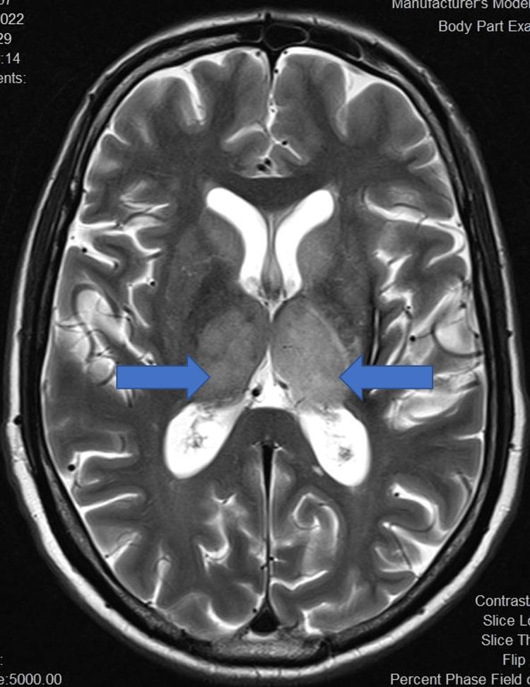
This diagnostic image is an axial T2-weighted magnetic resonance imaging (MRI) scan of the human brain. The focal pathology is highlighted by blue arrows, demonstrating bilateral, symmetrical enlargement and T2 hyperintensity of the thalami. These findings are characteristic of deep gray matter involvement in neuroinflammatory or infectious conditions, such as Japanese Encephalitis. The increased signal intensity within the thalami is distinct against the surrounding darker brain parenchyma. Other visible anatomical structures include the frontal and occipital horns of the lateral ventricles, which exhibit normal high-signal cerebrospinal fluid (CSF), and the cerebral cortex with clearly defined gyri and sulci. The imaging demonstrates a classic presentation of bilateral thalamic lesions, a key diagnostic feature for differentiating viral encephalitides and certain metabolic disorders. The educational value of this image lies in its clear demonstration of thalamic edema and signal changes on a standard T2 sequence.
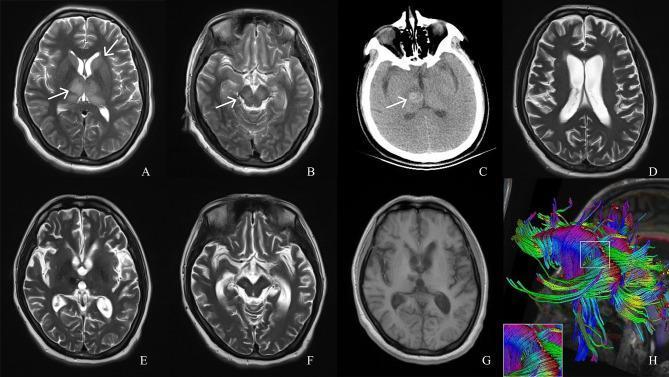
This longitudinal neuroimaging series tracks the progression of a brain injury, likely Japanese encephalitis, using T2-weighted MRI (A-B, D-F), non-contrast CT (C), T1-weighted MRI (G), and Diffusion Tensor Imaging (DTI) (H). Initial T2 MRI at day 6 (A, B) demonstrates symmetrical hyperintense signals in the bilateral thalamus, caudate nucleus, and cerebral peduncles (white arrows). By day 19, a CT scan (C) reveals a hyperdense region in the right thalamus consistent with an acute hemorrhage (arrow). Follow-up imaging at month 6 (D-F) and month 14 (G) shows the evolution into chronic sequelae: the primary lesions have reduced in size, but there is marked global brain atrophy characterized by prominent ventricles (ventriculomegaly), widened cortical sulci, and thinned gyri. The T1-weighted image (G) reinforces the presentation of diffuse atrophic changes. Finally, a DTI reconstruction (H) illustrates the integrity of white matter tracts, specifically highlighting a disruption or breakage in the corpus callosum fibers. This series serves as a pedagogical tool for understanding the natural history of viral encephalitis, from acute inflammatory edema and secondary hemorrhage to long-term neuroanatomical degeneration.
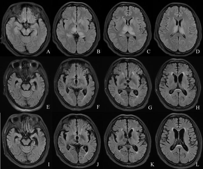
This diagnostic image is a longitudinal comparison of axial brain MRI scans (FLAIR sequence) across three time points: day 6 (A-D), month 6 (E-H), and month 14 (I-L). The initial scans (A-D) demonstrate acute bilateral, symmetrical hyperintense signals involving the thalami, caudate nuclei, lentiform nuclei, and hippocampi, characteristic of Japanese encephalitis or similar neuroinflammatory processes. In the subacute to chronic phase (E-H), these hyperintensities significantly regress; however, there is evidence of progressive global brain atrophy, characterized by compensatory enlargement of the lateral and third ventricles (ventriculomegaly) and widening of the cortical sulci. By month 14 (I-L), the atrophy has stabilized or slightly progressed, showing prominent ex-vacuo ventriculomegaly and advanced thinning of the brain parenchyma. The images illustrate the evolution from acute inflammatory lesions to chronic neurodegenerative changes and volume loss following severe viral encephalitis.
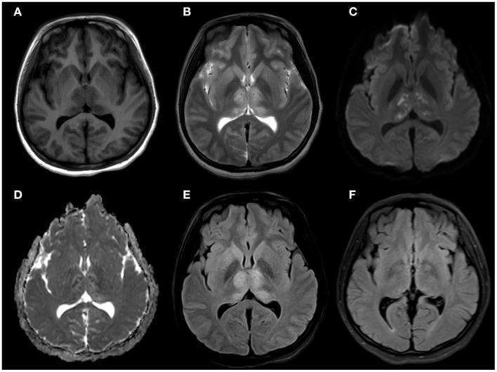
This composite diagnostic image displays six axial brain MRI sequences (A-F) documenting a case of viral encephalitis, specifically Japanese Encephalitis. Panels A-E represent the acute phase, showing symmetric involvement of the deep gray matter structures. (A) T1-weighted image reveals subtle hypointensity in the bilateral thalami, head of the caudate nuclei, and right lenticular nucleus. (B) T2-weighted imaging demonstrates corresponding hyperintensity in these regions, including the hippocampi. (C) Diffusion-weighted imaging (DWI) and (D) Apparent Diffusion Coefficient (ADC) map show bilateral punctate hyperintensity on DWI with corresponding hypointensity on ADC, indicating restricted diffusion and cytotoxic edema. (E) T2-FLAIR imaging confirms hyperintensity in the bilateral thalami and caudate heads without mass effect. (F) A follow-up T2-FLAIR image from two months later shows complete resolution of the previously seen signal abnormalities. The symmetrical involvement of the thalami and basal ganglia is a hallmark radiological finding for Flavivirus infections. This sequence serves as an educational example of deep gray matter restricted diffusion and its longitudinal progression in neuro-infectious diseases.
"Japanese encephalitis" AND treatment
Japanese Encephalitis (JE)
Overview
Etiology & Virology
- Caused by Japanese Encephalitis Virus (JEV), a member of the family Flaviviridae, genus Flavivirus (Group B arbovirus)
- Closely related to St. Louis encephalitis virus and West Nile virus
- Envelope glycoprotein contains specific and cross-reactive neutralizing epitopes
- Multiple lineages (I-V) exist with different geographic distribution, but all belong to a single serotype with similar virulence and host preference
- Lineage I and II strains are now endemic in northern Australia
Epidemiology
| Feature | Detail |
|---|---|
| Global burden | ~67,900 clinical cases/year; ~20,400 deaths annually |
| Case fatality rate | 20-30% |
| Neurological sequelae | 30-50% of survivors |
| Subclinical ratio | Only 1 in 200 infections (~0.5%) result in severe/clinical disease |
| Age group | Predominantly children < 15 years; ~10% cases in >60 years (waning immunity) |
| Geographic spread | Japan, China, Korea, Taiwan, India, Southeast Asia, Western Pacific, northern Australia |
- Vector: Culex tritaeniorhynchus (principal), also Aedes and Anopheles spp.
- Amplifying hosts: Pigs and aquatic birds (wading birds, ducks) - these are the reservoir hosts
- Dead-end hosts: Humans and horses (do not sustain adequate viremia for transmission)
- Transmission is seasonal, intensifying during the rainy season (flooded rice fields support breeding of Culex mosquitoes)
- Culex mosquitoes are outdoor resters (so indoor residual sprays are NOT effective)
Pathogenesis
- Bite of infected Culex mosquito → initial viral replication in skin
- Spread to draining lymph nodes and spleen
- Secondary viremia → end-organ dissemination, including spinal cord and brain
- CNS invasion - mechanism likely hematogenous (via blood-brain barrier breakdown)
- JEV causes apoptotic cell death + inflammatory immune responses in neural tissue
- Preferential involvement of thalamus, basal ganglia, brainstem, and cerebellum
Clinical Features
| Stage | Features |
|---|---|
| Subclinical | >99% of infections (especially in children in endemic areas) |
| Febrile headache syndrome | Fever, headache |
| Aseptic meningitis | Meningism without altered consciousness |
| Encephalitic form | See below (most severe) |
- High fever, severe headache
- Nausea, vomiting, dizziness, drowsiness
- Abdominal symptoms (especially in children)
- Meningoencephalitis with multifocal involvement:
- Cortical: Excitability, delirium, seizures (common in children)
- Extrapyramidal/subcortical: Expressionless ("Parkinsonian") facies, axial rigidity, limb tremors, myoclonus, other involuntary movements
- Bulbar: Cranial nerve palsies, bulbar palsy
- Cerebellar: Ataxia
- Spinal cord: Lower motor neuron weakness of arms, segmental sensory disturbances
- Erratic eye movements, hyperthermia, coma
- Death in 20-30%
- Neurological sequelae in 30-50% of survivors
Diagnosis
CSF Analysis
| Parameter | Finding |
|---|---|
| Pleocytosis | 10-500 WBC/µL (rarely up to 1000); early polymorphonuclear predominance, shifting to lymphocyte predominance later |
| Protein | Elevated: 50-100 mg/dL |
| Glucose | Normal |
Serology (Mainstay of Diagnosis)
- JEV-specific IgM antibody capture ELISA (MAC-ELISA) - WHO-recommended first-line rapid test
- IgM detectable in CSF by day 4 after symptom onset
- IgM detectable in serum by day 7 after symptom onset
- Sensitivity: 65-70% in CSF; Specificity: 89-100%
- Plaque Reduction Neutralization Test (PRNT) - gold standard for flavivirus diagnosis (distinguishes cross-reacting antibodies from dengue, Zika, etc.)
- Four-fold rise in IgG titers (acute vs. convalescent) - confirmatory when PRNT unavailable
Molecular
- RT-PCR on blood or CSF: useful in early/acute phase (virus cleared quickly when antibody appears)
- Virus isolation from CSF: possible in up to 1/3 of patients; blood isolation infrequent
Neuroimaging - MRI (Characteristic Pattern)
- Thalami (most characteristic)
- Basal ganglia (caudate nucleus, lentiform nucleus)
- Midbrain and cerebral peduncles
- Hippocampi
- Cerebellum
Complications: Thalamic hemorrhage on CT; follow-up imaging may show global brain atrophy, ventriculomegaly, and white matter tract disruption (DTI)
Treatment
- Interferon alfa-2a (10 million units/m² daily x 7 days) failed to reduce mortality or neurological sequelae in a randomized double-blind trial.
- Management is supportive and symptomatic:
- Anticonvulsants for seizures
- Antipyretics for hyperthermia
- Airway management and ventilatory support
- ICP management if raised intracranial pressure
- Nutritional support
Prevention & Control
Vaccination (Most Effective Measure)
| Vaccine type | Details |
|---|---|
| Inactivated whole-virus (Ixiaro/Jespect - Valneva) | Two 0.5 mL IM doses on Day 0 and Day 28; FDA-approved for ≥2 months of age; alternate 0+7 day schedule for ≥18 years; booster if >1 year from primary series |
| Live attenuated SA 14-14-2 strain | Used widely in endemic regions (China, India); highly efficacious even after single dose |
- Earlier inactivated mouse-brain-derived vaccines: 80-91% efficacy
- Indication for travelers: Persons spending ≥1 month in endemic areas during transmission season, especially in rural settings; second dose at least 1 week before travel
- In India: JE vaccination recommended for all children aged 1-15 years in endemic districts
Vector Control
- Larvivorous fish (e.g., Gambusia) in rice fields and water bodies
- Environmental management: draining stagnant water, flooding rotation
- Malathion outdoor fogging for outbreak control
- Indoor residual spraying is NOT effective (vectors rest outdoors)
Personal Protection
- Insecticide-treated bed nets
- Protective clothing (full-body cover), especially during dusk-to-dawn (peak biting time)
- Repellents (DEET-based)
- Keeping pigs away from human dwellings, especially during dusk-to-dawn
Surveillance & Public Health
- Sentinel surveillance sites at tertiary hospitals
- JE reported under Acute Encephalitis Syndrome (AES) umbrella in India
- Behavior change communication, health education
- Major outbreaks occur every 2-15 years
Sequelae (Long-term)
- Parkinsonism (extrapyramidal syndrome)
- Seizure disorders (epilepsy)
- Paresis (hemiplegia, monoplegia)
- Cognitive impairment / mental retardation
- Psychiatric disorders (behavioral changes, emotional lability)
Quick Summary Table
| Feature | Japanese Encephalitis |
|---|---|
| Virus | Flavivirus (JEV), Flaviviridae |
| Vector | Culex tritaeniorhynchus (main) |
| Reservoir hosts | Pigs + aquatic birds |
| Incubation | 6-16 days |
| Diagnostic test (1st line) | JEV-IgM ELISA (serum/CSF) |
| Gold standard serology | PRNT |
| MRI hallmark | Bilateral thalamic T2 hyperintensity |
| Treatment | Supportive (no specific antiviral) |
| CFR | 20-30% |
| Neurological sequelae | 30-50% of survivors |
| Vaccine (travelers) | Ixiaro: 2 doses, Day 0 + Day 28 |
| Prevention (vector) | Culex control; NO indoor residual spray |