
10 mark answer on surgical management of fibroosseous lesions of jaw
ossifying fibroma jaw mandible surgical enucleation
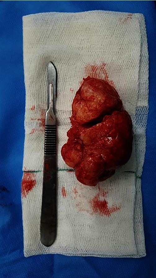
This clinical photograph displays a gross specimen of an enucleated lesion, likely a central ossifying fibroma, following surgical resection from the mandible. The specimen is a large, solid mass with a reddish-brown, hemorrhagic appearance and a multinodular, lobulated surface. It exhibits an irregular shape with areas of indentation and a moist, glistening texture. The lesion is placed on a sterile white surgical gauze pad, which shows minor blood staining. For scale and context, a stainless steel surgical scalpel with a curved blade and ribbed handle is positioned parallel to the specimen. This image illustrates the macroscopic features of a benign but expansive odontogenic tumor after successful enucleation and curettage, common in oral and maxillofacial surgery. The educational focus is on the gross morphology of resected jaw tumors and the clinical results of conservative surgical management for benign osseous lesions.
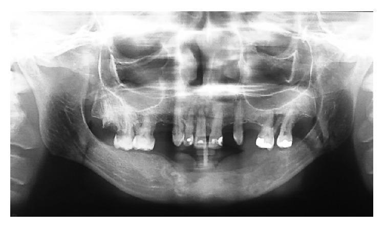
This diagnostic panoramic radiograph (orthopantomogram) displays a postoperative view of the mandible following the treatment of a central ossifying fibroma (COF). The image demonstrates a large, well-circumscribed lesion involving the anterior and left posterior mandible, extending from the right lateral incisor (tooth 41) to the left second molar region. The lesion exhibits a mixed radiopaque-radiolucent internal structure, characteristic of fibro-osseous lesions where mineralized tissue replaces normal bone. Key dental findings include evident radicular resorption of tooth 41 and the absence of several teeth in the affected mandibular quadrants. The radiograph, taken 12 months post-enucleation and curettage, shows the surgical site with no immediate signs of aggressive recurrence. The maxillary structures, including the maxillary sinuses and nasal cavity, appear within normal limits. This image serves as an educational example of the radiographic presentation and long-term follow-up of benign but expansive odontogenic tumors in oral and maxillofacial surgery.
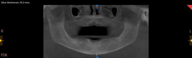
This diagnostic image is a reconstructed panoramic view from a postoperative cone-beam computed tomography (CBCT) scan, with a displayed slice thickness of 15.3 mm. The image provides a comprehensive view of the maxilla and mandible, focusing on the alveolar bone and remaining dentition following surgical intervention for an ossifying fibroma. Key findings include generalized bone loss and a prominent surgical void in the anterior maxilla where the lesion was excised. The trabecular bone throughout the jaw appears more coarse than normal, and the cortical plates show marginal thickening, which are characteristic radiographic features often associated with fibro-osseous conditions or Paget's disease. Multiple teeth are absent consistent with the postoperative state of debridement and extractions. The image is used in dental and oral surgery education to demonstrate postoperative healing, bone density changes in fibro-osseous lesions, and the use of CBCT reconstruction for monitoring surgical outcomes in the maxillofacial region.
fibrous dysplasia jaw ground glass radiology
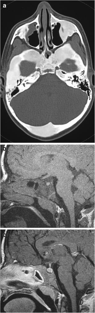
This composite educational image features three diagnostic views demonstrating polyostotic fibrous dysplasia in a pediatric patient with McCune-Albright syndrome. (a) Axial non-contrast CT scan of the skull showing classic 'ground glass' matrix within multiple expansile bony lesions, particularly involving the sphenoid and temporal bones, where normal marrow is replaced by fibro-osseous tissue. (b) Pre-contrast sagittal T1-weighted MRI illustrating significant expansion of the clivus with characteristic diffuse low signal intensity. (c) Post-contrast, fat-suppressed sagittal T1-weighted MRI demonstrating homogeneous, diffuse enhancement within the expanded clival lesion, indicating increased vascularity. The study highlights the imaging hallmarks used to identify fibrous dysplasia of the skull base, including skeletal expansion, hazy density on CT, and variable MRI signal patterns. This material is suitable for neuro-radiology education, focusing on craniofacial manifestations of systemic genetic disorders and the differentiation of fibro-osseous lesions.
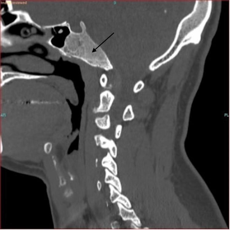
This diagnostic image is a non-contrast Computed Tomography (CT) scan of the skull base and cervical spine in a sagittal view. The focus is on a prominent, expansile lesion within the clivus, indicated by a black arrow. The lesion demonstrates a characteristic 'ground-glass' matrix, which is a hallmark radiologic feature of fibrous dysplasia. The mass measures approximately 3.8 cm (AP) by 2.2 cm (craniocaudal) and shows thinning of the overlying cortical bone without evidence of aggressive periosteal reaction or frank destruction. Anatomical landmarks include the sphenoid sinus anterior to the lesion and the cervical vertebrae (C1-C7) posteriorly. The clinical significance of this finding is the identification of benign fibrous dysplasia of the skull base, differentiating it from more aggressive clival pathologies such as chordoma or metastatic disease. The image is intended for medical education in radiology and neurosurgery, focusing on bone pathology and diagnostic imaging of the skull base.
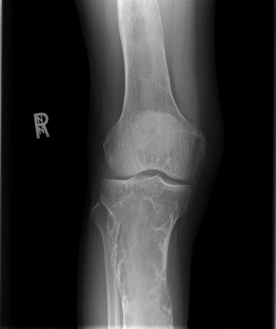
AP knee radiograph demonstrates features of polyostotic fibrous dysplasia in the distal femur and proximal tibia with characteristic ground-glass osteopathy. The lesions show diffuse sclerosis with soap-bubble trabeculation, endosteal scalloping, cortical thinning, and mild to moderate osseous expansion. The epiphyses are spared, and there is no periosteal reaction. These radiographic traits—intralesional lucencies and heterogeneous sclerosis within the medullary cavity—reflect abnormal fibrous tissue replacing normal cancellous bone. The imaging pattern matches Mazabraud syndrome when coupled with MRI-detected intramuscular myxomas in the vastus medialis near affected bones, pointing to a polyostotic FD with soft-tissue tumors. The diagnostic significance lies in distinguishing fibrous dysplasia from malignant bone processes and guiding management toward conservative bone-targeted therapies and orthopedic monitoring. Differential diagnoses include benign fibrous lesions and less likely entities such as osteosarcoma or Langerhans cell histiocytosis. Clinically, these findings explain hip and knee pain; radiographs serve as baseline for surveillance and MRI correlation for soft-tissue lesions. This case reiterates the Mazabraud spectrum: fibrous dysplasia with intramuscular myxomas, requiring multidisciplinary radiologic-pathologic correlation and longitudinal follow-up. Recognition of this pattern supports targeted MRI assessment of soft tissues and guides surveillance imaging intervals, biopsy decisions, and multidisciplinary planning for orthopedic oncology and radiology teams.
Surgical Management of Fibro-Osseous Lesions of the Jaw
Introduction
- Ossifying fibroma (OF) / Cemento-ossifying fibroma (COF)
- Juvenile / Aggressive ossifying fibroma (juvenile trabecular OF; psammomatoid OF)
- Fibrous dysplasia (FD)
- Periapical cemental dysplasia / Florid cemento-osseous dysplasia
1. Classification Relevant to Surgery
- Ossifying fibroma of odontogenic origin - affects exclusively tooth-bearing bone of the mandible (and rarely maxilla)
- Juvenile trabecular ossifying fibroma - mostly in the alveolar process of the maxilla in children
- Juvenile psammomatoid ossifying fibroma - predominantly fronto-orbital and ethmoid bones, characterized by psammoma body-like calcifications
- Scott-Brown's Otorhinolaryngology Head & Neck Surgery, Vol. 1
2. General Principles of Surgical Management
- Relieve symptoms (pain, swelling, airway compromise, nerve compression, cosmetic deformity)
- Prevent recurrence
- Preserve function and aesthetics
3. Surgical Management of Ossifying Fibroma (OF) / Cemento-Ossifying Fibroma (COF)
Key Surgical Feature: Shell-Out Principle
Procedure: Enucleation and Curettage
- Indication: Small-to-moderate sized, well-defined lesions in the mandible or maxilla
- Technique:
- Mucoperiosteal flap elevation to expose the overlying cortex
- Cortical window created (if not already thinned/perforated by the lesion)
- The lesion is enucleated in toto using periosteal elevators and curettes, exploiting the fibrous capsule
- Thorough curettage of the bony cavity walls
- Peripheral ostectomy of the cavity edges may be performed to reduce recurrence risk
- Primary closure of the mucoperiosteal flap
- Advantage: Conservative, preserves adjacent teeth and bone
- Recurrence rate: Generally low when complete enucleation is achieved
Resection for Large or Recurrent Lesions
- Large lesions causing significant bony destruction, cortical perforation, or root resorption may require marginal or segmental resection
- Segmental resection is followed by reconstruction with:
- Autogenous bone grafts (iliac crest, rib)
- Titanium reconstruction plates
- Distraction osteogenesis in select cases
- Immediate or delayed bone grafting restores mandibular continuity and function
Radiologic Follow-Up
4. Surgical Management of Juvenile / Aggressive Ossifying Fibroma
-
Surgical approach: More aggressive surgical excision than simple enucleation
-
Complete excision with wider margins (peripheral ostectomy or marginal resection) is recommended
-
Some lesions recur even after wide excision, necessitating repeat surgery
-
Despite local aggression, these lesions do not metastasize and most ultimately respond to conservative-to-moderate surgery
-
Close long-term follow-up (clinical + radiologic) is mandatory
-
Scott-Brown's Otorhinolaryngology Head & Neck Surgery, Vol. 1; Cummings Otolaryngology
5. Surgical Management of Fibrous Dysplasia
- It has no capsule (does not shell out)
- It is a genetic developmental anomaly, not a neoplasm
- Bone in FD transitions imperceptibly into adjacent normal bone
- The monostotic form tends to stabilize after puberty; polyostotic form may progress
Conservative Approach (First-Line)
- Asymptomatic patients with monostotic FD: observation and surveillance with serial clinical and radiologic review
- Medical therapy: Bisphosphonates (pamidronate, zoledronate) reduce bone pain, decrease the incidence of pathologic fractures, and slow disease progression. They do not eliminate the lesion but stabilize it.
Surgical Indications for Fibrous Dysplasia
- Significant cosmetic deformity (facial asymmetry, proptosis, malocclusion)
- Functional impairment (optic nerve compression, nasal obstruction, sinusitis)
- Progressive pain or pathologic fracture
- Suspected malignant transformation (rapid enlargement, severe pain)
Surgical Techniques
- Recontouring / Contouring osteoplasty: The primary surgical technique for FD of the jaw and craniofacial skeleton. Abnormal bone is shaved or contoured to restore facial symmetry and relieve compression. Bone is not fully resected (it blends into surrounding normal bone without a capsule).
- Timing: Ideally deferred until adolescence or after puberty, when the monostotic lesion stabilizes. However, functional compromise (optic nerve compression) demands urgent intervention regardless of age.
- Endoscopic sinus surgery: For paranasal sinus / orbital involvement, endoscopic approaches (with computer-assisted navigation where available) can improve sinus drainage and relieve orbital pressure.
- Combined craniofacial approach: For large lesions involving the orbital region, anterior skull base, or complex craniofacial bones.
- Resection and reconstruction: Radical resection is rarely performed for FD alone. It may be required if malignant transformation to osteosarcoma occurs (estimated rate ~0.4-0.5%).
Contraindication: Radiotherapy
- Scott-Brown's Otorhinolaryngology Head & Neck Surgery, Vols. 1 & 2; Cummings Otolaryngology
6. Management of Cemento-Osseous Dysplasias
- Periapical cemental dysplasia (periapical osseous dysplasia): Typically requires no surgical treatment. Teeth are vital; the lesion is self-limiting. Biopsy only if diagnosis uncertain.
- Florid cemento-osseous dysplasia: Generally observed. Surgery (debridement, sequestrectomy) only if secondary osteomyelitis develops, as operative intervention risks infective complications given the relatively avascular mature lesion.
7. Key Surgical Distinctions: OF vs. FD
| Feature | Ossifying Fibroma | Fibrous Dysplasia |
|---|---|---|
| Capsule | Present | Absent |
| Surgery of choice | Enucleation + curettage | Recontouring / contouring |
| Recurrence risk | Low (moderate for juvenile type) | Can regrow before puberty |
| Radiotherapy | Not indicated | Absolutely contraindicated |
| Malignant transformation | Rare | ~0.4-0.5% (to osteosarcoma) |
| Timing of surgery | When symptomatic | Ideally post-puberty (unless urgent) |
8. Complications of Surgery
- Damage to the inferior alveolar nerve (numbness/paresthesia of lip and chin)
- Damage to roots of adjacent teeth
- Recurrence (especially juvenile OF and FD before puberty)
- Infection / wound dehiscence
- Pathologic fracture of a weakened mandible if extensive curettage performed
- Malignant transformation (rare, not caused by surgery but must be monitored)
Summary
25 mark answer of fibrosseous lesions of jaw from neelima malik
Neelima Malik fibro-osseous lesions jaw classification fibrous dysplasia ossifying fibroma textbook oral maxillofacial surgery
https://www.oyschst.edu.ng/elib/dashboard/ebooks/7MiPkWHo.pd…
Neelima Malik fibrous dysplasia ossifying fibroma cemento-osseous dysplasia cherubism jaw clinical radiological histopathology treatment chapter 36
https://codental.uobaghdad.edu.iq/wp-content/uploads/sites/1…
fibrous dysplasia jaw bone histology woven bone Chinese letters pattern
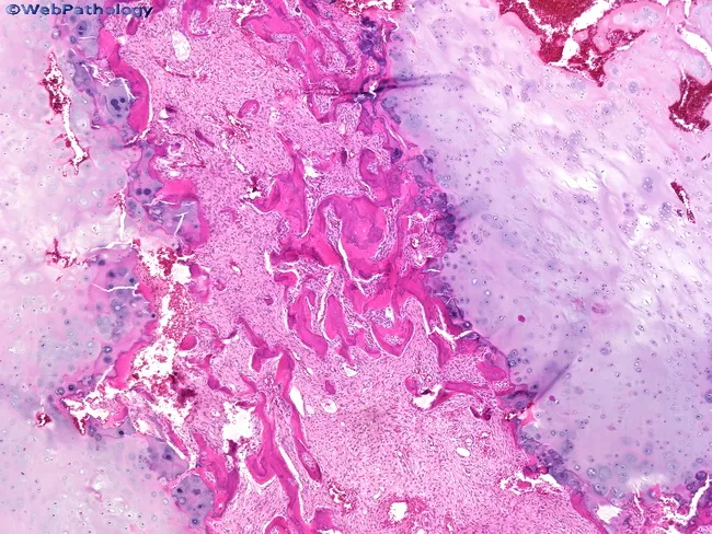
Histology of fibrous dysplasia. This hematoxylin and eosin stained bone biopsy section reveals a fibroblastic stroma populated by spindle-shaped cells and irregular, immature woven bone trabeculae. The trabeculae are variably sized, often curved or irregular in shape, resembling a Chinese character pattern, and they lack a consistent osteoblastic rim. The surrounding stroma is fibrous and moderately cellular, with a bland appearance and sparse mitotic activity. In this case, nodules of hyaline cartilage are present on either side of the osseous component, illustrating chondroid differentiation within the lesion. The chondroid component can be strikingly prominent in some fibrous dysplasia cases, contributing to a mixed fibro-osseous phenotype. Overall, the combination of woven bone within fibrous stroma and cartilaginous nodules supports a benign fibrous-osseous process rather than a malignant tumor; however, the cartilaginous elements may raise differential considerations such as chondromatous differentiation or low-grade chondrosarcoma if atypia were evident. Clinically, these features correspond to a slow-growing, benign lesion of bone that may cause deformity or structural weakness depending on location. This histologic pattern is classic for fibrous dysplasia and highlights heterogeneity within the lesion, including possible cartilage-rich variants and variable radiographic correlation. These features aid surgical planning and prognosis.
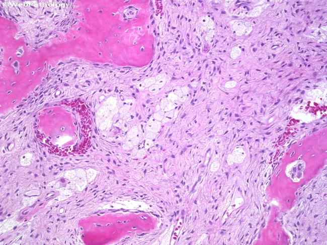
This histopathology image depicts a bone lesion analyzed by light microscopy after formalin fixation and hematoxylin and eosin staining. The cortical and trabecular bone shows irregular, curvilinear woven bone spicules embedded in a dense, moderately cellular fibroblastic stroma. The bony trabeculae lack osteoblastic rimming, a hallmark feature distinguishing fibrous dysplasia from normal lamellar bone and from reactive osseous processes. The surrounding stroma contains spindle-shaped fibroblasts with bland nuclei, and within the intertrabecular spaces there are clusters of lipid-laden macrophages (foam cells) and admixed adipocytes, consistent with marrow replacement by fibrous tissue with fatty elements. The overall pattern may resemble a Chinese-letter configuration in some fields but is variable; there is no significant atypia or mitotic activity. These features collectively support a benign fibro-osseous lesion with fibrous stroma and irregular woven bone, rather than a malignant sarcoma. Clinically, this histology is compatible with fibrous dysplasia of bone, often associated with adolescence or young adulthood and presenting as painless swelling or deformity; radiographs commonly show a ground-glass appearance with preserved/expanded cortex. Diagnostic significance lies in recognizing the combination of immature woven bone in fibrous stroma with absent osteoblastic rimming, which differentiates fibrous dysplasia from ossifying fibroma and low-grade osteosarcoma. Potential use includes guiding surgical planning, confirming fibro-osseous pathology, and correlation for diagnosis.
ossifying fibroma jaw panoramic radiograph radiolucent radiopaque mixed lesion mandible
This diagnostic panoramic radiograph (orthopantomogram) displays a postoperative view of the mandible following the treatment of a central ossifying fibroma (COF). The image demonstrates a large, well-circumscribed lesion involving the anterior and left posterior mandible, extending from the right lateral incisor (tooth 41) to the left second molar region. The lesion exhibits a mixed radiopaque-radiolucent internal structure, characteristic of fibro-osseous lesions where mineralized tissue replaces normal bone. Key dental findings include evident radicular resorption of tooth 41 and the absence of several teeth in the affected mandibular quadrants. The radiograph, taken 12 months post-enucleation and curettage, shows the surgical site with no immediate signs of aggressive recurrence. The maxillary structures, including the maxillary sinuses and nasal cavity, appear within normal limits. This image serves as an educational example of the radiographic presentation and long-term follow-up of benign but expansive odontogenic tumors in oral and maxillofacial surgery.
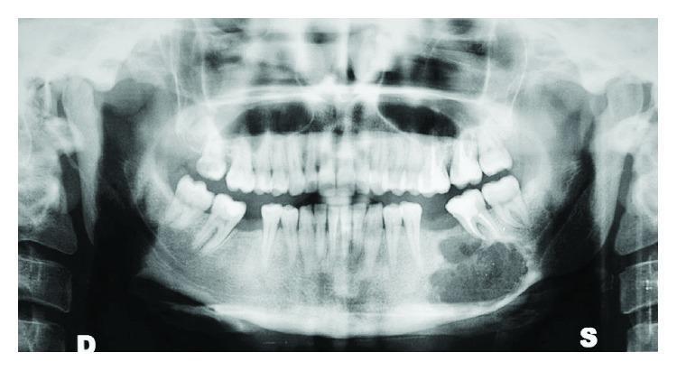
This diagnostic image is a panoramic radiograph (orthopantomogram) of the maxillofacial region. The primary finding is a large, well-circumscribed, mixed radiolucent-radiopaque lesion located in the left mandibular body, specifically extending from the premolar region to the molar region. The lesion exhibits characteristic features of an ossifying fibroma, including a well-defined sclerotic border and internal 'ground-glass' or granular radiopacity. There is significant downward expansion and thinning of the inferior border of the mandible. The lesion is in close proximity to the roots of the mandibular left second premolar and first and second molars, causing apparent root displacement and impingement on the alveolar bone. The rest of the dentition is visible, alongside typical anatomical landmarks including the maxillary sinuses, nasal cavity, and bilateral temporomandibular joints. This image is used in dental and maxillofacial surgery education to illustrate the radiographic presentation of benign fibro-osseous lesions and their impact on surrounding mandibular architecture.
cherubism jaw bilateral mandibular swelling radiograph multilocular radiolucency
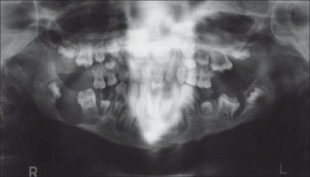
**Imaging Modality:** Panoramic radiograph (Orthopantomogram/OPG). **Anatomical Region:** Maxillofacial region including the mandible, maxilla, and temporomandibular joints. **Observed Pathology:** Large, well-defined multilocular radiolucent cystic lesions are visible bilaterally in the posterior mandible, extending from the molar regions into the ascending rami. These lesions demonstrate significant cortical thinning and medullary bone destruction. **Key Visual Features:** * **Mandibular Condyles:** Bilateral hypoplastic condyles characterized by reduced size and altered morphology. * **Dentition:** Mixed dentition is present with evidence of multiple impacted, displaced, and malpositioned permanent teeth secondary to the expansive cystic lesions. * **Bony Changes:** Extensive radiolucency indicates osteolytic activity, displacing the mandibular canal and compromising the structural integrity of the mandibular body and rami. **Differentiating Features:** The combination of bilateral multilocular radiolucencies in a pediatric/adolescent patient, associated with tooth displacement and condylar hypoplasia, is highly suggestive of systemic or genetic bone pathologies such as cherubism or Gorlin-Goltz syndrome. The symmetry and posterior mandibular involvement are hallmark diagnostic cues.
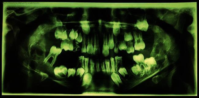
This diagnostic image is a panoramic radiograph (orthopantomogram) of a pediatric patient, demonstrating significant bilateral pathology within the mandible. The radiograph reveals extensive, well-defined, multilocular radiolucent lesions situated bilaterally. These ‘soap-bubble’ or ‘honeycomb’ appearing lesions originate in the primary molar regions and extend posteriorly to involve the entirety of the ascending rami. Notable structural changes include the marked expansion of the mandibular cortex, specifically along the inferior border and the alveolar crest. The lesions appear to displace several developing permanent tooth germs, which is characteristic of expansile jaw lesions in children, such as cherubism or giant cell granulomas. The maxillary structures and dentition are also visible, though the primary pathology is localized to the lower jaw. This visual is highly educational for dental and maxillofacial pathology, illustrating classic radiographic signs of expansile multilocular mandibular lesions and their impact on surrounding anatomical structures and dental development.
cemento-osseous dysplasia periapical florid jaw radiograph radiopaque
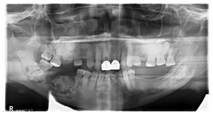
A panoramic dental radiograph (orthopantomogram) demonstrating extensive, multifocal pathology across all four quadrants of the jaws, characteristic of Florid Cemento-Osseous Dysplasia (FLCOD). The image shows a disorganized bone architecture with a classic mixed radiolucent-radiopaque appearance. Dense, sclerotic, cotton-wool-like radiopaque masses are predominantly located in the periapical regions of the mandible and maxilla. These lesions are often bordered by thin radiolucent rims. In the right posterior mandible, a particularly large, mottled, and dense calcified mass is visible. The patient exhibits several missing teeth, dental restorations (including radio-dense crowns on the maxillary central incisors), and impacted or displaced molars. There is evidence of significant alveolar bone remodeling and potential periapical involvement secondary to the dysplastic process. This diagnostic image serves as a clinical example of a benign fibro-osseous lesion commonly observed in middle-aged women of African descent.
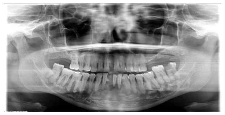
This orthopantomogram (panoramic radiograph) demonstrates a classic case of Florid Cemento-Osseous Dysplasia (FCOD) in a 48-year-old patient. The primary pathology consists of multiple, diffuse, and bilateral mixed radiolucent-radiopaque lesions involving both the right and left quadrants of the mandible. These lesions exhibit a varying 'cotton-wool' appearance with ill-defined borders, consisting of confluent dense sclerotic masses surrounded by thin radiolucent rims. The lesions are primarily located in the periapical and alveolar bone regions of the mandible. Key dental features include the absence of a visible lamina dura in affected areas, while the periodontal ligament spaces remain largely intact. Some teeth show evidence of restorative work and mild periodontal bone loss. The widespread, multifocal distribution across multiple quadrants without significant cortical expansion or tooth displacement is a hallmark of this fibro-osseous condition, common in middle-aged women of African descent. This diagnostic image serves as a high-level educational example for oral and maxillofacial pathology and dental radiology.
Fibro-Osseous Lesions of the Jaw
As per Prof. Dr. Neelima Anil Malik — Textbook of Oral and Maxillofacial Surgery (Chapter 36)
Definition
This terminology describes only a process, not a specific diagnosis. The histopathological picture of these lesions may be remarkably similar. A final diagnosis depends on clinical, radiographic, operative, and microscopic features considered together.
Classification
Neelima Malik's Classification (Table 36.2 / 36.3)
- Fibrous dysplasia of bone
- Cherubism (Inherited fibro-osseous bone disease)
- Ossifying fibroma
- Central giant cell granuloma
WHO Classification (Kramer, Pindborg, Shear 1992 - Table 36.4):
- Cemento-ossifying fibroma
- Fibrous dysplasia of the jaws
- Cemento-osseous dysplasias:
- a. Periapical cemento-osseous dysplasia
- b. Focal cemento-osseous dysplasia
- c. Florid cemento-osseous dysplasia
Common Fibro-osseous Lesions of the Jaws (Neelima Malik):
- Fibrous dysplasia of the jaws
- Cemento-osseous dysplasia (Periapical / Focal / Florid)
- Ossifying fibroma (including juvenile aggressive type)
- Cherubism
1. FIBROUS DYSPLASIA OF THE JAWS
Etiopathogenesis
- First described by von Recklinghausen (1891); term "fibrous dysplasia" introduced by Lichtenstein (1938)
- A developmental, tumor-like condition characterized by replacement of normal bone by proliferating cellular fibrous connective tissue with irregular bony trabeculae
- Results from a postzygotic activating mutation in the GNAS gene (chromosome 20q13), encoding the alpha subunit of stimulatory G-protein (Gs-alpha)
- Elevated cAMP levels affect transcription and expression of multiple downstream genes, resulting in the pathologic lesion
- Mutation has NOT been detected in ossifying fibroma or cemento-osseous dysplasia - distinguishing FD at a molecular level
Types
| Type | Description |
|---|---|
| Monostotic FD | Single bone involved; most common; often stabilizes at puberty |
| Polyostotic FD | Multiple bones; skull involved in >50% |
| Jaffe-Lichtenstein Syndrome | Polyostotic FD + cafe-au-lait spots (Coast of Maine irregular border) |
| McCune-Albright Syndrome | Polyostotic FD + cafe-au-lait spots + endocrinopathies (precocious puberty in girls, hyperthyroidism, acromegaly) |
| Craniofacial FD | Multiple craniofacial bone involvement; classified as monostotic |
Clinical Features
- Predominantly affects children and adolescents (first two decades of life)
- Painless, slowly progressive facial swelling and asymmetry
- Maxilla more commonly involved than mandible in FD (unlike ossifying fibroma)
- Lesion blends imperceptibly into surrounding normal bone (no capsule)
- Leontiasis ossea - lion-like facial deformity in severe maxillary FD
- Malocclusion, displacement of teeth, nasal obstruction
- In polyostotic form: sarcomatous transformation estimated at 0.4-0.5% (usually to osteosarcoma)
- Raised serum alkaline phosphatase in ~30% of polyostotic cases; serum calcium and phosphorus normal
Radiological Features (Figs 36.43A-C in Neelima Malik)
- Ground-glass (orange peel) appearance - most characteristic radiographic sign
- Lesion shows ill-defined borders; merges with surrounding bone without a capsule
- Three radiographic patterns depending on degree of mineralization:
- Pagetoid pattern - alternating radiolucent and radiopaque areas
- Sclerotic pattern - uniformly radiopaque
- Cystic pattern - radiolucent with fine granular ground-glass background
- Expansion of cortical plates without perforation (cortex maintained)
- In early stages: radiolucent; as mineralization increases: ground-glass to sclerotic appearance
Histopathological Features
- Replacement of normal cancellous bone by fibrous stroma arranged in a whorled pattern
- Irregular spicules of woven bone (trabeculae) arranged in a characteristic "Chinese letters" or "alphabet soup" pattern
- Absence of osteoblastic rimming around bone trabeculae - key distinguishing feature from ossifying fibroma
- Stromal fibroblasts are the predominant cells
Treatment
- Asymptomatic monostotic FD: Observation / surveillance; may stabilize at puberty
- Medical: Bisphosphonates (pamidronate/zoledronate) reduce bone pain, decrease fracture incidence, and slow progression
- Surgical indications: Cosmetic deformity, functional impairment (optic nerve compression, nasal obstruction, malocclusion), pathologic fracture, suspected malignant transformation
- Surgery of choice: Contouring / recontouring osteoplasty - bone is shaved to restore form and relieve compression; NOT simple enucleation (no capsule to exploit)
- Timing: ideally deferred until after puberty when lesion stabilizes; urgent if optic nerve or airway is compromised
- Radiotherapy is absolutely contraindicated - markedly increases risk of malignant transformation to osteosarcoma
- Long-term follow-up essential due to risk of malignant change
2. OSSIFYING FIBROMA (OF) / CEMENTO-OSSIFYING FIBROMA (COF)
Definition & Nature
- Ossifying fibroma (conventional / odontogenic origin)
- Cemento-ossifying fibroma (COF) - cementum-like material present
- Juvenile aggressive (active) ossifying fibroma - two variants:
- Juvenile trabecular ossifying fibroma (JTOF)
- Juvenile psammomatoid ossifying fibroma (JPOF)
Clinical Features
- Most common between ages 20-40 years; female predominance (F:M = 5:1)
- Mandible more commonly affected (premolar/molar region, 75% of cases)
- Slowly growing, painless, expansile swelling
- Expansion of buccal and lingual cortical plates; cortical perforation rare
- Displacement of adjacent teeth; root resorption rare; teeth remain vital
- Juvenile aggressive type: occurs in children and adolescents, predominantly affects maxilla; tends to recur
Radiological Features (Figs 36.44A and B in Neelima Malik)
- Well-circumscribed, well-defined unilocular lesion
- Initially radiolucent (fibrous stroma predominates)
- Progressive central calcification: evolves to mixed radiolucent-radiopaque
- Mature lesion: predominantly radiopaque with a narrow radiolucent rim (capsule)
- Roots of related teeth may be displaced
- A distinct sclerotic (eggshell) rim differentiates OF from fibrous dysplasia
Histopathological Features
- Fibrous capsule present (or well demarcated from surrounding bone)
- Collagenous stroma with uniform spindle or stellate cells
- Varying amounts of calcified tissue resembling bone, cementum, or both
- Two types of calcifications:
- Trabeculae of osteoid and bone
- Basophilic ovoid calcifications resembling cementum (cementicles)
- FOCAL PRESENCE OF OSTEOBLASTS lining bone deposits - key histological distinction from fibrous dysplasia
- As lesion matures: calcifications fuse into a dense mass with peripheral fibrous zone - this allows the lesion to shell out in one piece or several large pieces
Treatment
- Standard treatment: Enucleation and curettage - the fibrous capsule allows clean separation from surrounding bone
- Technique:
- Mucoperiosteal flap elevation
- Cortical window creation
- Enucleation exploiting the capsular plane
- Thorough curettage of the cavity walls
- Peripheral ostectomy to reduce recurrence
- Primary closure
- Large/recurrent lesions: Marginal or segmental resection, followed by reconstruction (autogenous bone graft / titanium plate)
- Juvenile aggressive OF: Wider excision margins; higher recurrence rate (30-58%); adjuvant interferon-alpha therapy for 1 year considered for rapidly growing lesions
- Prognosis: generally good; malignant transformation not documented
3. CEMENTO-OSSEOUS DYSPLASIAS (COD)
Subtypes
| Subtype | Location | Demographics |
|---|---|---|
| Periapical COD | Anterior mandible, around tooth root apices | Black women, middle age; most common |
| Focal COD | Posterior mandible, single quadrant | Similar demographics |
| Florid COD | Diffuse, bilateral, multiple quadrants | Middle-aged women; more common in African Americans |
Clinical Features
- Strong female predominance; more common in African Americans and Asians
- Usually asymptomatic - discovered incidentally on routine radiographs
- Teeth in affected area are vital (distinguishes from periapical pathology)
- Florid COD: predisposes to secondary osteomyelitis if exposed (especially after extractions in edentulous areas)
- No significant facial swelling or cortical expansion
Radiological Features
- Progresses through three stages:
- Osteolytic stage: Periapical radiolucency (may mimic periapical abscess - but teeth are vital)
- Mixed stage: Mixed radiolucent-radiopaque lesion
- Mature/radiopaque stage: Predominantly opaque lesion with a thin radiolucent rim
- Florid COD: multiple bilateral cotton-wool or cobblestone radiopaque masses throughout jaws
Histopathological Features
- Similar across all three subtypes
- Cellular fibrovascular connective tissue with scattered hemorrhage
- Variable mixture of: woven bone, lamellar bone, and cementum-like particles
- As lesions mature: ratio of fibrous tissue to mineralized material decreases
- Bony trabeculae become thick and curvilinear, resembling ginger root shapes
- No osteoblastic rimming in most areas (similar to FD - hence diagnostic challenge)
Treatment
- Generally NO surgical treatment needed for asymptomatic lesions
- Biopsy only when diagnosis is uncertain (to exclude malignancy)
- Avoid extractions in involved areas if possible (risk of osteomyelitis in mature radiopaque stage)
- If secondary osteomyelitis develops: debridement and sequestrectomy, antibiotics
- Regular clinical and radiological surveillance
4. CHERUBISM (Inherited Fibro-Osseous Bone Disease)
Etiology
- Autosomal dominant trait (mutation in SH3BP2 gene, chromosome 4p16.3)
- Due to variable penetrance: males more commonly affected (males 100%, females 70% penetrance)
- Onset: typically 2-5 years of age
Clinical Features
- Bilateral, painless swelling of the cheeks due to expansion of the posterior mandibular angles (gives the characteristic cherub-like appearance - upward gaze, chubby cheeks)
- Posterior maxillae may also be involved in severe cases
- Upward displacement of orbital floor causes eyes to appear to turn upwards - "eyes uplifted to heaven" appearance
- Interference with speech, mastication, and swallowing in severe cases
- Loosening and displacement of teeth; cervical lymphadenopathy (reactive hyperplasia)
- Rapid growth during childhood; lesions stabilize and may regress spontaneously at puberty
Radiological Features
- Bilateral multilocular radiolucencies (soap-bubble or honeycomb pattern)
- Predominantly affects the posterior mandible bilaterally
- Involvement of mandibular angles, rami, and posterior body
- Displacement and failure of eruption of permanent teeth
- Fine bony septae within the radiolucent lesions
- With age: increasing bony repair and partial fill of radiolucencies
Histopathological Features
- Early lesions: Resemble central giant cell granuloma
- Collections of multinucleated giant cells within cellular, vascular fibrous tissue
- Vessels may show a characteristic hyaline (eosinophilic perivascular) cuff - hallmark of cherubism
- Older lesions: Increasing bony repair; giant cells become fewer; fibrous stroma replaced by maturing bone
Treatment
- Primarily observation - many cases regress spontaneously at puberty
- Surgical treatment (curettage or paring down) is rarely needed; reserved for:
- Severe cosmetic deformity persisting after puberty
- Functional compromise (airway, mastication, vision)
- Failure of spontaneous regression
- Prognosis: variable; usually favorable with spontaneous improvement
Key Differentiating Features
| Feature | Fibrous Dysplasia | Ossifying Fibroma | Cemento-Osseous Dysplasia | Cherubism |
|---|---|---|---|---|
| Nature | Developmental anomaly | True neoplasm | Dysplastic process | Genetic hereditary |
| Gene | GNAS (Gs-alpha) | None identified | None identified | SH3BP2 |
| Age | 1st-2nd decade | 3rd-4th decade | Middle age (4th+) | 2-5 years |
| Sex | M=F | Female ++ | Female ++ (Black) | Males > Females |
| Site | Maxilla > mandible | Mandible ++ (premolar/molar) | Periapical, tooth-bearing | Bilateral posterior mandible |
| Capsule | Absent | Present | Absent | Absent |
| Radiograph | Ground-glass, ill-defined | Well-defined, mixed → opaque | 3 stages (lucent→opaque) | Bilateral multilocular |
| Histology | Woven bone, NO osteoblastic rimming | Osteoblastic rimming present, cementicles | Woven bone + cementum-like, ginger root | Giant cells + perivascular hyaline cuff |
| Surgery | Recontouring | Enucleation + curettage | Usually none | Curettage if severe |
| Recurrence | Can regrow pre-puberty | Low (high in juvenile type) | Low | Spontaneous regression |
| Radiotherapy | CONTRAINDICATED | Not indicated | Not applicable | Not indicated |
| Malignant change | 0.4-0.5% (osteosarcoma) | Not documented | Not documented | Not documented |
Differential Diagnosis
- Central giant cell granuloma (giant cells dominate; no perivascular cuff)
- Paget's disease (cotton-wool appearance; elevated alkaline phosphatase; older patient; hypercementosis)
- Hyperparathyroidism / Brown tumor (elevated PTH, calcium; multiple radiolucencies; reverses on treatment)
- Low-grade osteosarcoma (aggressive, rapid growth; periosteal reaction; malignant histology)
- Periapical abscess (tooth non-vital; response to vitality test distinguishes from COD)