
Brain stem anatomy
Reading File
Now let me search the medical library for brainstem anatomy content in parallel.
Finding Sources
Finding Sources
Good. Now let me search for more specific brainstem anatomy sections and also pull the main anatomy-focused content:
Finding Sources
Finding Sources
Reading File
Reading File
Finding Sources
Excellent - "Neuroanatomy through Clinical Cases" is a dedicated neuroanatomy textbook. Let me read it:
Reading File
Finding Sources
This is exactly what I need. Let me read the key sections - Main Components, Brainstem Sections, the Summary Table, Surface Features, and also grab the chapter intro images:
Reading File
Reading File
Excellent content. Let me now read the brainstem sections details and the summary table, plus surface features:
Reading File
Reading File
I have rich content. Let me now read the brainstem sections content (midbrain, pons, medulla details) and fetch the key images:
Reading File
Loading Image
Loading Image
Now let me fetch the key cross-section and surface anatomy images:
Loading Image
Loading Image
Loading Image
Excellent - I have outstanding labeled cross-section images. Now let me get the medulla sections and the reticular formation content:
Reading File
Loading Image
I now have comprehensive content from the textbook. Let me also quickly grab the medulla section image and surface anatomy image:
Loading Image
Loading Image
I now have everything needed for a comprehensive, image-rich answer. Let me compose it.
Brainstem Anatomy
Source: Neuroanatomy through Clinical Cases, 3rd Edition (Chapters 12 & 14)
Overview and Position
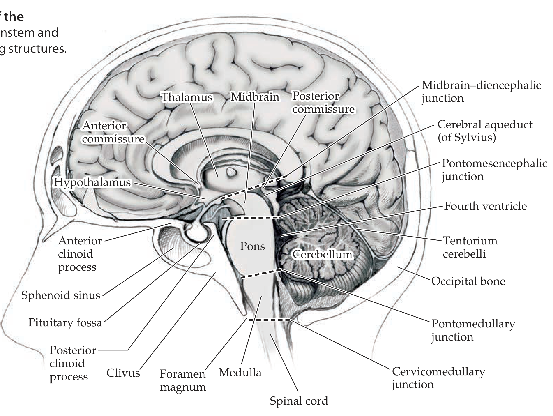
The brainstem is a compact, stalklike structure at the base of the cerebral hemispheres, lying within the posterior fossa of the cranial cavity. It carries nearly all information between the brain and the body. More than a simple conduit, it contains nuclei that control cranial nerves, level of consciousness, cerebellar circuits, muscle tone, posture, and cardiac/respiratory/autonomic functions.
Boundaries:
- Rostral: midbrain-diencephalic junction (meets thalamus/hypothalamus at the level of the tentorium cerebelli)
- Caudal: cervicomedullary junction at the foramen magnum (pyramidal decussation)
- The cerebellum attaches to its dorsal surface
Three divisions (rostral to caudal): Midbrain → Pons → Medulla oblongata
Midsagittal view showing brainstem in situ:
Four Main Functional Groupings
The brainstem can be organized into four functional categories (Table 14.1):
| Grouping | Key Structures |
|---|---|
| 1. Cranial nerve nuclei | Somatic motor (CN III, IV, VI, XII), Branchial motor (CN V, VII, IX-XI), Parasympathetic (Edinger-Westphal, salivatory nuclei, dorsal motor X), Sensory (trigeminal complex, vestibular, cochlear, nucleus solitarius) |
| 2. Long tracts | Corticospinal/corticobulbar tracts; posterior column-medial lemniscal system; anterolateral system; descending autonomic pathways |
| 3. Cerebellar circuitry | Superior/middle/inferior cerebellar peduncles; pontine nuclei; red nucleus; inferior olivary nucleus; central tegmental tract |
| 4. Reticular formation | Arousal systems; cholinergic, noradrenergic (locus ceruleus), serotonergic (raphe), dopaminergic nuclei; pain modulation (PAG); autonomic, respiratory, cardiovascular control |
Three Subdivisions: Key Anatomy
Terminology: Tectum, Tegmentum, Basis
- Tectum ("roof"): dorsal to the cerebral aqueduct in the midbrain; consists of the superior and inferior colliculi
- Tegmentum ("covering"): ventral to the aqueduct in midbrain, and ventral to the 4th ventricle in pons/medulla; the main bulk of brainstem nuclei and reticular formation
- Basis: most ventral portion; contains the large corticospinal and corticobulbar tracts
1. Midbrain
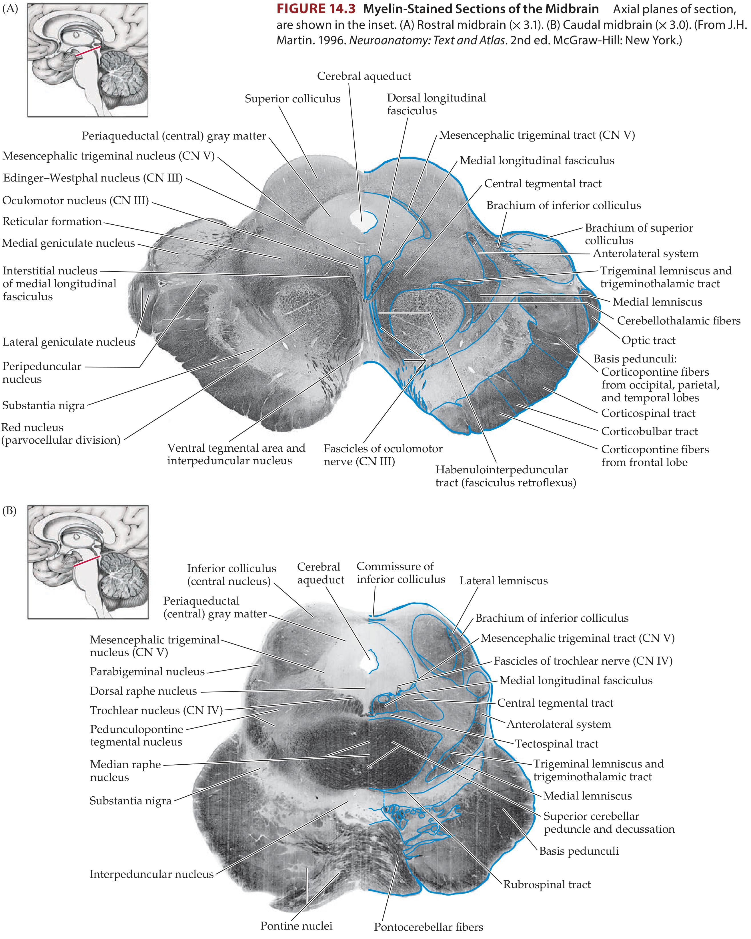
The midbrain contains the cerebral aqueduct (of Sylvius) centrally. Motor nuclei lie more ventrally, sensory nuclei more dorsally (separated by the sulcus limitans).
Key structures:
- Tectum: Superior colliculi (visual reflexes) and inferior colliculi (auditory relay)
- Cerebral peduncles = substantia nigra + basis pedunculi (corticospinal, corticobulbar, and corticopontine fibers)
- Periaqueductal gray (PAG): pain modulation
- Oculomotor nucleus (CN III) and Edinger-Westphal nucleus (parasympathetic to pupil)
- Trochlear nucleus (CN IV) - only CN to exit dorsally
- Red nucleus: receives superior cerebellar peduncle; sends rubrospinal tract and projects via central tegmental tract to inferior olive
- Substantia nigra: dopaminergic; involved in motor control
- Medial longitudinal fasciculus (MLF): coordinates eye movements
- Medial lemniscus and anterolateral system: ascending sensory tracts
Midbrain cross-sections (myelin-stained):
2. Pons
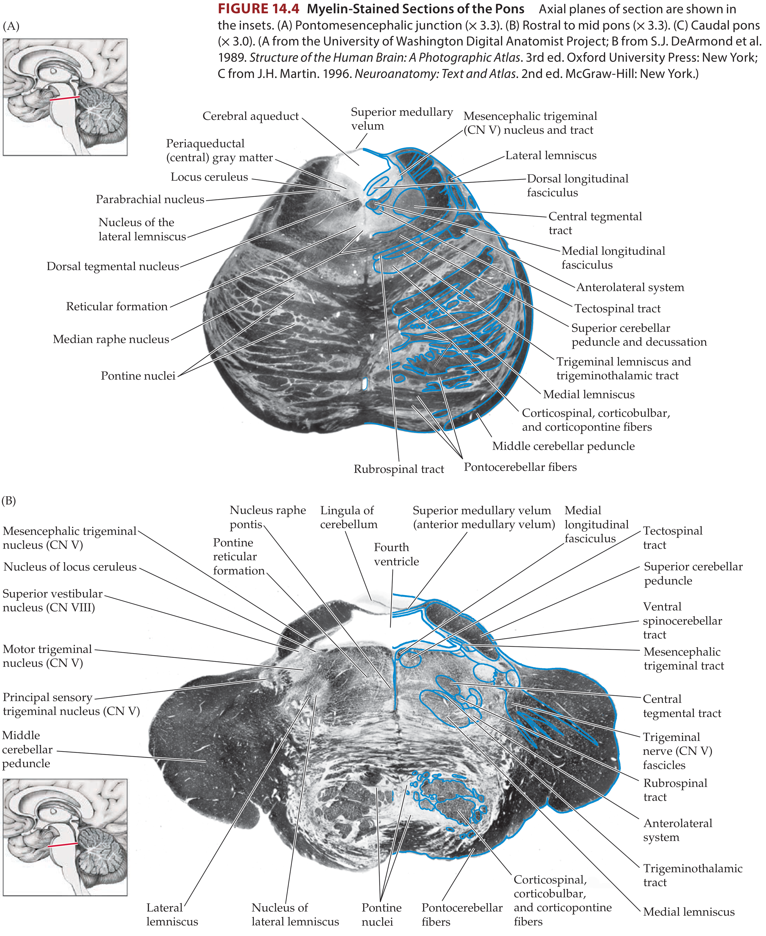
The pons (Latin: "bridge") gets its name from the large middle cerebellar peduncles visible laterally, which bridge to the cerebellum.
Key structures:
- Basis pontis (ventral pons): corticospinal, corticobulbar tracts + pontine nuclei (relay to cerebellum via middle cerebellar peduncle)
- Pontine tegmentum (dorsal): major nuclei and pathways
- Fourth ventricle: separates pontine tegmentum from cerebellum
- Trigeminal nuclear complex (CN V): principal sensory nucleus (discriminative touch, face), motor trigeminal nucleus (mastication), mesencephalic nucleus (proprioception), spinal trigeminal nucleus (pain/temperature)
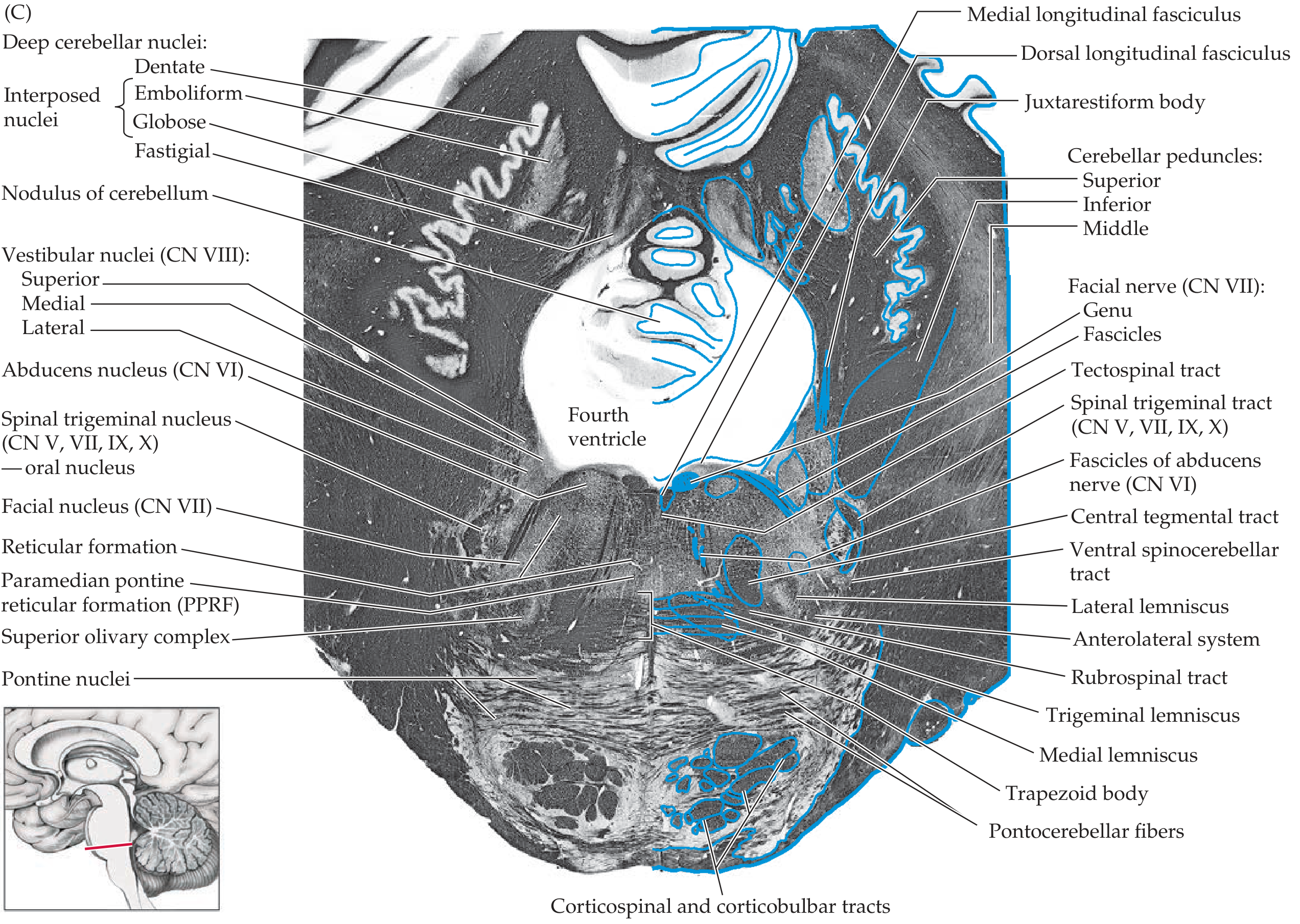
- Abducens nucleus (CN VI): horizontal gaze; the facial nerve (CN VII) loops around it forming the facial colliculus
- Facial nucleus (CN VII)
- Vestibular nuclei (CN VIII): superior, medial, lateral, inferior
- Cochlear nuclei (CN VIII): dorsal and ventral
- Superior olivary complex + trapezoid body + lateral lemniscus: auditory pathway
- Paramedian pontine reticular formation (PPRF): horizontal gaze center
- Locus ceruleus: noradrenergic nucleus; major source of norepinephrine to CNS
- Raphe nuclei: serotonergic; modulate sleep, mood, pain
Pons cross-sections (myelin-stained) at three levels:
3. Medulla Oblongata
Key structures:
- Pyramids (ventral): corticospinal tracts; at the caudal end they decussate (pyramidal decussation), marking the medulla-spinal cord boundary
- Inferior olivary nucleus (rostral medulla): cerebellar circuitry; receives red nucleus input via central tegmental tract; sends climbing fibers to cerebellum via inferior cerebellar peduncle
- Nucleus gracilis and nucleus cuneatus: receive posterior column (fine touch/proprioception) input; their axons cross in the medulla as internal arcuate fibers to form the medial lemniscus
- Nucleus tractus solitarius (NTS): receives visceral (CN VII, IX, X) and gustatory afferents; integrates autonomic and eating-related signals
- Dorsal motor nucleus of CN X: parasympathetic output to thoracoabdominal viscera
- Nucleus ambiguus: motor for CN IX, X, XI (swallowing, phonation)
- Hypoglossal nucleus (CN XII): tongue motor
- Spinal trigeminal nucleus (extends from pons through medulla into upper cervical cord): pain/temperature from face, CN V, VII, IX, X
- Vestibular nuclei (also at ponto-medullary level)
- Area postrema: chemoreceptor trigger zone (lacks normal BBB; mediates vomiting); on the dorsal surface of the caudal medulla
- Inferior cerebellar peduncle: connects medulla to cerebellum
Surface Anatomy and Cranial Nerve Attachments
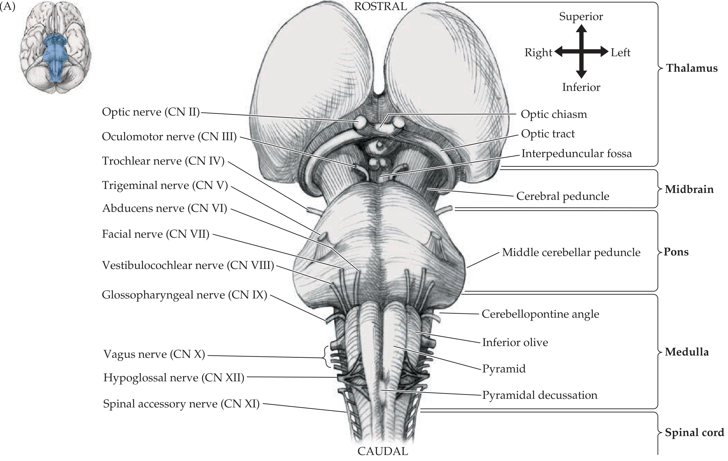
Ventral (anterior) surface:
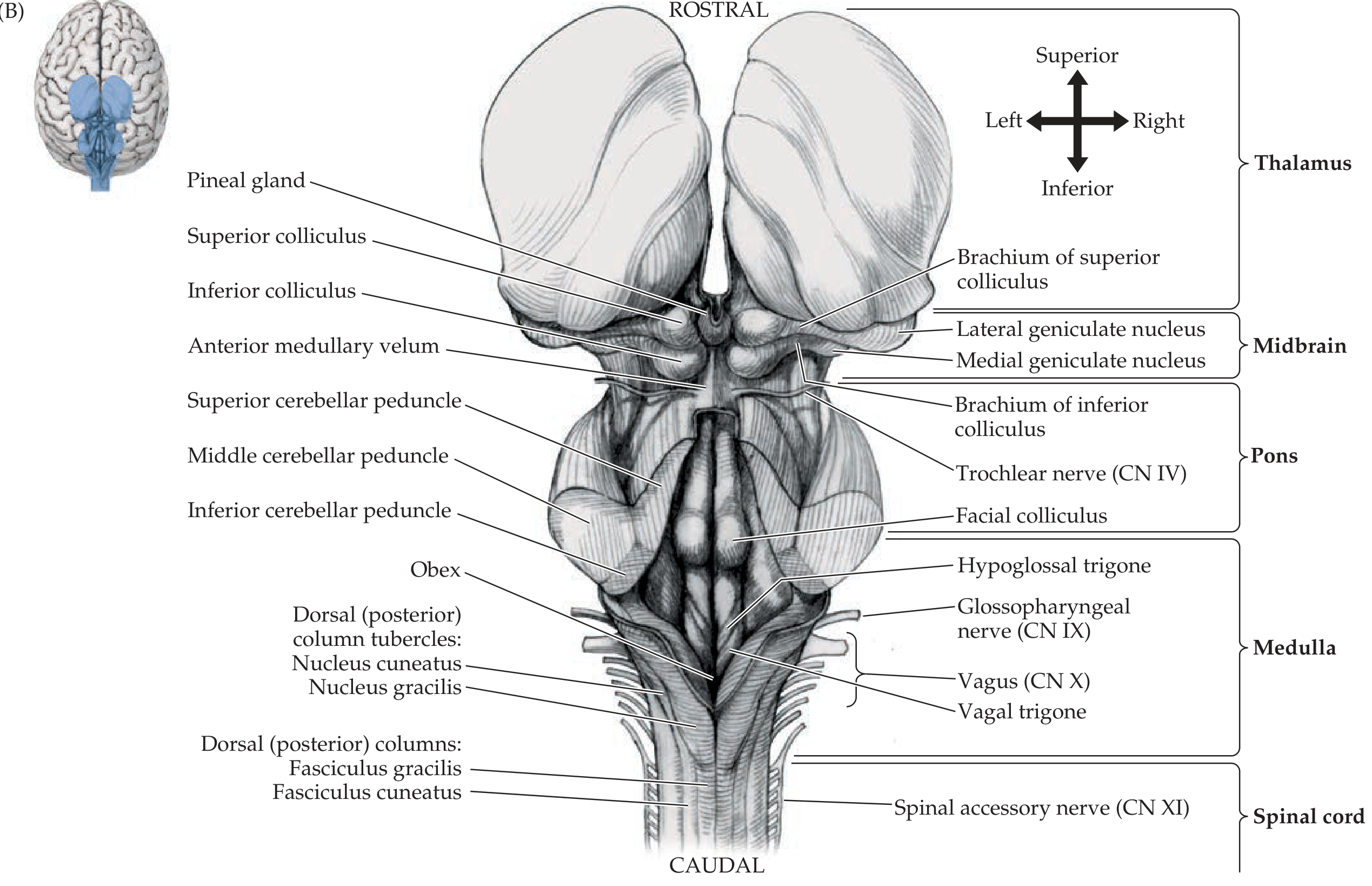
Dorsal (posterior) surface:
Cranial nerve attachments by level:
- Midbrain: CN III (oculomotor, ventral), CN IV (trochlear, dorsal - only CN to exit dorsally)
- Pons: CN V (trigeminal, lateral), CN VI (abducens, pontomedullary junction), CN VII (facial), CN VIII (vestibulocochlear)
- Medulla: CN IX (glossopharyngeal), CN X (vagus), CN XI (spinal accessory), CN XII (hypoglossal)
Reticular Formation
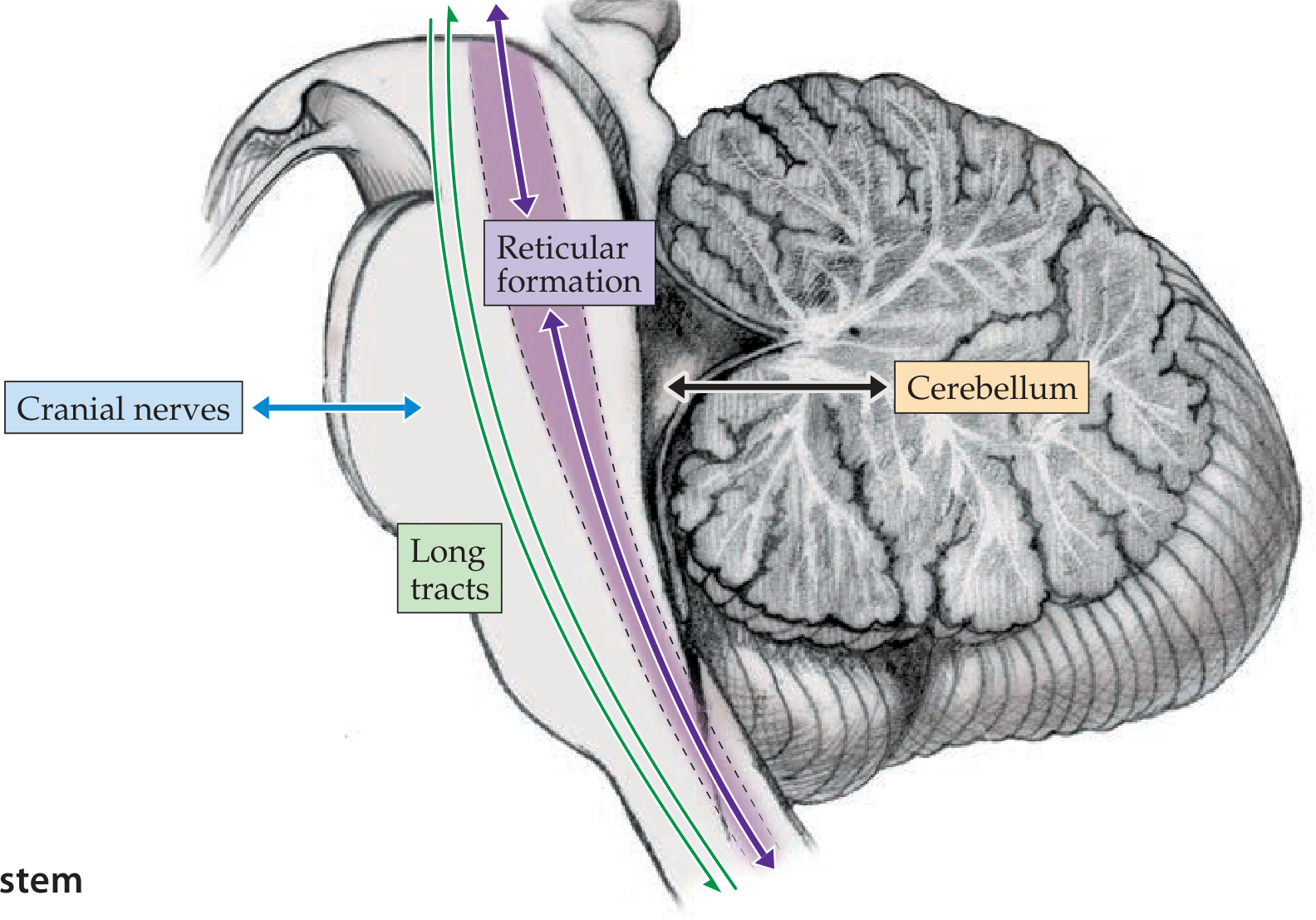
The reticular formation is a continuous core of nuclei embedded in the tegmentum, running the entire length of the brainstem. It merges rostrally with the diencephalon and caudally with the spinal cord intermediate zone.
Main components of the reticular formation:
Functional division:
- Rostral (midbrain/upper pons): works with diencephalon to maintain arousal and conscious state - the ascending reticular activating system (ARAS)
- Caudal (pons/medulla): controls respiration, cough, hiccup, sneeze, swallowing, nausea/vomiting, cardiovascular function, and sphincter control
Named nuclei within/related to reticular formation:
| System | Nucleus | Location | Neurotransmitter | Function |
|---|---|---|---|---|
| Cholinergic | Pedunculopontine tegmental nucleus | Caudal midbrain | Acetylcholine | Arousal, REM sleep, locomotion |
| Noradrenergic | Locus ceruleus | Dorsal pons | Norepinephrine | Attention, arousal, stress |
| Serotonergic | Raphe nuclei | Midline throughout | Serotonin | Sleep-wake, mood, pain modulation |
| Dopaminergic | Ventral tegmental area; substantia nigra | Midbrain | Dopamine | Reward, motor control |
| Pain modulation | Periaqueductal gray (PAG) | Midbrain | Multiple | Endogenous opioid pain control |
Cerebellar Peduncles
The cerebellum connects to the brainstem via three pairs of peduncles:
| Peduncle | Level | Main Contents |
|---|---|---|
| Superior (brachium conjunctivum) | Midbrain | Output from cerebellum to red nucleus and thalamus |
| Middle (brachium pontis) | Pons | Input from pontine nuclei (corticopontocerebellar pathway) |
| Inferior (restiform body) | Medulla | Input from spinal cord (spinocerebellar), inferior olive; output via juxtarestiform body to vestibular nuclei |
Long Tracts Passing Through
| Tract | Location | Function |
|---|---|---|
| Corticospinal tract | Basis (ventral) | Voluntary limb motor |
| Corticobulbar tract | Basis (ventral) | Voluntary motor to cranial nerve nuclei |
| Medial lemniscus | Tegmentum | Fine touch, vibration, proprioception (body); formed in medulla, migrates ventrally as it ascends |
| Anterolateral system | Lateral tegmentum | Pain, temperature, crude touch (body) |
| Trigeminothalamic tract | Lateral tegmentum | Pain/temperature from face |
| MLF | Dorsal tegmentum | Coordinates conjugate eye movements |
| Descending autonomic fibers | Lateral tegmentum | Sympathetic output to spinal cord (Horner syndrome if disrupted) |
Clinical Relevance Summary
Small brainstem lesions produce devastating deficits because all four functional categories are crowded into a tiny space. Classic signs include:
- Cranial nerve deficits (ipsilateral to lesion)
- Long-tract signs (contralateral hemiplegia or hemisensory loss)
- Ipsilateral Horner syndrome (descending sympathetic fibers in lateral tegmentum)
- Ataxia (cerebellar peduncle involvement)
- Impaired consciousness (reticular formation/ARAS damage)
The crossed pattern - ipsilateral face + contralateral body - is the hallmark of a lateral brainstem lesion (e.g., lateral medullary/Wallenberg syndrome from posterior inferior cerebellar artery occlusion).
Neuroanatomy through Clinical Cases, 3rd Ed. - Chapters 12 & 14 (Blumenfeld)
This is a shared conversation. Sign in to Orris to start your own chat.