
Givevme Rappid advance revision for DM Neurology student on Internal capsule
internal capsule anatomy MRI diagram showing anterior limb posterior limb genu
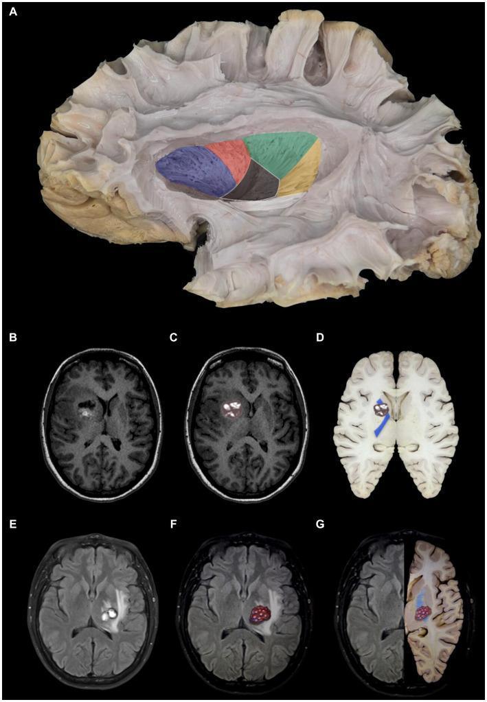
This composite educational graphic details the neuroanatomy of the internal capsule and its clinical correlation with cerebral cavernomas. Panel A features a gross anatomical white matter dissection (lateral view) with color-coded segmentation: anterior limb (blue), genu (red), posterior limb (green), retrolenticular portion (yellow), and sublenticular portion (white), alongside a partially resected lentiform nucleus. Panels B–D illustrate a cavernoma in the anterior limb of the internal capsule using axial T1-weighted MRI and anatomical overlays. The lesion demonstrates mixed signal intensity and is situated lateral to the caudate head and medial to the lentiform nucleus. Panels E–G depict a cavernoma in the posterior limb of the internal capsule via axial T2-weighted MRI and side-by-side anatomical comparisons. This lesion exhibits a heterogeneous, predominantly hyperintense signal and is localized medial to the lentiform nucleus and lateral to the thalamus. The collection serves to bridge gross anatomical segments of the internal capsule with diagnostic radiological findings and clinical pathology.
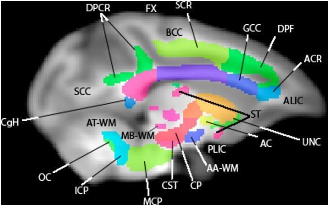
This diagnostic image shows a sagittal slice of a human brain white matter atlas overlaid on a T1-weighted MRI template. The visualization utilizes color-coded regions of interest (ROIs) to define specific neuroanatomical tracts. Superiorly, the corpus callosum is segmented into the green genu (GCC), light-green body (BCC), and pink splenium (SCC). The internal capsule is delineated by the light-blue anterior limb (ALIC) and the dark-blue posterior limb (PLIC). Other prominent structures include the green corticospinal tract (CST) descending through the brainstem, the cerebral peduncle (CP) in pink, and the middle cerebellar peduncle (MCP) in light green. Deep structures such as the fornix (FX), stria terminalis (ST), and anterior commissure (AC) are also identified. This anatomical diagram serves as a reference for fiber tract orientation and localization, demonstrating the relationship between major commissural, projection, and association fibers in the rhesus macaque or human-approximate brain for educational and research-based neuroimaging analysis.
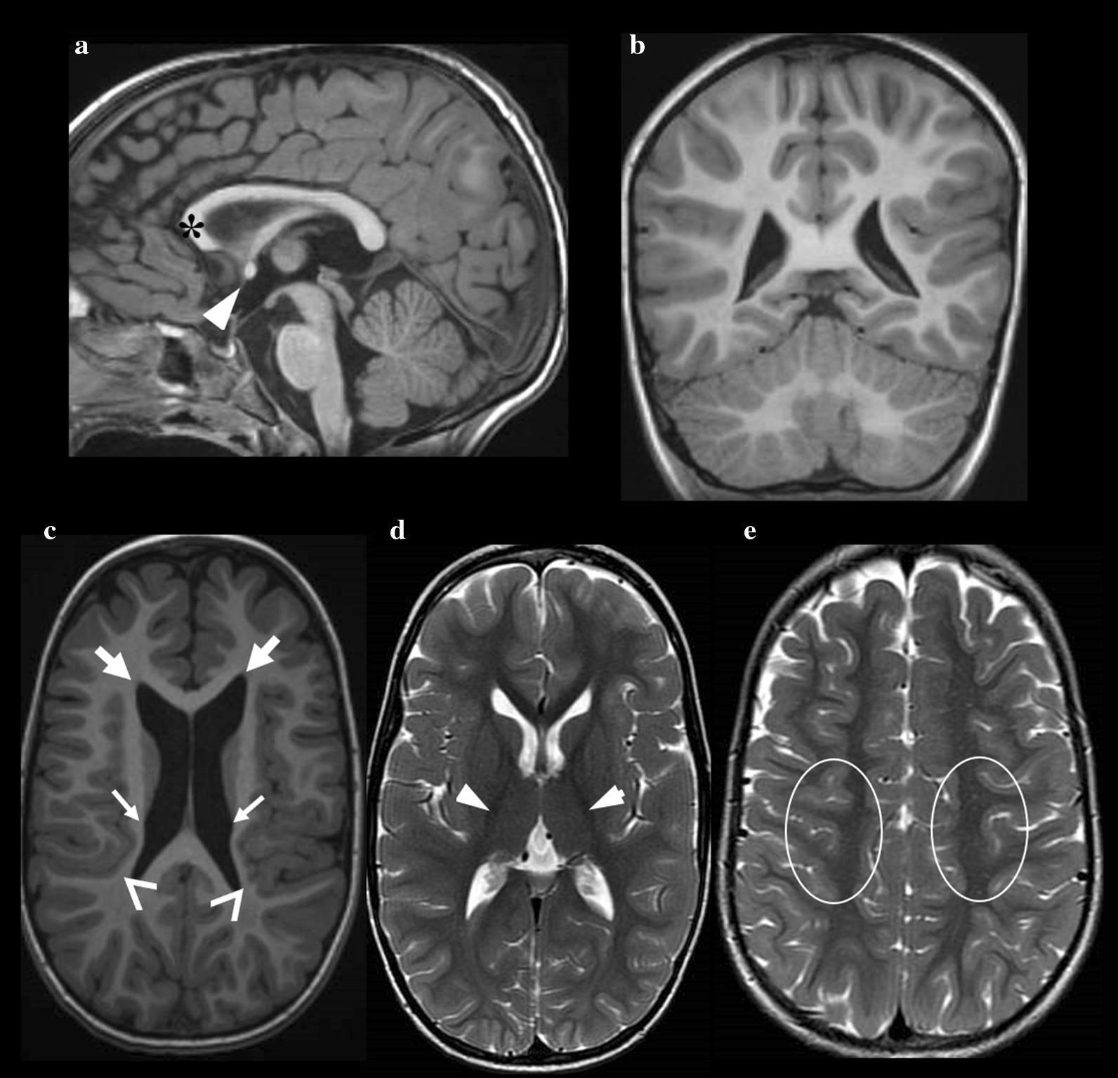
This composite of brain MRI scans demonstrates several diagnostic features associated with a pediatric neurodevelopmental condition. The sagittal T1-weighted image (a) reveals a thinning of the genu of the corpus callosum (asterisk) and relative hypertrophy of the anterior commissure (white arrowhead). A scaphocephalic skull morphology is also visible. Sagittal and coronal T1-weighted views (a, b) show a normal cerebellum. The axial T1-weighted image (c) highlights dysmorphic lateral ventricles, specifically showing dilatation of the frontal horns (thick arrows) and irregular, scalloped ventricular walls (thin arrows). A reduction in insulo-temporal white matter volume is also marked (chevron). In contrast, axial T2-weighted images (d, e) demonstrate preserved signal intensity within the white matter of the posterior limb of the internal capsule (white arrowheads) and the paracentral region (ovals). These findings are characteristic of neuroradiological manifestations in specific types of Congenital Disorders of Glycosylation (CDG), illustrating structural brain anomalies despite relatively preserved white matter signal.
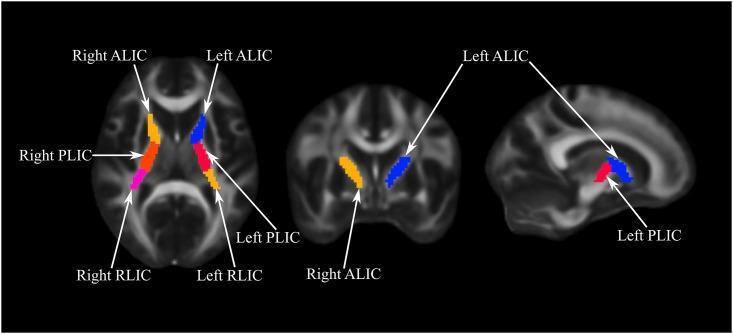
This diagnostic neuroimaging figure displays three planes of a brain MRI—axial (left), coronal (middle), and sagittal (right)—highlighting the sub-regions of the internal capsule using color-coded Regions of Interest (ROIs). The image serves as an anatomical guide for white matter localization. In the axial view, the internal capsule is segmented into the Anterior Limb (ALIC), Posterior Limb (PLIC), and Retrolenticular Limb (RLIC). On the right hemisphere, these are color-coded yellow/orange, red/orange, and magenta, respectively. On the left hemisphere, the ALIC is colored blue and the PLIC is colored red. The coronal and sagittal views further demonstrate the 3D spatial orientation: the ALIC is situated anteriorly and superiorly relative to the PLIC. These ROIs are mapped onto a grayscale structural template, likely for Fractional Anisotropy (FA) or Mean Diffusivity (MD) analysis in psychiatric or neurological research. Key landmarks visible include the ventricles and the thalamic/striatal boundaries surrounding the V-shaped internal capsule structure.
pure motor hemiplegia lacunar infarct posterior limb internal capsule MRI
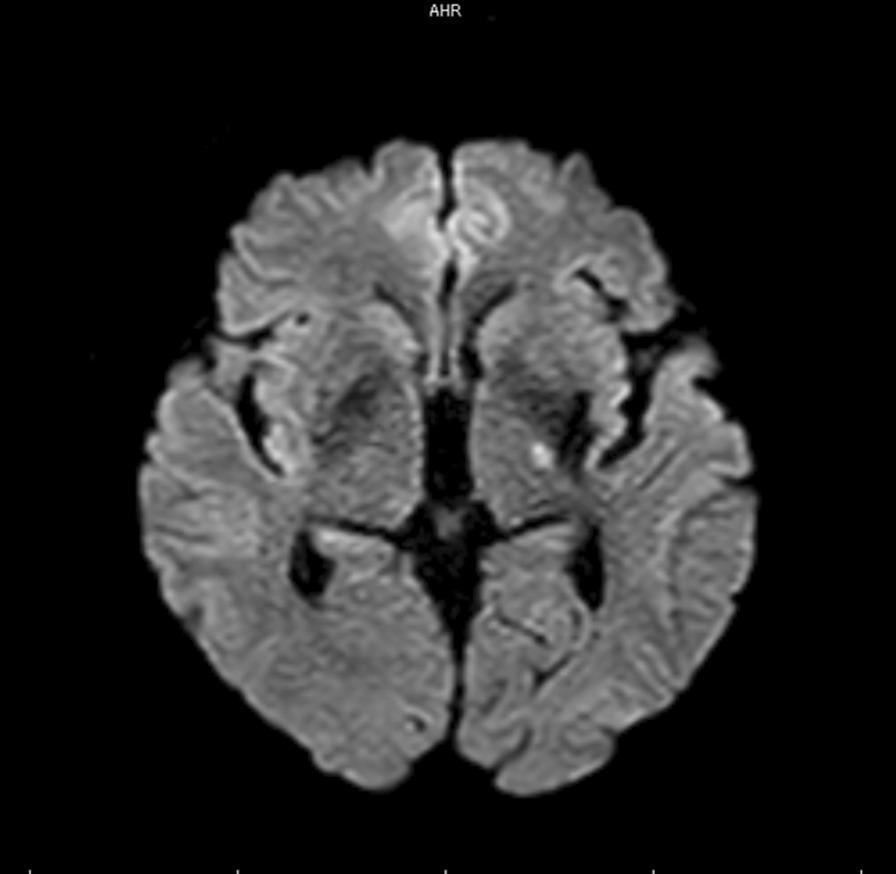
This diagnostic image is an axial section of a brain using Magnetic Resonance Imaging (MRI), specifically Diffusion-Weighted Imaging (DWI). The scan demonstrates a focal area of restricted diffusion, appearing as a hyperintense (bright) signal located in the left posterior limb of the internal capsule. This finding is characteristic of an acute lacunar infarct, specifically attributed to an occlusion of a lenticulostriate artery (LSA) perforator branch. The anatomical region shown includes the basal ganglia and thalamus, with the hyperintensity clearly demarcated against the surrounding normointense parenchyma. This visual evidence correlates with clinical presentations of acute stroke, such as hemiparesis or sensory deficits. The image serves as a key educational example of neuroimaging in acute ischemic stroke, illustrating the sensitivity of DWI sequences in detecting small-vessel perforator territory infarctions shortly after symptom onset.
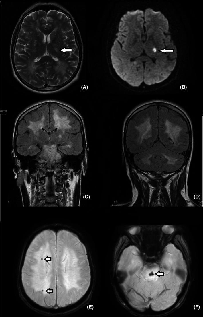
Six-panel brain MRI study demonstrating acute and chronic neurovascular findings. (A) Axial T2-weighted and (B) Diffusion-Weighted Imaging (DWI) show a focal hyperintense lesion (arrows) in the posterior limb of the left internal capsule, diagnostic of an acute lacunar ischemic stroke. (C, D) Coronal Fluid-Attenuated Inversion Recovery (FLAIR) images reveal extensive, confluent white matter hyperintensities (WMH) involving both supratentorial and infratentorial regions, representing severe small vessel disease or leukoaraiosis. (E, F) Susceptibility-Weighted Imaging (SWI) sequences identify multifocal hypointense 'blooming' artifacts (arrows) located in the centrum semiovale and the midline of the pons. These signal voids are consistent with chronic hemosiderin deposits or microbleeds. The combination of findings—acute lacunar infarct, diffuse leukoencephalopathy, and microhemorrhages—is highly characteristic of cerebral small vessel disease and hypertensive vasculopathy.
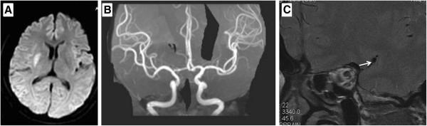
This composite diagnostic image illustrates clinical findings in a patient with a cerebral vascular event. Panel A is an axial MRI Diffusion-Weighted Image (DWI) showing a focal, hyperintense (bright) lesion in the region of the right posterior limb of the internal capsule, consistent with an acute lacunar infarct. Panel B displays a Magnetic Resonance Angiogram (MRA) of the Circle of Willis, which shows no gross occlusion or high-grade stenosis in the major branches, including the middle cerebral arteries (MCA). Panel C is a high-resolution MRI (HR-MRI) focusing on the proximal segment of the right MCA. A white arrow highlights an eccentric, T2-weighted hypointense thickening on the arterial wall, identified as an atherosclerotic plaque. This image series demonstrates how HR-MRI can detect vulnerable or non-stenotic atherosclerotic plaques as an underlying etiology for ischemic stroke when conventional MRA appears normal. The content is suitable for intermediate to advanced neurology and radiology trainees.
INTERNAL CAPSULE - Rapid Advance Revision for DM Neurology
1. OVERVIEW & DEFINITION
"The internal capsule, a compact lamina of white matter, representing the largest projection fiber tract, contains afferent and efferent nerve fibers passing to and from the brainstem to the cerebral hemispheres." - Localization in Clinical Neurology, 8e
2. ANATOMY & SPATIAL RELATIONSHIPS
| Medial to IC | Lateral to IC |
|---|---|
| Caudate nucleus (always) | Lentiform nucleus - Putamen + Globus pallidus (always) |
| Thalamus (always) |
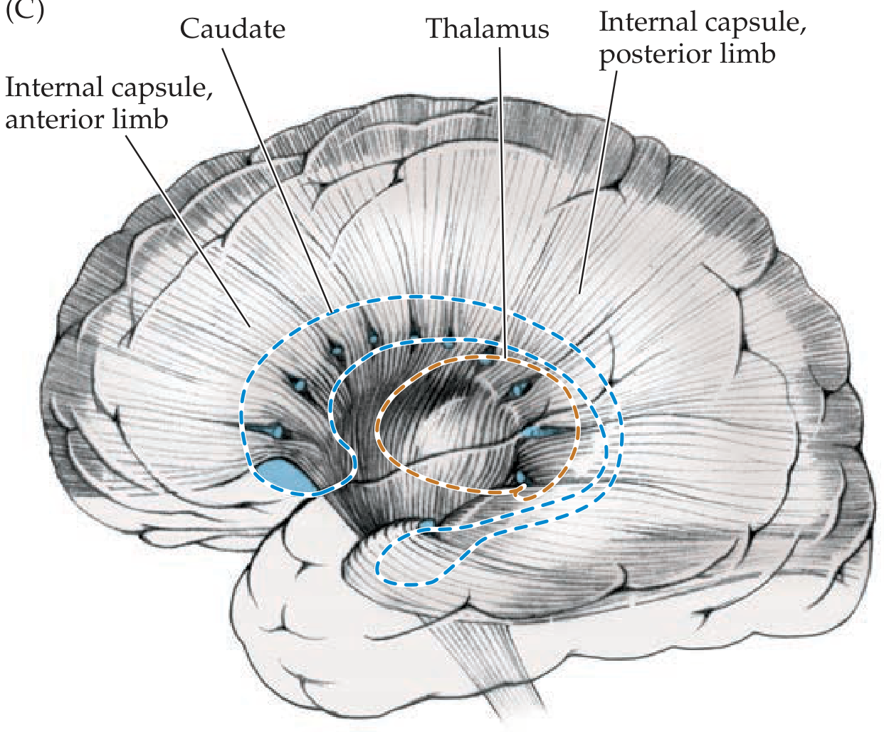
3. THE FIVE SEGMENTS - Anatomy and Fiber Contents
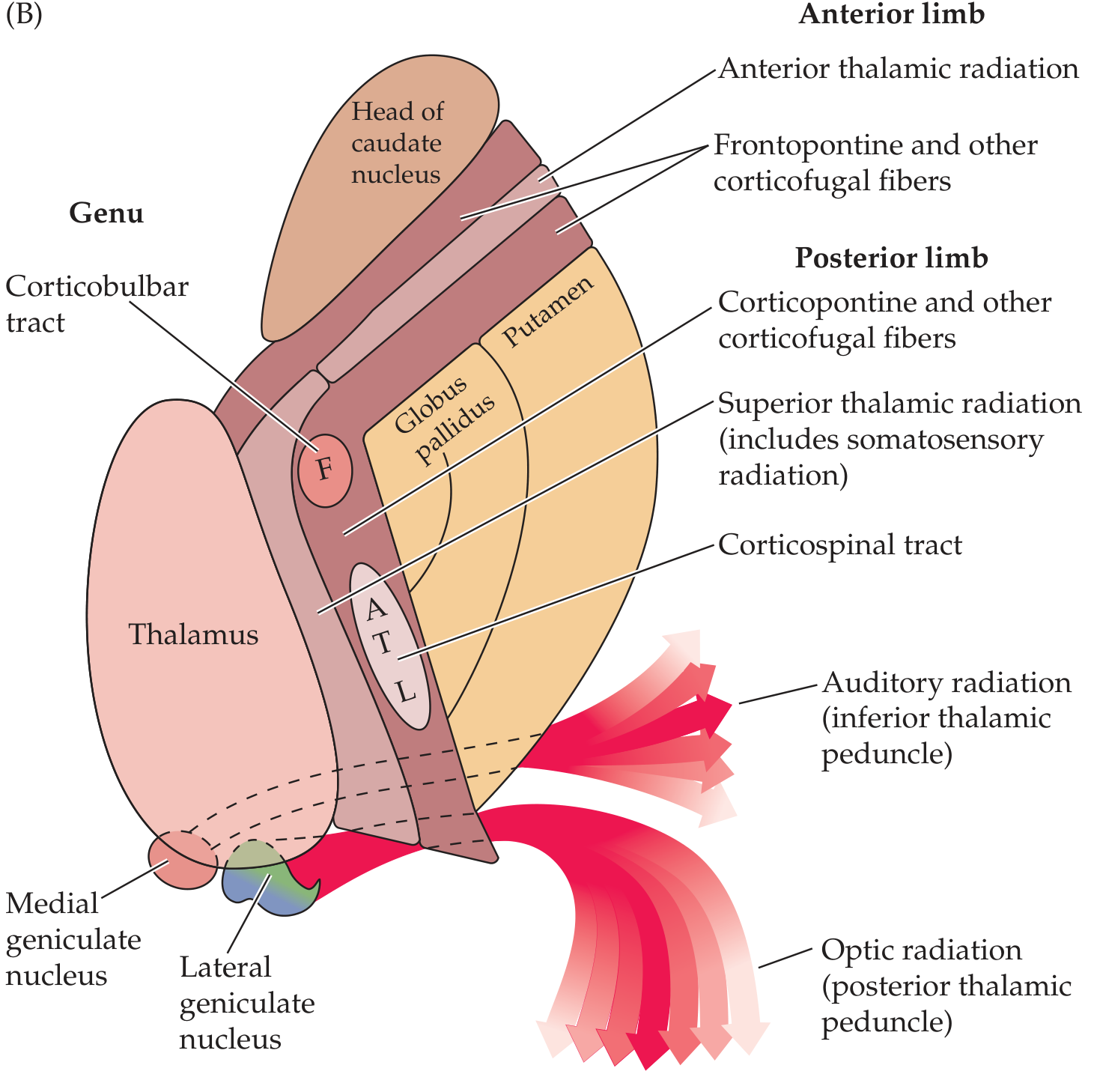
A. ANTERIOR LIMB (lenticulocaudate segment, ~2 cm)
- Separates lentiform nucleus (lateral) from caudate head (medial)
- Fibers:
- Frontopontine fibers (corticopontine from frontal lobe)
- Anterior thalamic radiation (connecting frontal lobe and anterior/dorsomedial thalamic nuclei)
- Corticothalamic and thalamocortical fibers (frontal-thalamus reciprocal)
- Caudate-putaminal fibers
B. GENU ("knee," at level of foramen of Monro)
- Transition between anterior and posterior limbs
- Prominence of the curve projects between caudate and thalamus
- Fibers:
- Corticobulbar tract (motor fibers to cranial nerve nuclei in brainstem)
- Corticoreticular fibers (descend to both sides of brainstem reticular formation)
- Motor corticopontine fibers (perhaps)
Genu lesion = Capsular genu syndrome: facial and lingual hemiparesis with MILD limb involvement (corticobulbar-dominant picture)
C. POSTERIOR LIMB (lenticulothalamic segment, ~3-4 cm)
- Separates lentiform nucleus (lateral) from thalamus (medial)
- Fibers (anterior to posterior within posterior limb):
- Corticopontine and other corticofugal fibers (anterior)
- Superior thalamic radiation (includes somatosensory radiation - VPL → parietal cortex)
- Corticospinal tract (CST) - main motor pathway
- Corticoreticular fibers
- Corticorubral fibers
- Corticothalamic fibers
Face (F) → Arm (A) → Trunk (T) → Leg (L)
D. RETROLENTICULAR SEGMENT (posterior to lentiform nucleus)
- Posterior thalamic radiation (visual fibers)
- Optic radiation (from LGN to primary visual cortex V1 via Meyer's loop)
- Parietooccipital and occipitotemporal corticopontine fibers
E. SUBLENTICULAR SEGMENT (below lentiform nucleus)
- Auditory radiation (inferior thalamic peduncle, from MGB to Heschl's gyrus)
- Temporopontine fibers
- Anterior part of optic radiation
4. FIBER CONTENT - QUICK SUMMARY TABLE
| Segment | Key Fibers | Clinical Relevance |
|---|---|---|
| Anterior limb | Frontopontine, anterior thalamic radiation | Frontal lobe-thalamus circuit |
| Genu | Corticobulbar, corticoreticular | Face/tongue paresis |
| Posterior limb | CST (F-A-T-L), somatosensory radiation, corticopontine | Pure motor hemiplegia |
| Retrolenticular | Optic radiation, posterior thalamic radiation | Homonymous hemianopia |
| Sublenticular | Auditory radiation | Contralateral auditory deficit |
5. BLOOD SUPPLY
| Segment | Artery |
|---|---|
| Anterior limb | Lateral lenticulostriate arteries (from MCA) + Recurrent artery of Heubner (ACA) |
| Genu | Anterior choroidal artery (AChA) + lenticulostriate arteries |
| Posterior limb (anterior 2/3) | Lateral lenticulostriate arteries (MCA branches) |
| Posterior limb (posterior 1/3) | Anterior choroidal artery (AChA) |
| Retrolenticular | Anterior choroidal artery |
| Sublenticular | Anterior choroidal artery |
6. CLINICAL SYNDROMES
6A. POSTERIOR LIMB LESIONS (most common)
- Complete contralateral face + arm + leg weakness, NO sensory loss, NO cortical signs
- Most common lacunar syndrome (~50%)
- Caused by small infarct in posterior limb or basis pontis
- Lesion of CST at a level where all fibers are packed tightly together
- Prognosis: generally good
- Contralateral hemiparesis + hemisensory loss
- Posterior limb (posterior portion) - CST + somatosensory radiation involved together
6B. ANTERIOR CHOROIDAL ARTERY (AChA) OCCLUSION SYNDROME
- Hemiplegia (posterior limb - CST)
- Hemisensory loss (posterior limb - somatosensory radiation)
- Homonymous hemianopia (retrolenticular/sublenticular - optic radiation)
"Lateral and Posterior IC lesion: Contralateral hemiparesis, diminished pain and temperature, dysarthria, homonymous hemianopia usually with a tongue of sparing along the horizontal meridian, memory impairment; with right-sided lesions - visuoperceptual abnormalities" - Bradley and Daroff's Neurology
6C. GENU SYNDROME (Capsular Genu Syndrome)
- Facial and lingual hemiparesis (corticobulbar predominant)
- Dysarthria, contralateral lower facial weakness
- Mild limb involvement (corticospinal largely spared)
- Blood supply: AChA and lenticulostriate branches
6D. INTERNAL CAPSULAR HEMORRHAGE
- Most common cause: hypertension (lenticulostriate artery rupture)
- Small hemorrhages in genu or posterior limb → PMH or mixed sensorimotor syndrome
- Large hemorrhages → dense contralateral hemiplegia ± hemisensory loss ± hemianopia
- Putaminal hemorrhage typically extends to involve IC (most common site of hypertensive ICH)
- Decorticate rigidity (flexion of arm, extension of leg) after IC hemorrhage - small arteries of IC especially prone to rupture or thrombotic occlusion in hypertension
- Bilateral posterior limb hemorrhages (rare) → paraparesis
7. IMAGING
MRI of Posterior Limb Internal Capsule - Acute Lacunar Infarct
Internal Capsule Segments on MRI (Axial)
IC Anatomy with Cavernoma - Color Dissection
8. LACUNAR SYNDROMES LINKED TO IC
| Lacunar Syndrome | Location | Key Features |
|---|---|---|
| Pure Motor Hemiplegia | Posterior limb IC or pons | Face + arm + leg weakness, no sensory, no cortical signs |
| Mixed Sensorimotor | Posterior limb IC (posterior) | Hemiparesis + hemisensory loss |
| Dysarthria-Clumsy Hand | Genu IC or pons | Dysarthria + ipsilateral hand clumsiness |
| Ataxic Hemiparesis | Posterior limb IC or pons | Mild hemiparesis + ipsilateral cerebellar ataxia |
| Pure Sensory Stroke | Thalamus (VPL) NOT IC | Hemisensory only |
Note: A contralateral supratentorial stroke (e.g., thalamic extending into posterior limb of IC, or stroke restricted to posterior limb of IC) may mimic a contralateral cerebellar infarct by producing ipsilateral limb ataxia - Localization in Clinical Neurology, 8e
9. KEY EXAM POINTS - HIGH YIELD
-
Five segments: Anterior limb - Genu - Posterior limb - Retrolenticular - Sublenticular (anterior to posterior/inferior)
-
Corticobulbar = Genu; Corticospinal = Posterior limb
-
Somatotopy in posterior limb: Face → Arm → Trunk → Leg (anterior to posterior) - "FATL"
-
AChA occlusion = hemiplegia + hemisensory + hemianopia (classic triad)
-
Lenticulostriate arteries supply anterior limb + most of posterior limb (MCA branches); AChA supplies the posterior third of posterior limb + retrolenticular + sublenticular segments
-
Decorticate posturing = lesion at or above red nucleus (IC/internal capsule level or above) - arm flexion, leg extension
-
Capsular genu syndrome = predominantly facial/lingual paresis with mild limb weakness (corticobulbar > corticospinal)
-
Optic radiation passes through retrolenticular segment (→ homonymous hemianopia with IC lesions)
-
Auditory radiation passes through sublenticular segment
-
Pure motor hemiplegia - most common lacunar syndrome, most often at posterior limb IC or basis pontis. Distinguished from cortical stroke by ABSENCE of cortical signs (no aphasia, no neglect, no hemianopia, no agnosia).
-
Small perforating arteries of IC are highly susceptible to hypertension - most common site of hypertensive ICH is putamen/IC region.