
If i pull my hair frontal part Am getting around 10 hair follicles onto my hand and i noticed am having hair fall more these days Is it normal or any abnormal Explain hair fall stages
hair pull test normal abnormal how many hairs is normal hair fall stages anagen telogen catagen
hair growth cycle stages anagen catagen telogen diagram follicle
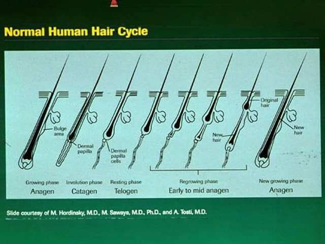
This educational diagram depicts the Normal Human Hair Cycle in a longitudinal schematic of a pilosebaceous unit from the scalp. Modality: Educational illustration; not an in vivo imaging study. Technique: Digital vector illustration with color-coded phases to aid rapid recognition. Anatomy shown includes the scalp hair follicle, hair shaft, hair bulb, dermal papilla, outer root sheath, bulge region, and associated sebaceous gland. Phases represented are Anagen (growth phase) with active matrix proliferation and elongation of the hair shaft; Catagen (transitional phase) with follicular involution and brief regression; Telogen (resting phase) with a quiescent follicle and club hair waiting for shedding; and Exogen (shedding phase) illustrating release of a mature hair and initiation of a new anagen cycle. Notable features emphasize the continuous cyclic renewal of the pilosebaceous unit, directional Bow-like angles of hair emanation, and the temporal sequence that governs production of long continuous hair versus short sparse hair. Clinical significance includes the ability to explain common hair disorders: telogen effluvium from premature shift to telogen, androgenetic alopecia from shortened anagen duration, and anagen effluvium from abrupt matrix disruption. Contextual applications span medical education, patient counseling, and research into hair biology, growth factors, and therapeutic interventions aimed at prolonging anagen or modulating cycle duration.
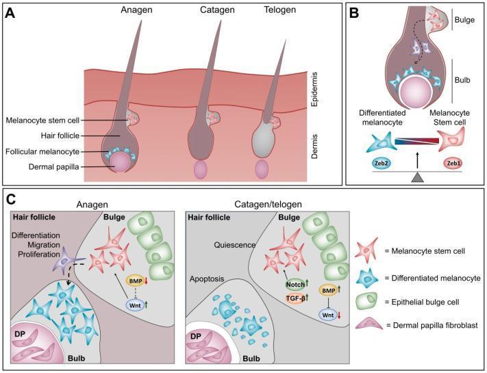
This medical illustration depicts the hair follicle cycle and the molecular mechanisms governing melanocyte homeostasis across three panels. Panel A shows the morphological progression of the hair follicle through the anagen (growth), catagen (regression), and telogen (resting) phases, highlighting the migration of melanocyte stem cells and the presence of follicular melanocytes in the hair bulb. Panel B demonstrates the cellular differentiation gradient within the hair follicle, where ZEB1 expression is associated with quiescent melanocyte stem cells in the bulge, and ZEB2 expression drives differentiation into mature, pigment-producing melanocytes in the bulb. Panel C details the micro-environmental signaling cues. During anagen, low BMP and high Wnt signaling promote stem cell proliferation, migration, and differentiation. In contrast, the catagen/telogen phases are characterized by high BMP, Notch, and TGF-beta signaling alongside low Wnt activity, leading to stem cell quiescence and follicular melanocyte apoptosis. This schematic serves as an educational resource for understanding the pathophysiology of hair pigmentation and the role of the ZEB transcription factor family in dermatological science.
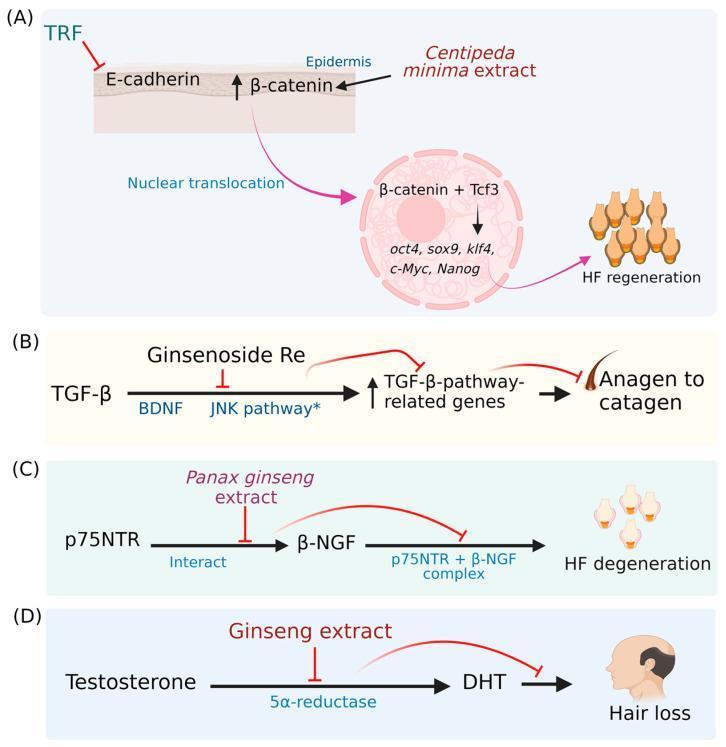
This pathophysiology diagram illustrates four distinct molecular mechanisms (A-D) through which various phytochemicals influence the hair growth cycle and prevent hair loss. Panel A focuses on the Wnt/β-catenin pathway: Tocotrienol-rich fraction (TRF) inhibits E-cadherin, while Centipeda minima extract activates β-catenin, facilitating its nuclear translocation and binding with Tcf3 to induce transcription factors (oct4, sox9, klf4, c-Myc, Nanog) for hair follicle (HF) regeneration. Panel B shows the TGF-β pathway: Ginsenoside Re inhibits the activation of TGF-β-pathway-related genes (stimulated by BDNF and JNK), thereby preventing the transition from anagen to catagen phase. Panel C depicts the neurotrophin pathway: Panax ginseng extract inhibits the interaction between p75NTR and β-NGF, preventing the formation of a complex that leads to HF degeneration. Panel D illustrates the androgenic pathway: Ginseng extract inhibits the enzyme 5α-reductase, blocking the conversion of testosterone to dihydrotestosterone (DHT) to mitigate hair loss. The diagram uses standard notation where arrows signify activation and T-bars signify inhibition.
Is Pulling Out ~10 Hairs Normal?
| Threshold | Interpretation |
|---|---|
| 2 or fewer hairs | Normal (2017 evidence-based guideline, McDonald et al., JAAD) |
| Up to 10% of grasped hairs (5-6 hairs) | Borderline - warrants monitoring |
| More than 10% (>6 hairs) | Positive - active hair shedding (abnormal) |
The 4 Stages of the Hair Growth Cycle
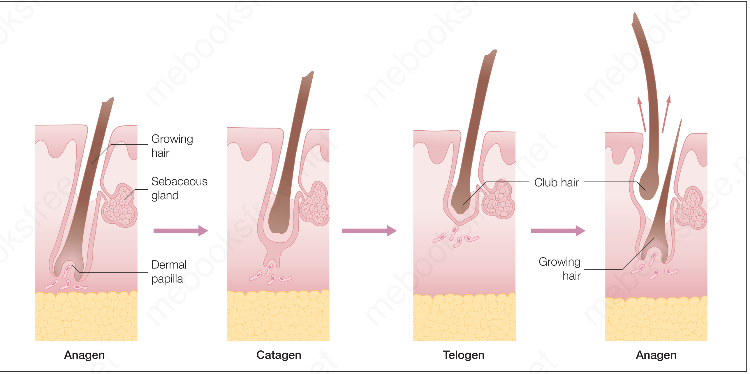
Stage 1 - Anagen (Growth Phase)
- Duration: 2-7 years on the scalp
- What happens: The follicle is fully active. The hair bulb produces new cells, the dermal papilla is engaged, and the hair shaft grows actively at about 0.37-0.4 mm/day (roughly 1 cm/month).
- How many hairs: Normally 85-90% of all scalp hairs are in anagen at any given time. This is what allows your hair to look full and long.
- Key fact: The longer the anagen phase, the longer the hair can grow. This phase shortens with age and in androgenetic (pattern) hair loss.
- Source: Andrews' Diseases of the Skin; Goldman-Cecil Medicine
Stage 2 - Catagen (Transition Phase)
- Duration: About 2-3 weeks
- What happens: Cell division in the hair matrix stops. The follicle shrinks and regresses. The dermal papilla separates from the hair bulb and moves upward. The hair detaches from its blood supply but stays in the follicle.
- How many hairs: Only about 1-2% of hairs are in catagen at any moment.
- Histology tip: Catagen hairs show many apoptotic (dying) cells in the outer root sheath. - Andrews' Diseases of the Skin
Stage 3 - Telogen (Resting Phase)
- Duration: About 3-5 months on the scalp
- What happens: The follicle is completely at rest. The old hair becomes a club hair (non-pigmented, with a shaggy lower border - visible as a white bulb when it falls). A new anagen hair begins forming beneath it.
- How many hairs: Normally 10-15% of scalp hairs are in telogen at any time.
- Daily shedding: Since these club hairs eventually shed, losing 50-100 hairs/day is considered normal.
- Source: Goldman-Cecil Medicine, Andrews' Diseases of the Skin
Stage 4 - Exogen (Shedding Phase)
- Duration: Overlaps with late telogen / early new anagen
- What happens: The old telogen club hair is actively shed (released from the follicle) as the new anagen hair pushes up from beneath. This is the phase where the hair physically falls out.
- Normal daily loss: 50-100 hairs/day
- The cycle then restarts at Anagen.
What Could Be Causing Your Increased Hair Fall?
| Cause | Details |
|---|---|
| Telogen Effluvium | Most common - triggered by fever/illness, surgery, stress, significant weight loss, or nutritional deficiency. A large batch of hairs shift into telogen at once, then shed 2-4 months later. |
| Androgenetic Alopecia | Genetic pattern hair loss; anagen phase progressively shortens. Frontal region is typically first affected in men. |
| Nutritional Deficiency | Iron, zinc, biotin, vitamin D deficiencies are common culprits. |
| Hormonal Changes | Thyroid disorders, post-pregnancy, or hormonal imbalance. |
| Alopecia Areata | Autoimmune patchy hair loss; pull test may show "pencil point" tapered hairs. |
| Medications | Many drugs (blood thinners, chemotherapy, some antihypertensives) can cause anagen or telogen effluvium. |
What You Should Do
- See a dermatologist - A pull test done properly (in 3 areas: front, crown, temporal), along with a scalp examination, is the first step.
- Blood tests often ordered: CBC, ferritin (iron stores), thyroid function (TSH, T3/T4), vitamin D, zinc, hormonal profile.
- Track the pattern - Is it diffuse all over, or mostly frontal/temporal? Is it in patches? This helps narrow the cause.
- Do not pull or tug your hair repeatedly as a self-test - this can itself accelerate shedding.